


INTRODUCTION
Primary ocular complaints prompted nearly two million emergency department (ED) visits in the United States in 2010, comprising approximately 1.5% of total ED visits.Reference Channa, Zafar and Canner1 Acute loss of vision is alarming for the patient and can represent an emergent condition requiring prompt subspecialty treatment. For many ocular conditions, accurate diagnosis depends on obtaining a detailed clinical history and performing a thorough physical examination, which can be challenging in a busy ED. Many physicians are uncomfortable or unskilled with ophthalmoscopy, and prompt ophthalmology evaluation may not be possible in some regions.Reference Mackay, Garza, Bruce, Newman and Biousse2 We present a case in which point-of-care ultrasound (POCUS) was used as an adjunct during initial evaluation to diagnose a submacular hemorrhage, an uncommon cause of acute painless vision loss.
CASE
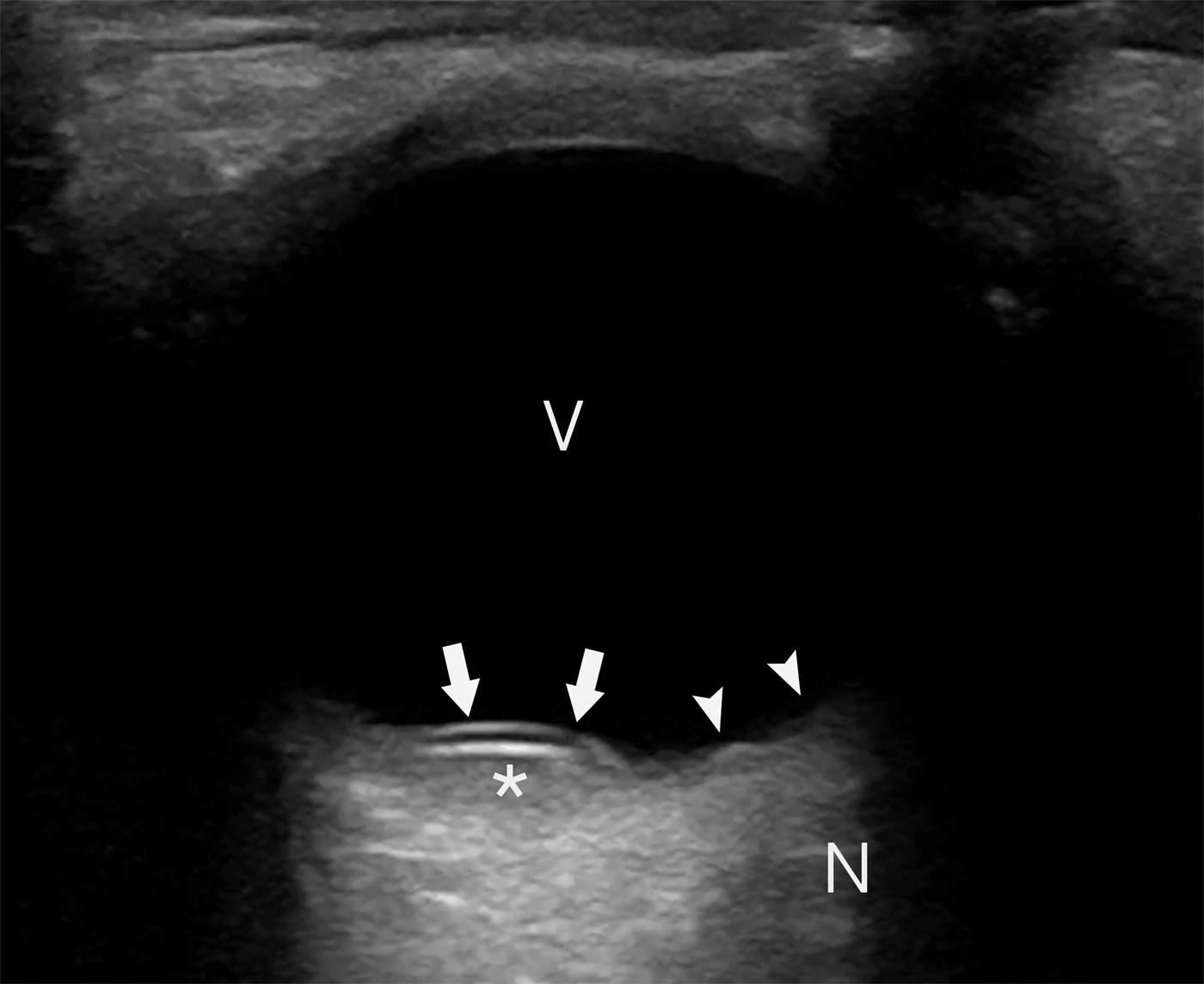
A 43-year-old, left-handed man presented with approximately 24 hours of right eye vision loss. He noticed an acute onset of blurry vision, as well as a “red cloud,” that appeared after waking up at 4:00 a.m. and brushing his teeth. There was no precipitating trauma, infection, or other event that he could recall. He had decreased vision in the right eye and was unable to make out any movement. He denied headache, dizziness, lightheadedness, loss of consciousness, extremity weakness, or extremity numbness. His exam was notable for symmetric and reactive pupils, without an afferent or efferent pupillary defect. Vision was 20/200 in the right eye and 20/30 in the left eye. He had no history of previous vision deficits or need for corrective lenses. Bedside ocular ultrasound was performed, and a still image from the clip is shown below (Figure 1) that revealed a non-mobile hyperechoic lesion along the posterior retina that did not follow the normal contour of the posterior chamber. There was a mixed echogenic signal posterior to the lesion concerning for hemorrhage. The identified lesion did not appear to involve the optic nerve. No mobile retinal flap or posterior chamber debris was identified. Ophthalmology was consulted, and a dilated fundoscopic exam was performed, confirming the diagnosis of a large submacular hemorrhage (SMH) in addition to scattered smaller retinal hemorrhages. A hypercoagulable workup was initiated given concern for a thromboembolic event such as a central retinal vein occlusion as the possible etiology of this localized hemorrhage. The patient was referred to the subspecialty retina clinic. The hypercoagulable workup was non-contributory. While conservative management was initially recommended, the central vision did not improve, and a vitrectomy with possible internal limiting membrane peeling was scheduled.
Ocular ultrasound showing the vitreous (V), optic nerve (N), retina (arrowheads), and non-mobile retinal contour abnormality (arrows). Also noted is a mixed echogenic material (asterisk) posterior to the retinal lesion indicating the presence of hemorrhage.
DISCUSSION
SMH is a vision-threatening condition that can occur spontaneously and result in significant morbidity even after treatment is initiated.Reference Hochman, Seery and Zarbin3 This condition is because of the accumulation of blood between the retinal layers and can arise from the choroid or retinal blood supply.Reference Hochman, Seery and Zarbin3 Non-traumatic SMH can be related to age-related macular degeneration, but it may also be related to arterial microaneurysms, trauma, or other primary ophthalmological pathologies.Reference Hochman, Seery and Zarbin3,Reference Ozkaya, Erdogan and Tarakcioglu4 Patients typically present with an acute onset of blurry vision, decreased visual acuity, or scotomas.Reference Hochman, Seery and Zarbin3 Currently, there is no gold standard for treatment of this condition, with options including tissue plasminogen activator, anti-vascular endothelial growth factor, and pneumatic decompression.Reference Hochman, Seery and Zarbin3,Reference Ozkaya, Erdogan and Tarakcioglu4
The differential diagnosis for acute painless vision loss includes central retinal artery occlusion, central retinal vein occlusion, and retinal detachment. This case presentation is not entirely consistent with those diagnoses. A fundoscopic exam is a valuable component of the initial diagnostic workup for acute non-traumatic vision loss. However, this procedure is difficult to learn and even more difficult to employ when not consistently practised. One study of medical students, residents, and faculty physicians showed that confidence in performing an appropriate fundoscopic exam was poor across all levels including faculty.Reference Wu, Fagan, Reinert and Diaz5 Furthermore, performing a non-dilated exam can severely limit a physician's ability to identify and interpret ocular findings. In one study of 500 patients, 38% of conditions that required additional treatment or intervention were not evident on non-dilated exams.Reference Siegel, Thompson, Yolton, Reinke and Yolton6
POCUS in the ED is a rapidly growing diagnostic modality in both adult and pediatric settings.Reference Marin, Abo and Arroyo7,Reference Blaivas, Theodoro and Sierzenski8 The normal anatomy of the eye is easily identifiable with ultrasound (Figure 2), and the pathology shown in this case can be easily identified (Figure 1). Ocular ultrasound is used in the point-of-care setting to assess the posterior chamber, and studies have shown that POCUS after minimal training has sensitivities and specificities for retinal detachment of 91–100% and 83–96%, respectively.Reference Lahham, Shniter and Thompson9–Reference Shinar, Chan and Orlinsky12 Traditional evaluation and diagnosis of SMH relies on a direct fundoscopic exam, but, as previously shown, this can be difficult to perform. This case is an example of the ability of POCUS to diagnose less common ocular pathologies and direct further workup.
Normal ocular exam showing the vitreous (V), optic nerve (N), retina (arrowheads), lens (L), iris (chevrons), anterior chamber (A), and cornea (arrow).

Guidelines published by both the Canadian Association of Emergency Physicians (CAEP) and the American College of Emergency Physicians (ACEP) state that the scope of practice for POCUS by emergency physicians includes ocular imaging.Reference Henneberry, Hanson and Healey13,14 Furthermore, the American College of Graduate Medical Education (ACGME) mandates POCUS education for all emergency medicine residents. This case highlights the use of ocular POCUS to reveal a rare condition that requires urgent ophthalmology follow-up yet is traditionally difficult to diagnose by non-specialty trained physicians.
CONCLUSION
As the use of POCUS increases, emergency providers should seek to become comfortable with its various uses, including ocular evaluation. Ocular POCUS is a valuable diagnostic tool in the ED, and this case highlights the ability of this modality to identify a condition that requires urgent ophthalmological consultation and management, in addition to expanding the growing body of literature showing that POCUS can identify ophthalmologic conditions not commonly considered sonographic diagnoses.
Competing interests
This research received no specific grant from any funding agency, commercial, or not-for-profit sectors. None declared.


