

Introduction
Jungite was first characterised as a new secondary phosphate mineral from the Hagendorf Süd pegmatite, Bavaria, by Moore and Ito (Reference Moore and Ito1980). They gave the formula as [Ca2(H2O)6][Zn4Fe3+8(PO4)9(OH)9]·10H2O, and from single crystal diffraction studies they reported the symmetry as orthorhombic, with possible space groups Pcmm, Pcm21 or Pc2m and with unit-cell parameters a = 11.98, b = 20.17, c = 9.95 Å and Z = 2. The authors noted that the diffraction patterns showed pronounced streaking parallel to b* and suggested that the mineral had a layer structure, parallel to (010), with stacking fault defects. Despite intensive studies on ∼20 different crystals, they were unable to find a crystal that diffracted cleanly enough to obtain data for a structure analysis. They reported the powder diffraction pattern but were unable to obtain a satisfactory indexing of the diffraction lines based on their unit cell.
In an effort to resolve the crystal structure, we took jungite crystals from the type locality to the Australian Synchrotron microfocus beamline MX2 to obtain diffraction data. The diffraction data for several crystals indexed with an orthorhombic cell, 12.0 × 24.7 × 9.9 Å, having the same a and c parameters as the Moore cell but with the b parameter increased by ∼20%. A structure solution was readily obtained in space group Aba2 using SHELXT (Sheldrick, Reference Sheldrick2015), which showed that the structure comprised (010) heteropolyhedral double layers composed of ZnO4 and PO4 tetrahedra and edge-shared Fe2O10 octahedral dimers, with hydrated Ca-centred polyhedra located between the layers.
Indexing of the diffraction data for other crystals from the same specimen consistently gave a triclinic cell, with a ≈ 11.95, b = 10.05, c = 9.9 Å, α ≈ 86.7, β =89.9 and γ = 83.4° which approximates to the Moore and Ito (Reference Moore and Ito1980) cell parameters with a halved b parameter. The diffraction patterns for these crystals were complex due to superposition of patterns from differently orientated and twinned components. It was possible, however, to obtain a structure solution from data corresponding to one of the contributing components. This showed that it had identical heteropolyhedral layers to the orthorhombic crystals, but the interlayer Ca atoms were coordinated predominantly to the layers, corresponding to a partially dehydrated form of the mineral. We report here the details of the structure determination and refinement for the fully hydrated, orthorhombic mineral, with comments on the dehydration.
Specimen description
The jungite specimen was collected in the late 1970s. It is located in the Erich Keck collection now in the Mineralogical State Collection Munich (MSM), Germany. The mineral was found associated with altered phosphophyllite in a heavily corroded triphylite nodule at the 67 m level of the Hagendorf mine, Bavaria. Phosphophyllite, Zn2(Fe,Mn)(PO4)3·4H2O, heavily altered and corroded, is the oldest mineral remaining in the nodule, together with numerous other secondary Zn-bearing phosphate minerals that have grown on and from the phosphophyllite (Grey et al., Reference Grey, Keck, MacRae, Glenn, Mumme, Kampf and Cashion2018).
The extremely thin flaky crystals of jungite on the specimen measure up to 3 mm, but most are much smaller. They are often aggregated in radiating groups. The crystals are greenish yellow in colour and are transparent to opaque. Accompanying minerals are olive green mitridatite and deep red flurlite in accordion-like aggregates of tabular crystals as well as jahnsite-(CaMnZn) and wilhelmgümbelite.
Crystallography of orthorhombic jungite
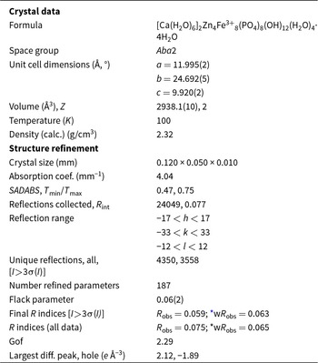
A jungite flake, measuring 0.120 × 0.050 × 0.010 mm, was used for a data collection at the Australian Synchrotron microfocus beamline MX2 (Aragao et al., Reference Aragao, Aishima, Cherukuvada, Clarken, Clift, Cowieson, Ericsson, Gee, Macedo, Mudie, Panjikar, Price, Riboldi-Tunnicliffe, Rostan, Williamson and Caradoc-Davies2018). Intensity data were collected using a Dectris Eiger 16M detector and monochromatic radiation with a wavelength of 0.7107 Å. The crystal was maintained at 100 K in an open-flow nitrogen cryostream during data collection. The diffraction data were collected using a single 36 second sweep of 360° rotation around phi. The resulting dataset consists of 3600 individual images with an approximate phi angle of each image being 0.1 degrees. The raw intensity dataset was processed using XDS software (Kabsch, Reference Kabsch2010) to produce data files that were analysed using SHELXT (Sheldrick, Reference Sheldrick2015) and JANA2006 (Petříček et al., Reference Petříček, Dušek and Palatinus2014).
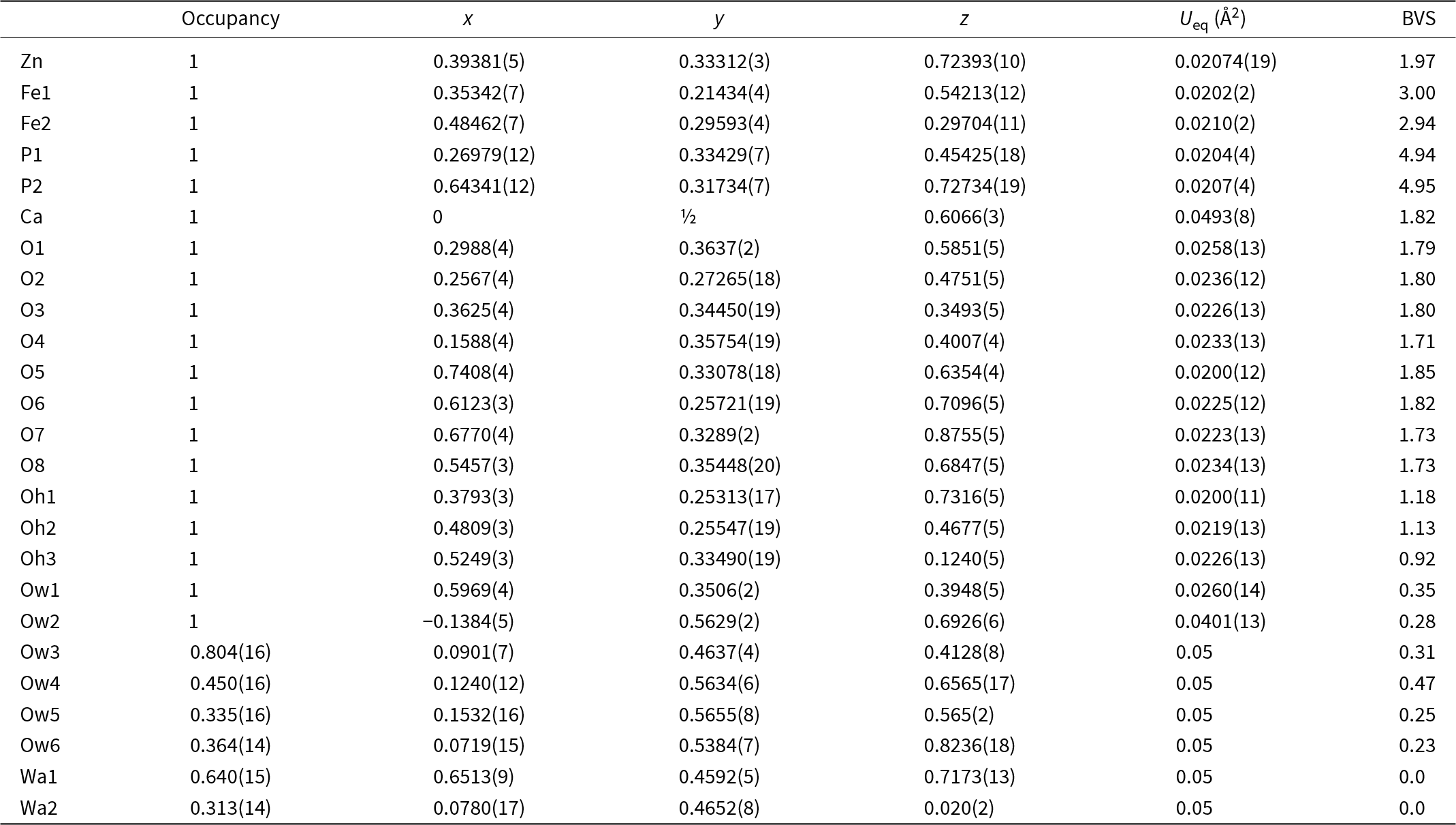
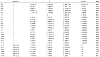
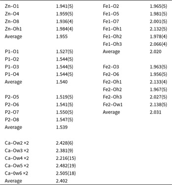
Automatic indexing of the frame diffraction data gave a face-centred orthorhombic unit cell with systematic extinctions consistent with Cmca (SG #64) or C2ca (SG #41, standard setting Aba2 with a and c interchanged). Application of SHELXT gave a structure solution in the non-centrosymmetric space group Aba2. Refinement of the model in JANA2006 confirmed that Zn was fully ordered at a tetrahedral site whereas Fe, as Fe3+, was ordered at two octahedral sites. The Ca site was located halfway between the heteropolyhedral layers, and the anions coordinated to the Ca were disordered and partially occupied. For these sites the isotropic displacement parameter was fixed at 0.05 and the occupancies were refined. Refinement with anisotropic displacement parameters for the layer atoms and Ca converged at wR obs = 0.063 for 3558 observed reflections with I > 3σ(I). Other details of the data collection and refinement are given in Table 1. Refined coordinates, displacement parameters and calculated bond valence sums (BVS, Gagné and Hawthorne, Reference Gagné and Hawthorne2015) are reported in Table 2. Polyhedral bond distances are given in Table 3.
Crystal data and structure refinement results for orthorhombic jungite

* w = [σ2(ǀF oǀ)+(0.01Fo)2]–1
Coordinates, site occupancies, equivalent isotropic displacement parameters and bond valence sums (BVS in valence units) for model for orthorhombic jungite
Polyhedral bond distances (Å) for orthorhombic jungite
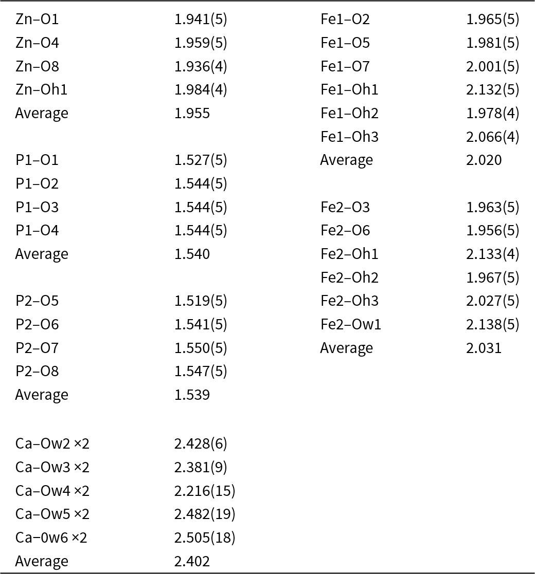
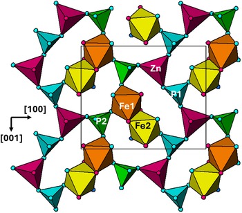
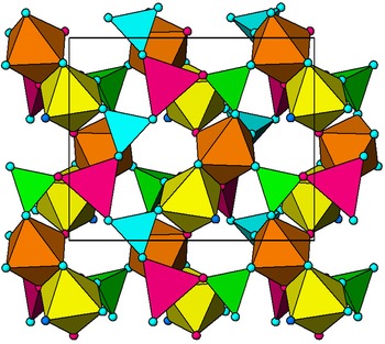
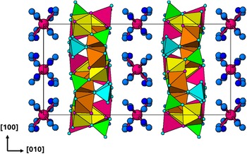
A view of the heteropolyhedral layer in jungite is shown in Fig. 1. It is unusual in that it is built from seven-member rings of corner-connected ZnO4 and PO4 tetrahedra and dimers of Fe-centred octahedra. Each ZnO4 tetrahedron corner-shares its basal anions with three separate PO4 tetrahedra. The P1O4 tetrahedron shares its basal anions with two ZnO4 and the Fe2-centred octahedron whereas the P2O4 tetrahedron shares its basal anions with one ZnO4 and two Fe1-centred octahedra. The Fe2-centred octahedron has one unshared vertex, occupied by H2O (Ow1 in Table 2), whereas the Fe1-centred octahedron share all vertices with other polyhedra. Pairs of layers are fused together into double layers by octahedral–octahedral and tetrahedral–octahedral corner connections as shown in Fig. 2. The corner-connection of the octahedral dimers gives sinusoidal chains along [001].
[010] view of heteropolyhedral layer in orthorhombic jungite. The unit cell is shown. Red circles are OH, light blue, oxygen and dark blue, H2O. Drawn using Atoms (Dowty, Reference Dowty2004).

Double heteropolyhedral layer in orthorhombic jungite. Atom labels and axes as in Fig. 1. Drawn using Atoms (Dowty, Reference Dowty2004).

A projection of the structure along [001] is shown in Fig. 3. This figure shows the location of Ca atoms, halfway between the heteropolyhedral layers, which are coordinated only to H2O molecules, Ow2 to Ow6 in Table 2. The sites Ow2 and Ow3 are fully occupied or almost so, whereas the sites Ow4 to Ow6 are less than half-occupied. Distances Ow4–Ow5 (1.01 Å) and Ow4–Ow6 (1.98 Å) are too short for these pairs of sites to be occupied at the same time. Summing the partial occupancies at Ow2 to Ow6 gives 5.91(3) H2O per Ca, so the Ca is essentially six-coordinated. A similar situation occurs in the structure of tomsquarryite, [Mg(H2O)6]NaAl3(PO4)2(OH)6(H2O)2, in which six-coordinated hydrated Mg2+ ions are located between heteropolyhedral layers, and the coordinated H2O are disordered over three partially occupied sites (Elliott et al., Reference Elliott, Grey, Mumme, MacRae and Kampf2022).
[001] projection of the structure for orthorhombic jungite, showing hydrated Ca2+ (red circles) in interlayer region. Medium blue circles are coordinated H2O and dark blue circles are free H-bonded H2O. Drawn using Atoms (Dowty, Reference Dowty2004).

In addition to the [Ca(H2O)6]2+ hydrated cations, the interlayer region of jungite contains uncoordinated H2O molecules at sites Wa1 and Wa2. Both sites are partially occupied (see Table 2) with the sum of their occupancies approximating to 4H2O per formula unit.
Triclinic jungite
As mentioned in the Introduction, crystals from the same specimen that provided the orthorhombic crystals were indexed by a triclinic cell with a b value half that reported by Moore and Ito (Reference Moore and Ito1980). In contrast to the clean diffraction displayed by the orthorhombic crystals, the diffraction frames for the triclinic crystals were complex with many split spots due to both differently orientated components and to twinning. The data were reprocessed several times, as multi-component twins, using CrysalisPro (Rigaku Oxford Diffraction, 2024). The best quality dataset was obtained from a single component when the data was processed as a two-component twin; the twin components had occupancies of 0.65:0.35 and were related by a rotation of ∼180° around [010]. The resulting intensity data were very weak, with less than one third of the intensities having I > 3σ(I). Nevertheless, independent processing of the MX2 frame diffraction data by two authors using CrysAlisPro produced intensity data that gave the same structural solution in space group P1 using Superflip (Palatinus and Chapius, Reference Palatinus and Chapuis2007) and SHELXT. Although a satisfactory refinement of the model was not possible (an anisotropic refinement resulted in nearly all atoms having non-positive displacement parameters), the validity of the model was confirmed by the fact that it had an identical topology of the double heteropolyhedral layer with almost the same bond distances as found for the orthorhombic structure. The structural parameters for the partially refined triclinic model have been deposited as supplementary material (see below).
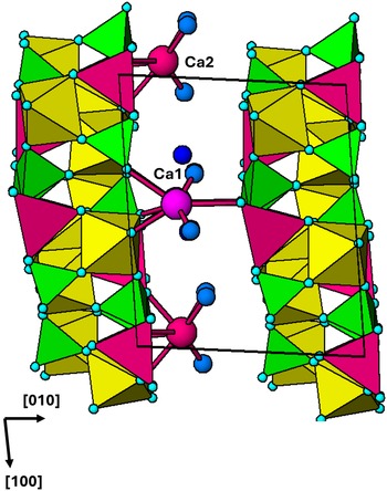
The major difference from the orthorhombic model is the interlayer Ca coordination. In the triclinic structure, the Ca is displaced from the midpoint between the layers towards one layer, resulting in coordination to the layer anions. There are two independent Ca sites in the triclinic structure, as shown in Fig. 4. Ca1 coordinates to four layer-anions plus two H2O, and Ca2 coordinates to four layer-anions plus three H2O. A [001] projection of the structure of triclinic jungite showing the Ca coordination is shown in Fig. 4. In addition to the H2O coordinated to Ca, the interlayer region also contains two H2O per formula unit as free, H-bonded groups.
[001] projection of the structure for triclinic jungite. Medium blue circles are coordinated H2O and dark blue circles are free H-bonded H2O. Drawn using Atoms (Dowty, Reference Dowty2004).

Discussion
The BVS values in Table 2 allow unambiguous assignment of O, OH and H2O to the anions in orthorhombic jungite, as well as confirming the valence states of the metal atoms. The resulting formula for orthorhombic jungite is [Ca(H2O)6]2Zn4Fe3+8(PO4)8(OH)12(H2O)4·4H2O, with a calculated water content of 22.2 wt.%. The formula is in generally good agreement with that reported by Moore and Ito (Reference Moore and Ito1980), apart from the higher water content and having 8 PO4 (plus 3OH) rather than 9 PO4. The higher PO4 content in Moore and Ito’s (Reference Moore and Ito1980) ideal formula is most likely due to impurity phases, such as jahnsite-(CaMnZn) (Grey et al., Reference Grey, Keck, MacRae, Glenn, Mumme, Kampf and Cashion2018), in the 500 mg specimen used for their wet chemical analyses, that they reported were composed of “fairly pure flakes”.
For the triclinic mineral, the corresponding formula, based on the structure shown in Fig. 4 is [Ca2(H2O)5]Zn4Fe3+8(PO4)8(OH)12(H2O)4·2H2O, with a calculated water content of 15.7 wt.%. The triclinic phase thus corresponds to a partially dehydrated form of jungite. The dehydration is accompanied by a shrinking of the heteropolyhedral layer separation from 12.35 Å to 10.0 Å. The collapse of the layer separation involves disorder in the layer stacking, resulting in poor diffracting quality of the dehydrated crystals. The situation is analogous to that reported for the heteropolyhedral layer structure mineral galeaclolusite, [Al6(AsO4)3(OH)9(H2O)4]·8H2O, that dehydrates to bulachite, [Al6(AsO4)3(OH)9(H2O)4]·2H2O, with a decrease in the layer spacing from 9.9 Å to 7.7 Å. (Grey et al., Reference Grey, Favreau, Mills, Mumme, Bougerol, Brand, Kampf, MacRae and Shanks2021).
The unit cell for the jungite specimen reported by Moore and Ito (Reference Moore and Ito1980) has b = 20.37 Å, corresponding to a layer spacing of 10.18 Å, so on the basis of our study, they were working with a partially dehydrated jungite specimen, which probably explains why they were unable to get a cleanly diffracting crystal for a structure solution. Moore and Ito (Reference Moore and Ito1980) reported a chemical analysis for water, giving 17.8 wt.% H2O, which is intermediate between the values for the orthorhombic and triclinic minerals, but much closer to the latter.
Supplementary material
The supplementary material for this article can be found at https://doi.org/10.1180/mgm.2025.10145.
Acknowledgements
Thanks to Vaclav Petříček and Jakub Plasil for their interest in processing the MX2 frame data for triclinic jungite (by MD).
Competing interests
The authors declare none.


 Open access

