


Introduction
The pterygopalatine fossa (PPF) is a pyramidal cavity between the maxillary, sphenoid and palatine bones. Tumours that originate in or affect the PPF are extremely rare, making up just 0.5 per cent of all head and neck tumours.Reference Bao, Ni, Zhang, Li, Mo and Guo1 Pterygopalatine fossa schwannoma (PPFS) is a rare primary benign tumour that typically presents with nonspecific symptoms such as facial pain, numbness and nasal congestion, which can be challenging for clinicians to diagnose. Despite the availability of advanced radiological tests, some patients still present with a large PPFS.
The traditional treatment approach for PPFS involves surgical resection. However, the intricate and complex anatomy of the PPF poses significant challenges during these procedures.Reference Cappello, Arbor and Potts2 To address these difficulties, surgeons began utilizing endoscopic techniques, which offer improved visualization and access with less invasiveness.Reference Behairy, Barsem and Eldemerdash3 Despite these advantages, endoscopic techniques require extensive training and still present certain complications,Reference Yang, Hu, Zhao, Zhang, Liu and Wang4–Reference Zoli, Sollini, Zaccagna, Fabbri, Cirignotta and Rustici8 necessitating alternative minimally invasive treatment modalities, such as gamma knife radiosurgery (GKRS), for treating such challenging-to-reach lesions.Reference Niranjan, Faramand, Raju, Lee, Yang and Nabeel9–Reference Peker12 Patients may also need GKRS as salvage therapy in cases where subtotal resection is performed.Reference Park, Hong, Kim, Hong, Woo and Yun5
Herein, we present the first case report of a PPFS patient treated with GKRS and followed in our clinic for 15 years after the treatment.
Case report
A 21-year-old female presented with a history of progressive left-sided facial numbness over the past year. Initially diagnosed with a PPFS by a referring clinic and followed for 1 year, the patient showed tumour progression on further magnetic resonance imaging (MRI), leading to referral to our clinic for further treatment as the patient did not want to undergo surgery. The patient had no significant medical history and was not on any regular medications. She reported no history of trauma, infections, or surgeries in the affected region. On physical examination, no masses were detectable intra-orally or on facial inspection. Neurological examination was largely unremarkable, with no deficits in cranial nerve functions. The patient’s visual acuity and ocular movements were normal. The initial workup included a contrast-enhanced MRI, which revealed a well-defined, homogeneously enhancing mass in the left pterygopalatine fossa, exhibiting characteristics consistent with a schwannoma.
After a multidisciplinary discussion, the patient was offered GKRS as the primary treatment modality, considering the tumour’s location and the patient’s preference to avoid open surgery. The radiosurgical procedure was conducted under local anesthesia with the patient in a stereotactic frame to ensure precise targeting. High-resolution thin-slice MRI was used to delineate the tumour margins. A marginal dose of 14 Gy was delivered to the 50 per cent isodose line, encompassing the entire tumour volume in a single session (Figure 1). The procedure was completed without complications, and the patient was monitored in the recovery area before being discharged the same day.
PreGK (a,b,c,d) and postGK (e,f,g) MR images of the patient.
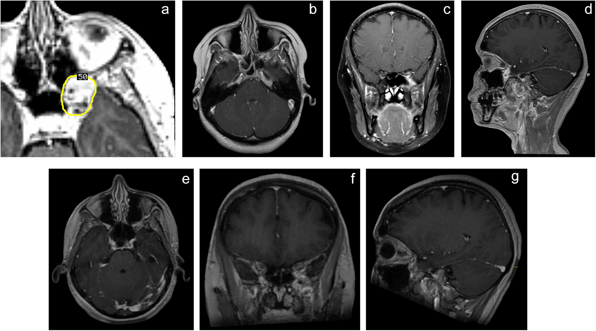
The patient was monitored with regular follow-up visits and imaging. At the six-month follow-up, the patient reported significant improvement in facial numbness. Neurological examination remained normal. At this time, MRI showed stable tumour size with no evidence of progression. The 12-month follow-up MRI showed further tumour shrinkage, with a total reduction of 35 per cent in size from the initial measurement. The patient remained symptom-free, and no adverse effects of the treatment were noted. At the 15-year follow-up, the MRI confirmed further tumour shrinkage and stable findings with continued symptom relief (Figure 1).
Discussion
PPFSs are exceedingly rare, accounting for a small fraction of all schwannomas. Their rarity, coupled with the complex anatomical region they inhabit, presents unique challenges in diagnosis and management. As in other schwannomas, this case highlights the efficacy and safety of GKRS in treating PPFSs. The precise delivery of high-dose radiation minimizes damage to adjacent critical structures, offering a favorable outcome.
Traditional management of schwannomas involves surgical resection, especially for tumours causing significant mass effects or in cases where a definitive histopathological diagnosis is required. Tumours in this region have been excised using an open lateral or anterior surgical corridor. However, these techniques are highly invasive, requiring extensive facial incisions, craniotomies, or osteotomies. Endoscopic approaches with image guidance have become integral to the skill set of skull base surgeons, offering improved cosmetic outcomes and quality of life. On the other hand, these techniques require extensive training and are considered an advanced procedure that should be performed in specialized referral centres. Despite this, complications can still occur as surgical access is technically challenging in the PPF due to the region’s intricate anatomy. The PPF is filled with adipose tissue, specifically the pterygopalatine extension of the buccal fat pad. It contains the maxillary nerve and its branches, the pterygopalatine ganglion, the nerve of the pterygoid canal and the pterygopalatine segment of the maxillary artery, along with its branches and corresponding veins.Reference Cappello, Arbor and Potts2 Thus, surgical approaches, whether open or endoscopic, may lead to significant morbidity.Reference Porras, Rowan and Mukherjee13 Post-operative cerebrospinal fluid (CSF) leak is the most common complication following endoscopic approaches and can lead to complications such as meningitis, pneumocephalus or acute subdural hemorrhage.Reference Lai, Trooboff, Morgan and Harvey14 Additionally, CSF leaks can result in increased hospital stays and a higher risk of readmission. Injury to the internal carotid artery is a rare but serious complication of the endoscopic endonasal approach, with an incidence ranging up to 1 per cent and a mortality rate of up to 10 per cent.Reference Iranmehr, Sarpoolaki, Sadrehosseini, Tabari and Zeinalizadeh15, Reference Chin, Ghosh, Fang, Baredes, Liu and Eloy16 In addition to major complications, decreased olfaction, sensory changes, nasal crusting, nasal obstruction, rhinosinusitis and mucocele formation can also be observed.Reference de Almeida, Witterick and Vescan17 Dry eye, V2 hypesthesia and trismus are also site-specific complications.Reference Plzák, Kratochvil, Kešner, Šurda, Vlasák and Zvěřina18
GKRS has emerged as a valuable alternative for treating benign tumours like schwannomas, particularly in complex anatomical regions like the PPF or those with contraindications to surgery. The procedure is minimally invasive, typically performed on an outpatient basis, and associated with minimal recovery time and morbidity. The efficacy of GKRS in treating schwannomas, including vestibular and non-vestibular types, has been well-documented.Reference Bin-Alamer, Abou-Al-Shaar, Peker, Samanci, Pelcher and Begley19–Reference Langlois and Mathieu21 Studies have shown high rates of tumour control and symptom relief with no to minimal complications. Although this is the only case report of PPFS managed with GKRS, Niranjan et al.Reference Niranjan, Faramand, Raju, Lee, Yang and Nabeel9 reported an overall tumour control rate of 94.5 per cent in a cohort of 309 patients with trigeminal schwannomas, with only 9 per cent experiencing peri-lesional edema. The authors reported symptom improvement in 45 per cent of the patients at the last follow-up. In this case report, the patient experienced significant symptomatic relief, substantial tumour reduction, continued symptom relief and no new neurological deficits at the 15-year follow-up.
• This paper documents the first successful treatment of PPFS with GKRS.
• GKRS is an effective and safe minimally invasive treatment for PPFS.
• GKRS has long-term success in local tumour control and symptom relief.
Conclusion
In conclusion, this case report is the first in the literature to document the successful treatment of a PPFS using GKRS. The patient’s positive outcome, with significant tumour reduction and symptom relief, underscores the potential of this non-invasive technique as a primary treatment modality for schwannomas in challenging anatomical locations. Further studies are needed to validate these findings and establish standardized treatment protocols.
Acknowledgements
None
Financial disclosure
This research received no specific grant from any funding agency, commercial or not-for-profit sectors.
Competing interests
The authors declare none.


 Open access