


Introduction
The human brain functions through an intricate network of electrical and chemical signals, where neurons communicate via rapid ion fluxes and neurotransmitter release (Bean, Reference Bean2007; Kress and Mennerick, Reference Kress and Mennerick2009; Südhof, Reference Südhof2012; Kaeser and Regehr, Reference Kaeser and Regehr2014; Zhang et al., Reference Zhang, Jiang and Yetisen2021). Understanding these processes is fundamental to unraveling the mechanisms of cognition, memory, behavior, and the pathophysiology of neurological disorders (Kandel et al., Reference Kandel, Dudai and Mayford2014; D’Esposito and Postle, Reference D’Esposito and Postle2015; Gao et al., Reference Gao, van den Brink, Pfeffer and Voytek2020). However, the complexity of brain activity spans multiple spatial and temporal scales, from millisecond ion transients at individual synapses to long-range neuromodulation across entire circuits, posing significant challenges for direct observation and quantitative analysis (Tavakolian-Ardakani et al., Reference Tavakolian-Ardakani, Hosu, Cristea, Mazloum-Ardakani and Marrazza2019; Xu et al., Reference Xu, Wu, Yu and Mao2019).
To address these challenges, a range of analytical methods has been developed. Traditional microdialysis coupled with high-performance liquid chromatography (HPLC) or mass spectrometry (MS) provides comprehensive, multi-analyte chemical qualification and quantification on a minute timescale (Zestos and Kennedy, Reference Zestos and Kennedy2017; Wells et al., Reference Wells, Bain, Valenta, Lenhart, Steyer and Kennedy2024). In contrast, electrochemical techniques offer subsecond temporal resolution, can be miniaturized into implantable micro-/nanoelectrodes for in vivo and in situ studies, and operate without genetic modification, making them a uniquely versatile and complementary platform to optical and MS-based approaches for monitoring dynamic neurotransmitter signaling. For example, fast-scan cyclic voltammetry (FSCV) has been integrated with scanning electrochemical microscopy (SECM), ion conductance microscopy (ICM), and carbon nanoelectrodes to achieve subcellular and even single-vesicle neurotransmitter detection with millisecond resolution (Wang et al., Reference Wang, Rodriguez, Alden, Choi, Alanis, Srinivasan and Baker2024; Subedi et al., Reference Subedi, Mohan Kumar, Weese-Myers and Ross2025). Recently, label-free biosensors, such as enzyme-functionalized microelectrodes or aptamer-based field-effect transistors (aptamer-FETs) (Xu et al., Reference Xu, Wu, Yu and Mao2019; Wang et al., Reference Wang, Zhao, Wang, Yang, Cheng, Liu, Yu, Lin, Zhao, Cheung, Lin, Hojaiji, Weiss, Stojanović, Tomiyama, Andrews and Emaminejad2022), have pushed detection limits into the low-picomolar range.
Recent advancements in nanoelectrodes (Liu et al., Reference Liu, Cao, Zhou, Li, Du, Xue, Li and Mao2025), scanning probe microscopy (SPM) (Zhang et al., Reference Zhang, Zhu, Lang, Fu and Li2020; Putnam et al., Reference Putnam, Santiago-Carboney, Qian and Rodríguez-López2025), and multimodal nanosensors (Ahmed Taha et al., Reference Ahmed Taha, Addie, Saeed, Haider, Chaudhary and Arsad2024; Ria et al., Reference Ria, Eladly, Masvidal-Codina, Illa, Guimerà, Hills, Garcia-Cortadella, Duvan, Flaherty, Prokop, Wykes, Kostarelos and Garrido2025) have further enabled in situ measurements of ion concentrations, neurotransmitter flux, and redox dynamics in living neural tissues at highly localized scales. These tools not only reveal the heterogeneity of neurochemical microenvironments but also contribute to the development of brain–machine interfaces and diagnostic platforms for neurological diseases. In particular, dual-mode probes, which enable simultaneous detection of chemical and electrical signals, provide a more comprehensive insight into complex neural activities. Despite these advances, persistent technical challenges remain, including limited sensor selectivity, mechanical invasiveness, and across-scale data integration barriers. The development of artificial intelligence (AI), especially machine learning (ML), promises to address these limitations by accelerating the design of novel electrode interfaces, enhancing signal processing, suppressing noise, and deconvolving multi-analyte signals (Cui et al., Reference Cui, Yue, Zhang, Zhang and Zhou2020; Flynn and Chang, Reference Flynn and Chang2024).
In this perspective, we focus on recent micro- and nanoscale electrochemical innovations for real-time brain chemistry monitoring, including the adaptation of miniaturized, label-free probes for in vivo and in situ neurochemical analysis, and the transformative role of ML in driving the rapid development of this field. Furthermore, we attempt to offer an outlook on potential directions for future research. Although not exhaustive, this review highlights key advances in the electrochemical detection of representative neurotransmitters and neuromodulators over the past decade.
Electrochemical techniques for in situ and in vivo analysis of brain signaling molecules
Over the last decade, real-time in situ monitoring of neurotransmitter release has achieved single-vesicle resolution, yielding unprecedented insights into exocytotic kinetics. Meanwhile, surface engineering of micro- and nanoelectrodes has been optimized to accommodate diverse neuromodulators, enhance sensitivity, reduce detection limits, and improve antifouling properties, thereby enabling extended in situ and in vivo applications.
Single-vesicle electrochemistry sheds light on neurotransmitter release dynamics during exocytosis
Dopamine (DA), a crucial monoamine neurotransmitter in the central nervous system (CNS), underlies reward processing, motor control, mood regulation, and cognition (Derosiere et al., Reference Derosiere, Shokur and Vassiliadis2025; Speranza et al., Reference Speranza, Miniaci and Volpicelli2025). Dysregulation of DA signaling is closely implicated in neuropsychiatric and neurodegenerative disorders, including Parkinson’s disease (PD), depression, schizophrenia, and addiction.
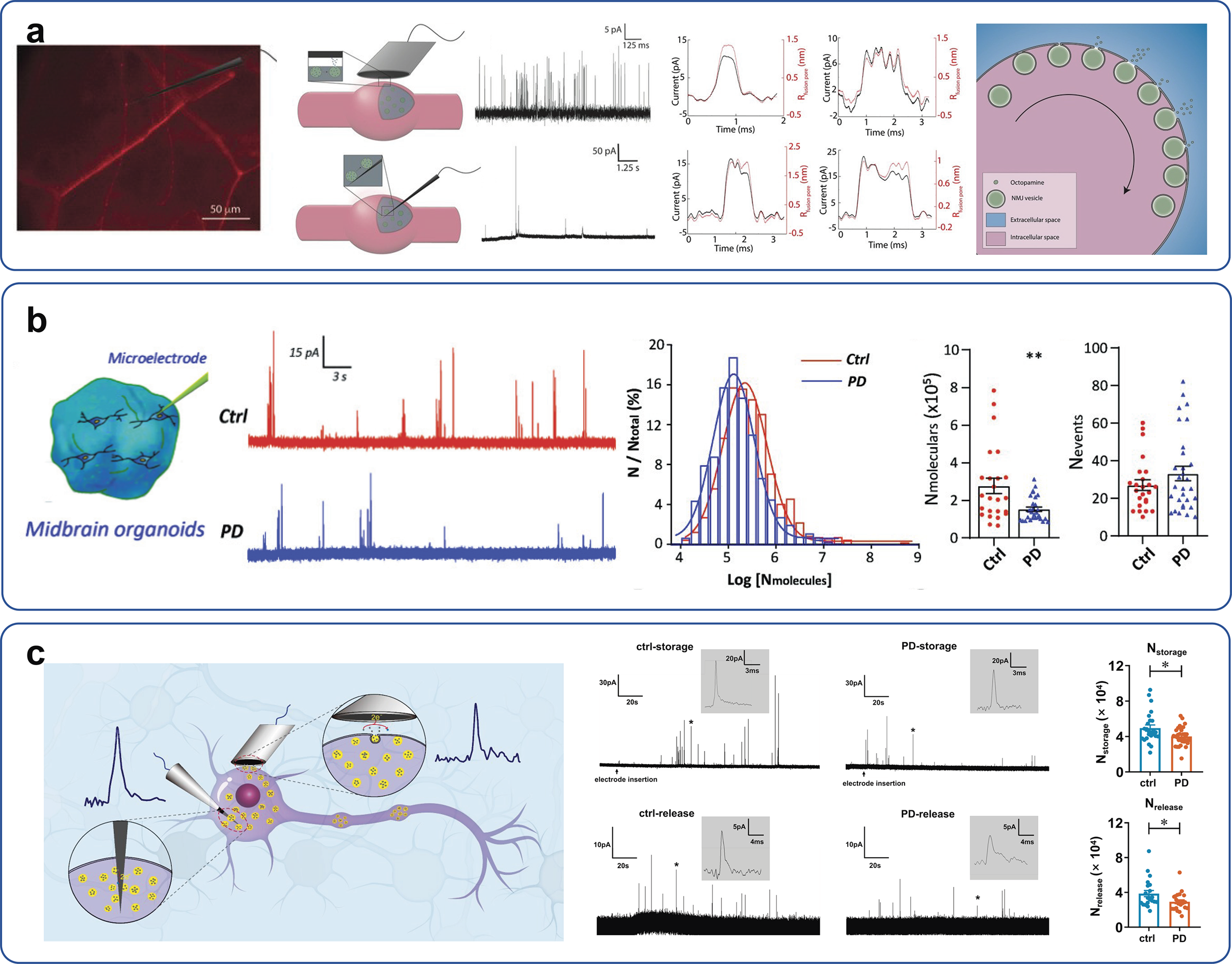
Single-cell amperometry (SCA), a well-established method, enables direct detection of monoamines released during exocytosis using micro-/nanoelectrodes (Kuhr et al., Reference Kuhr, Ewing, Caudill and Wightman1984). On the other hand, Ewing’s group pioneered intracellular vesicle impact electrochemical cytometry (IVIEC), which allows for quantification of neurotransmitter content in vesicles (Li et al., Reference Li, Majdi, Dunevall, Fathali and Ewing2015, Reference Li, Dunevall and Ewing2016a, Reference Li, Dunevall, Ren and Ewing2017, Reference Li, Ren, Dunevall, Ye, White, Edwards and Ewing2018; Liu et al., Reference Liu, Du, Wang, Zhang, Liu and Li2021). Studies employing SCA and IVIEC have shown that exocytosis often releases only a fraction of the stored neurotransmitter in model systems (Zhou et al., Reference Zhou, Zhang, Cao, Liu, Liu, Liu and Li2023; Cao et al., Reference Cao, Zhang, Li, Zhou, Liu, Liu and Li2024; Nan et al., Reference Nan, Wang, Du, Liu, Cao, Zhou, Liu and Li2024; Li et al., Reference Li, Dunevall and Ewing2016b; Chen et al., Reference Chen, Liu, Cao, Zhou, Nan, Li, Liu, Xiong, Jiang, Dai, Yuan and Li2026), implying the complexity and plasticity of synaptic transmission. For instance, in Drosophila neuromuscular (type II) neurons, combined SCA and IVIEC revealed that ‘simple’ fusion events release <5% of vesicular octopamine content, while flickering-pore events release nearly 11%, both far less than the ~60% typically released in PC12 and adrenal chromaffin cell models (Figure 1a) (Larsson et al., Reference Larsson, Majdi, Oleinick, Svir, Dunevall, Amatore and Ewing2020). Mathematical modeling of these events according to fusion pore dynamics predicted intravesicular transmitter release for four types of events, including simple open and closed exocytosis, different levels of pore opening, flickering, and closing during complex events. Extending these findings, Zhu et al. demonstrated that α-synuclein overexpression in both neurons and organoids from young-onset PD (YOPD) patients impairs vesicular DA storage, revealing a direct link between synaptic vesicle dysfunction and disease pathology (Figure 1b) (Zhu et al., Reference Zhu, Tao, Hong, Wu, Chu, Zheng, Han, Zhu, Xu, Ewing, Guo and Liu2022).
SVE quantifies vesicular neurotransmitter content and release dynamics. (a) Typical image, schematic diagram, and amperometric traces of SCA and IVIEC quantifying octopamine molecules at neuromuscular varicosities. Overlaid traces compare exocytotic events recorded by SCA (black) with mathematically modeled fusion pore radius variations (red), revealing release mode–dependent differences. Reprinted from Larsson et al. (Reference Larsson, Majdi, Oleinick, Svir, Dunevall, Amatore and Ewing2020) with permission from Wiley. (b) Schematic diagram and typical traces of vesicular neurotransmitter content in midbrain organoids derived from a control iPSC line (H9) and a young-onset Parkinson’s disease (YOPD) patient iPSC line (PD52). Histograms show normalized frequency distributions of vesicle dopamine (DA) content, average DA molecules per vesicle, and event counts. Reprinted from Zhu et al. (Reference Zhu, Tao, Hong, Wu, Chu, Zheng, Han, Zhu, Xu, Ewing, Guo and Liu2022) with permission from Royal Society of Chemistry. (c) Schematic of an SVE platform combining IVIEC and SCA for measuring vesicular neurotransmitter storage and exocytotic release in soma and varicosities in fresh brain slices. Representative current–time (i–t) traces of IVIEC and SCA in somas of the substantia nigra pars compacta (SNc) are shown, with insets displaying magnified asterisk-marked spikes (*). Reprinted from Liu et al. (Reference Liu, Cao, Zhou, Li, Du, Xue, Li and Mao2025) with permission from American Chemical Society.

In addition to DA, the combination of SCA and IVIEC also contributes to deepening the understanding of the release pattern of serotonin (5-hydroxytryptamine (5-HT)). Gu et al. applied these techniques to investigate vesicular serotonin storage and release in the ventral nerve cords of Drosophila larvae (Gu et al., Reference Gu, Wang, Yeoman, Patel and Ewing2024). They found that serotonin exocytotic events follow a bimodal Gaussian distribution, dominated by small-amplitude events alongside a comparable or slightly higher incidence of large-amplitude events. Pharmacological modulation of exocytosis, either enhancing or suppressing release, resulted in vesicle release fractions ranging from partial (13–18%) to complete (100%). These results suggest that a single-vesicle pool can release highly variable fractions of its transmitter load during exocytosis, indicating a potential mechanism for regulating exocytosis and neuronal signaling.
In situ single-vesicle electrochemistry (SVE) developed by us recently permits measurement of monoamine neurotransmitter dynamics in brain slices with high spatiotemporal resolution (Liu et al., Reference Liu, Cao, Zhou, Li, Du, Xue, Li and Mao2025). Positioning carbon-fiber microelectrodes (CFMEs) in or adjacent to the soma and varicosities of neurons across multiple brain regions captures current spikes from individual vesicles, enabling precise quantification of vesicular neurotransmitter storage, exocytotic release amount, and kinetics. Notably, SVE has enabled the first in situ quantification of monoamine transmitter at the single-vesicle level in fresh brain slices, advancing our understanding of the pathological mechanisms underlying PD at the vesicle level (Figure 1c).
Taken together, the above advances indicate how the analysis of neurotransmitters at the vesicle level is gradually contributing to a more complete understanding of their transmission in intact neural circuits of the brain.
Advances in electrode design for in situ monitoring of non-electroactive species and intracellular signal molecules
In addition to monoamine neurotransmitters, glutamate (Glu), a key excitatory amino acid neurotransmitter in the mammalian CNS, plays critical roles in synaptic transmission, plasticity, learning, and memory. Dysregulation of glutamatergic signaling has been implicated in a wide range of neurological disorders (Zhou and Danbolt, Reference Zhou and Danbolt2014; O’Donovan et al., Reference O’Donovan, Sullivan and McCullumsmith2017; Biswas et al., Reference Biswas, Shahriar, Bachay, Arvanitis, Jamoul, Brunken and Agalliu2024; Lai et al., Reference Lai, Pritišanac, Liu, Liu, Gong, Li, Lu, Qi, Xu, Forman-Kay, Shi, Wang and Yin2024). In response, recent advances in chemical biology have facilitated the development of innovative biosensing platforms that enable high-sensitivity, real-time monitoring of Glu dynamics. Nanostructured electrodes have significantly enhanced the sensitivity of Glu sensors. Recently, a nonenzymatic Glu sensor has been developed by synthesizing copper oxide nanostructures and physically mixing them with multiwall carbon nanotubes (MWCNTs) onto a screen-printed carbon electrode (Ali et al., Reference Ali, Knight and Howlader2023). In parallel, enzymatic electrochemical sensors have been refined by integrating nanomaterials, such as graphene, CNTs, and Pt/Au nanoparticles, that offer high surface-to-volume ratios and excellent conductivity, as well as provide scaffolds for enzyme immobilization. When integrated with Glu oxidase systems, these nanostructures improve electron transfer and sensor stability, enabling quantification of Glu content in individual vesicles (Figure 2a, b) (Wang et al., Reference Wang, Fathali, Mishra, Olsson, Keighron, Skibicka and Cans2019a, Reference Wang, Mishra, Bergman, Keighron, Skibicka and Cans2019b; Yang et al., Reference Yang, Zhang, Wu, Tang, Yan, Liu, Amatore and Huang2021). Additionally, the emergence of aptamer-based detection strategies has provided promising enzyme-free alternatives, capitalizing on the high specificity and chemical robustness of aptamers to overcome the limitations of conventional enzymatic approaches (Wu et al., Reference Wu, Barkova, Komarova, Offenhäusser, Andrianova, Hu, Kuznetsov and Mayer2022). Future Glu sensors are expected to integrate nanostructured materials to maximize sensitivity and selectivity.
Nanostructured electrode modifications enable quantitative intracellular analysis of neurochemicals. (a) Fabrication and scanning electron microscopy (SEM) images of Pt/GluOx-modified SiC@C nanowire electrodes. Depending on the placement, these nanoelectrodes enable amperometric monitoring of Glu exocytotic release from single varicosities (I) or vesicular Glu content by IVIEC (II) in a hippocampal neuron. Reprinted from Yang et al. (Reference Yang, Zhang, Wu, Tang, Yan, Liu, Amatore and Huang2021) with permission from Wiley. (b) Ultrafast Glu sensor modified with AuNPs (red hemispheres) and a thin GluOx coating (yellow hemispheres) enables monitoring vesicle rupture and release of Glu at the sensor surface. Typical amperometric trace and an illustration of six different current spike types associated with Glu release in the nucleus accumbens of a brain slice. Reprinted from Wang et al. (Reference Wang, Fathali, Mishra, Olsson, Keighron, Skibicka and Cans2019a) with permission from American Chemical Society. (c) The fabrication process of SiC@Pt nanowire electrode and its application for quantitative monitoring of freshly produced ROS/RNS in individual phagolysosomes during a single IVIEC event. Reprinted from Qi et al. (Reference Qi, Jiang, Wu, Zhang, Tian, Fan, Liu, Amatore and Huang2022) with permission from American Chemical Society. (d) Characterization of PEDOT/PB/CFNE electrodes, including SEM, cyclic voltammograms, sensitivity, stability, and selectivity. Experimental setup for detecting H2O2 using PEDOT/PB/CFNE upon following MPP+ injection, along with a representative amperometric response obtained in SH-SY5Y cells. Reprinted from Zhang et al. (Reference Zhang, Qin, Cheng, Zhang, Gao and Zhang2023) with permission from Wiley.

Acetylcholine (ACh), another crucial neurotransmitter, modulates cortical and hippocampal circuits by regulating neuronal excitability and synaptic strength, thereby influencing attention, learning, and memory (Curtis and Andersen, Reference Curtis and Andersen1962; Picciotto et al., Reference Picciotto, Higley and Mineur2012; Chevy and Kepecs, Reference Chevy and Kepecs2018; Monk and Hussain Shuler, Reference Monk and Hussain Shuler2019). Early amperometric biosensors co-immobilized acetylcholinesterase (AChE) and choline oxidase (ChO) at an optimized ratio on AuNP-deposited CFMEs, achieving sub-millisecond resolution monitoring of single-vesicle ACh release during exocytosis (Keighron et al., Reference Keighron, Wigström, Kurczy, Bergman, Wang and Cans2015; Wang et al., Reference Wang, Pradhan, Gupta, Hanrieder, Zetterberg and Cans2024). Subsequently, multimodal platforms combining electrochemical and optical fibers or flexible AuNP/MWCNT-coated threads further improved the spatiotemporal resolution of cholinergic signaling dynamics during behaviorally relevant tasks (Amirghasemi et al., Reference Amirghasemi, Soleimani, Bawarith, Tabassum, Morrel and Mousavi2023).
Meanwhile, interfaces between two immiscible electrolyte solutions (ITIESs) provide a nanoscale electrode architecture for detecting both redox-active and redox-inactive analytes. Its selectivity originates from ion-transfer energetics, as each ion possesses a characteristic transfer potential defined by its Gibbs free energy of transfer across the aqueous/organic boundary. Differences in solvation and hydrophobicity give ACh a distinct potential relative to other quaternary ammonium ions, which can be further tuned by the choice of organic phase and supporting electrolyte. Miniaturization to the nanoscale reduces capacitive background and improves resolution, thereby enabling reliable discrimination of ACh. Using this strategy, Shen et al. demonstrated nanoscale ITIES electrodes that successfully monitored synaptic ACh concentrations and release dynamics in living Aplysia neurons (Shen et al., Reference Shen, Qu, DesLaurier, Welle, Sweedler and Chen2018; Jetmore et al., Reference Jetmore, Anupriya, Cress and Shen2022; Ribeiro et al., Reference Ribeiro, Silva, Girault and Pereira2024; Jiang et al., Reference Jiang, Liu, Xiong, Liu, Wang, Liu, Cao, Zhou, Li, Yuan, Mao and Li2026).
Reactive oxygen species (ROS) and reactive nitrogen species (RNS), important neuromodulators, and signaling molecules participate in diverse processes of neural regulation. Recent advances in nano- and microelectrode-based electrochemical sensors have made it possible to monitor ROS/RNS with both high temporal and subcellular resolutions (Figure 2c) (Zhao et al., Reference Zhao, Zang, Zhang, Liu, Wang, Cai, Durkan, Xie and Wang2021; Qi et al., Reference Qi, Jiang, Wu, Zhang, Tian, Fan, Liu, Amatore and Huang2022; Gu et al., Reference Gu, Gu, Locker and Ewing2024; Gu et al., Reference Gu, Gu, Du Toit, Yu, Chen, Struckman, Silva, Dai and Ewing2025). Qi et al. pioneered the use of time-stepped amperometry with microelectrodes positioned on cellular surfaces to simultaneously distinguish release fluxes of H₂O₂, NO, and ONOO− at the single-cell level, providing direct electrochemical evidence of oxidative bursts in neuroimmune cells. To enhance specificity for RNS, Meiller et al. introduced fluorinated hydrogel- and nickel–porphyrin-modified CFMEs, which effectively suppress electrochemical interference and enable dynamic in vivo detection of NO·in the brain (Meiller et al., Reference Meiller, Sequeira and Marinesco2020).
Li et al. created a biocompatible, antifouling polydopamine/Prussian blue/CNT/carbon-fiber microelectrode (PDA/PB/CNT/CFE) sensor by electrodepositing PB onto CNT-assembled CFEs and further coating them with a self-polymerized PDA membrane for selective in vivo H₂O₂ monitoring (Li et al., Reference Li, Liu, Qiu and Zhang2016). Zhang et al. then adapted this design to a ring-disk configuration with a PB/poly(3,4-ethylenedioxythiophene (PEDOT)-modified carbon-fiber disk for H₂O₂ sensing and a concentric gold ring for DA detection, enabling simultaneous, interference-free measurements (Zhang et al., Reference Zhang, Feng, Zhang and Zhang2019). They subsequently demonstrated that PEDOT/PB/carbon-fiber nanoelectrode (CFNE) shows enhanced H₂O₂ sensing upon nicotinamide adenine dinucleotide (NADH) treatment (Figure 2d) (Zhang et al., Reference Zhang, Qin, Cheng, Zhang, Gao and Zhang2023).
Advances in real-time in vivo monitoring of neurochemicals
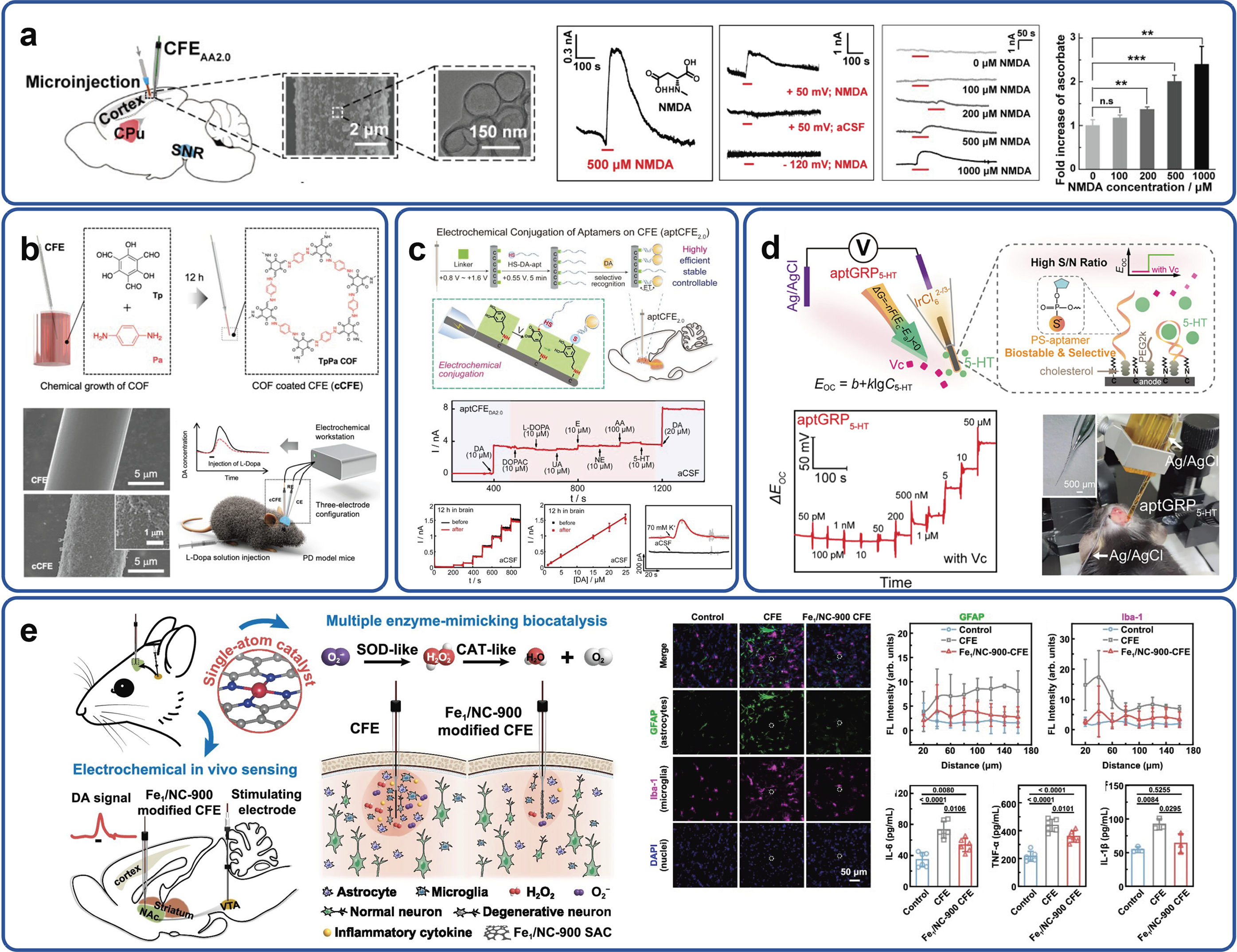
Complementary to in situ approaches, in vivo techniques allow direct access to neurochemical signals in living animals (Nan et al., Reference Nan, Li, Dai, Zhou, Cao, Liu, Chen, Xiong, Jiang and Li2026). Ascorbic acid (AA; vitamin C) has been recognized not only as the principal antioxidant in the brain but also as a neuromodulator that regulates synaptic transmission by modulating both Glu and gamma-aminobutyric acid (GABA) release and receptor function (Ballaz and Rebec, Reference Ballaz and Rebec2019). During synaptic activation, AA inhibits glucose transporter 3 (GLUT3)–mediated glucose uptake, shifting the metabolic substrate from glucose to lactate to sustain neuronal activity. Changes in AA concentrations in the brain are closely linked to oxidative stress levels in neurodegenerative diseases. Studies have shown that higher serum AA levels are associated with slower progression of Alzheimer’s disease in elderly populations, suggesting a potential role in cognitive function preservation (Kocot et al., Reference Kocot, Luchowska-Kocot, Kiełczykowska, Musik and Kurzepa2017; Appiah et al., Reference Appiah, Ingabire-Gasana, Appiah and Yang2024). Xiao et al. employed CNT-sheathed CFMEs to achieve the first in vivo imaging of AA oscillations during spreading depolarization in living rats with high selectivity and spatiotemporal resolution (Xiao et al., Reference Xiao, Wang, Wei, Yu, Jiang and Mao2019). Following this, Jin et al. developed a molecularly tailored ‘CFEAA2.0’ implantable sensor, using AuNPs and an optimized enzyme layer modification, which enabled in situ observation of AA efflux induced by cytotoxic edema. They further demonstrated that blockade of edema completely abolishes AA efflux, providing a valuable tool for studying the pathology of brain injury (Figure 3a) (Jin et al., Reference Jin, Ji, Li, Zhao, Wu, Wei, Ma, Jiang and Mao2020).
Modified CFEs and aptamer-based sensors enable selective in vivo monitoring of AA, DA, and 5-HT. (a) Selective monitoring of AA in the rat cortex using the CFEAA2.0 sensor. Current responses are shown following local microinjection of artificial cerebrospinal fluid (aCSF) and 500 μM NMDA at applied potentials of +50 mV and − 120 mV, respectively. Reprinted from Jin et al. (Reference Jin, Ji, Li, Zhao, Wu, Wei, Ma, Jiang and Mao2020) with permission from American Chemical Society. (b) Schematic illustration of TpPa covalent organic framework (COF) growth on CFEs, showing pore structure and SEM images of bare and COF-modified CFE. Integration with a closed-loop feedback system enables diagnosis and therapeutic intervention for PD. Reprinted from Zhou et al. (Reference Zhou, Yang, Li, Dong, Zhu, Wang, Lin and Su2023) with permission from American Chemical Society. (c) Two-step DA-mediated electrochemical conjugation strategy got aptamer-functionalized DA sensor (aptCFEDA2.0). Typical amperometric responses and calibration curves in response to successive DA additions in aCSF, both pre- and post-12-h in vivo implantation in the striatum, along with real-time DA sensing upon localized KCl or aCSF injection (2 μL min−1 for 30 s). Reprinted from Li et al. (Reference Li, Jin, Zhu, Liu, Jiang, Jiang and Mao2022) with permission from Wiley. (d) Phosphorothioate aptamer–based galvanic redox potentiometric sensor (aptGRP5-HT) for 5-HT detection. Potential responses toward successive 5-HT additions in aCSF and illustration of in vivo monitoring of 5-HT in the rodent brain are presented. Reprinted from Zhu et al. (Reference Zhu, Liu, Sun, Ni and Jiang2025) with permission from Wiley. (e) Schematic diagram of inflammation-free in vivo electrochemical sensing using Fe1/NC-modified CFEs. Experimental results demonstrate the anti-inflammatory properties of the modified electrodes. Reprinted from Gao et al. (Reference Gao, Wei, Ma, Wu, Ji, Mao, Yu and Mao2024) with permission from Springer Nature.

Substantial progress has also been made in DA monitoring in vivo. Novel material–based electrode modifications including conductive polymers (e.g. PEDOT/graphene oxide (GO) (Taylor et al., Reference Taylor, Robbins, Catt, Cody, Happe and Cui2017) and carbon nanomaterials (e.g. graphene quantum dots/multiwalled CNTs (GQDs-MWCNTs) (Huang et al., Reference Huang, Lin, Tong and Tong2020) have markedly improved the specificity, sensitivity, and in vivo stability of DA detection. These coatings enhance electrocatalytic activity and analyte adsorption while suppressing interferent signals, pushing DA detection limits from the micromolar down to the nano-/picomolar range. Further innovations in electrode design, including 3D-printed CNT yarns (Yang et al., Reference Yang, Cao, Puthongkham, Lee, Ganesana, Lavrik and Venton2018; Shao et al., Reference Shao, Puthongkham, Hu, Jia, Mirkin and Venton2020), nano-/micro-architectural optimizations (metal–organic frameworks (MOFs) and covalent–organic frameworks (COFs)) (Figure 3b) (Zhou et al., Reference Zhou, Yang, Li, Dong, Zhu, Wang, Lin and Su2023), and nanopore electrode arrays (Fu et al., Reference Fu, Han, Ma and Bohn2017), improve mechanical flexibility, signal fidelity, and chronic biocompatibility. Li et al. also developed chemoselective aptamer–carbon interfaces using electrochemical catechol grafting on CFMEs to generate quinone intermediates that undergo rapid reaction with thiol-modified oligonucleotides, yielding an interface with exceptional sensitivity and stability for continuous DA monitoring in vitro and in vivo (Figure 3c) (Li et al., Reference Li, Jin, Zhu, Liu, Jiang, Jiang and Mao2022).
Beyond conventional amperometry, the combination of FSCV with organic electrochemical transistors (OECTs) has produced a highly sensitive approach for in vivo DA monitoring in the rat brain (Li et al., Reference Li, Jin, Xiong, Yu and Mao2022). This system utilizes fast-scanning potential (FSP) as a gating mode and transconductance (g m) as a sensing parameter to achieve nM detection limits, excellent reproducibility, enhanced sensitivity (0.899S M−1), and real-time monitoring of both basal and electrically stimulated DA release in vivo. Moreover, multimodal systems integrating electrochemical sensing with optogenetics, miniature fluorescence imaging, and electrophysiological recording offer multidimensional insights into DA signaling and its relationship with neural circuit function and pathology (Liu et al., Reference Liu, Zhao, Cai, Xie, Wang, Cheng, Li, Li, Deng, Ding, Lv, Zhao, Liu, Zou, Feng, Sun, Yin and Sheng2020; Stuart et al., Reference Stuart, Jeang, Slivicki, Brown, Burton, Brings, Alarcón-Segovia, Agyare, Ruiz, Tyree, Pruitt, Madhvapathy, Niemiec, Zhuang, Krishnan, Copits, Rogers, Gereau, Samineni, Bandodkar and Gutruf2023).
Monitoring 5-HT, however, remains technically challenging due to rapid electrode fouling by its oxidation products. Although inherently electroactive, 5-HT suffers from poor signal reliability. Mao’s group pioneered galvanic redox potentiometry (GRP), a near-zero-current method yielding Nernstian potentials insensitive to electrode surface area. This unique mechanism of GRP minimizes biofouling, making it ideally suitable for sensitive 5-HT detection in living biosystems (Zhu et al., Reference Zhu, Xue, Ji, Li, Ma, Yu, Jiang and Mao2023).
To further overcome signal interference from AA and sensor biofouling, a self-powered aptamer-engineered GRP sensor (aptGRP5-HT) was recently developed using phosphorothioate-modified aptamers and redox potentiometric readouts. This sensor couples phosphorothioate-modified aptamers with a redox potentiometric readout, eliminating the need for any pretreatment steps. Compared to conventional devices, aptGRP₅-HT eliminates the need for pretreatment, achieves a 21.5-fold improvement in selectivity, and offers a 98.3-fold increased sensitivity against AA, enabling direct, real-time monitoring of 5-HT dynamics in a rodent model of psychosocial stress (Figure 3d) (Zhu et al., Reference Zhu, Liu, Sun, Ni and Jiang2025).
The implantation of microelectrodes into the brain inevitably triggers inflammatory responses, leading to excessive ROS production and secondary injuries, which can exacerbate the inflammatory response. An in vivo sensing platform capable of eliminating ROS would allow for the monitoring of neurochemicals more precisely. Gao et al. utilized the properties of antioxidant enzymes and their mimics to mitigate the negative effects of ROS and implantation-induced inflammation. Using a temperature-controlled pyrolysis strategy, they achieved atomic-level modulation of metal active sites to create dual-functional iron single-atom catalysts (Fe SACs) that mimic antioxidant enzymes while preserving electrochemical activity for neurochemical sensing. The porous nitrogen-doped carbon-supported Fe SACs (i.e. Fe1/NC SACs), featuring FeN4 active sites, exhibit remarkable natural antioxidative enzymes, such as catalase (CAT)–like and superoxide dismutase (SOD)–like properties and •OH scavenging capabilities. This innovation supports ROS suppression while maintaining sensitivity for DA detection in vivo (Figure 3e) (Gao et al., Reference Gao, Wei, Ma, Wu, Ji, Mao, Yu and Mao2024).
Integrated electrophysiology–electrochemistry for simultaneous monitoring of neural activity
Neural communication relies on tightly coordinated chemical and electrical signaling. This necessitates dual-mode probes capable of simultaneously monitoring neurotransmitter dynamics and electrophysiological activity in vivo (Xu et al., Reference Xu, Zhao, Xu, Zhang, Sun, Sun, Wang and Pei2022). Electrochemical methods such as FSCV provide subsecond resolution for tracking DA, 5-HT, Glu, and other neurochemicals. When combined with simultaneous recordings of local field potentials (LFPs) or action potentials, these platforms elucidate causal links between neurotransmitter release and neuronal firing.
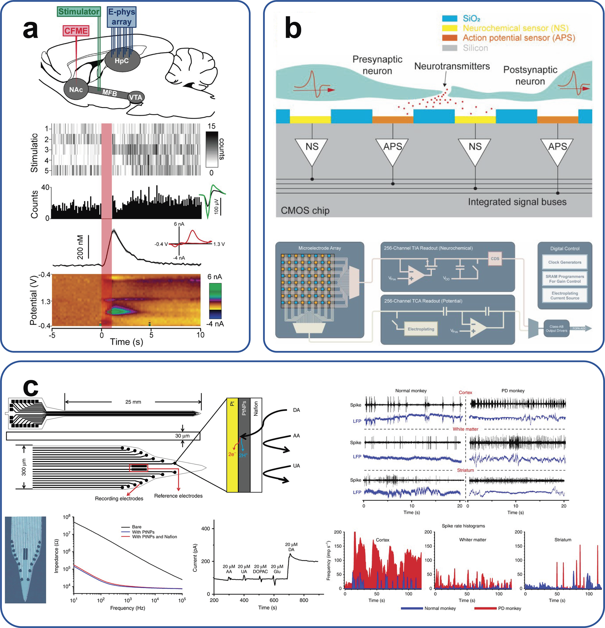
Over the past decade, various integrated electrochemical–electrophysiological systems have emerged for simultaneous neurochemical and electrical recordings in the living brain (Figure 4a, b) (Wei et al., Reference Wei, Song, Wang, Zhang, Luo, Xu and Cai2015; Parent et al., Reference Parent, Hill, Crown, Wiegand, Gies, Miller, Atcherley, Heien and Cowen2017; Mulberry et al., Reference Mulberry, White, Crocker and Kim2023). These include CFMEs, enzyme-coated electrodes, and multichannel microelectrode arrays (MEAs) using FSCV or amperometry (Wei et al., Reference Wei, Song, Wang, Zhang, Luo, Xu and Cai2015; Shin et al., Reference Shin, Wang, Borgus and Venton2019). For example, Shen et al. modified a silicon MEA with Glu oxidase to simultaneously record Glu dynamics and LFP/single-unit spikes (Wei et al., Reference Wei, Song, Wang, Zhang, Luo, Xu and Cai2015). Atcherley et al. developed the ‘DANA’ platform, combining high-density electrode arrays with a CFME to monitor single neuronal firing and DA levels simultaneously in the mouse brain (Parent et al., Reference Parent, Hill, Crown, Wiegand, Gies, Miller, Atcherley, Heien and Cowen2017). In primates, Zhang et al. used nanoengineered MEAs to achieve the first simultaneous recording of neuronal spiking, field potentials, and DA release in the monkey’s striatum (Figure 4c) (Zhang et al., Reference Zhang, Song, Wang, Xiao, Gao, Li, Tao, Zhuang, Yue, Chan and Cai2018).
Integrated electrophysiological and neurochemical sensors for real-time in vivo monitoring of neuronal activity and neurochemicals. (a) Simultaneous measurement of single-unit action potentials and DA release following medial forebrain bundle stimulation. DA levels were monitored with a CFME in the nucleus accumbens, while spike recording was performed in the contralateral hippocampus. Raster plots and peri-event histograms of neuronal responses, average DA concentration changes with characteristic voltammograms, and false-color time–voltage current plots were shown. Reprinted from Parent et al. (Reference Parent, Hill, Crown, Wiegand, Gies, Miller, Atcherley, Heien and Cowen2017) with permission from American Chemical Society. (b) Conceptual schematic and circuit block diagram of a dual-mode device enabling co-recording of electrophysiological and electrochemical signals. Reprinted from Mulberry et al. (Reference Mulberry, White, Crocker and Kim2023) with permission from MDPI. (c) Implantable microelectrode array (MEA) probe fabricated using silicon-on-insulator (SOI) substrates via microelectromechanical systems (MEMS) technology. Typical recordings illustrate signal distributions from three brain regions in both healthy and PD monkeys. Reprinted from Zhang et al. (Reference Zhang, Song, Wang, Xiao, Gao, Li, Tao, Zhuang, Yue, Chan and Cai2018) with permission from Springer Nature.

Moreover, architectures include flexible, dual-sided carbon-based probes capable of long-term electrical stimulation and neurotransmitter detection at nanomolar concentrations (Nimbalkar et al., Reference Nimbalkar, Castagnola, Balasubramani, Scarpellini, Samejima, Khorasani, Boissenin, Thongpang, Moritz and Kassegne2018). High-density complementary metal oxide semiconductor (CMOS) chip arrays have integrated hundreds of channels for electrochemical and electrophysiological recordings on a single chip (Mulberry et al., Reference Mulberry, White, Crocker and Kim2023). However, signal decoupling remains a critical challenge. To address this, spectral separation methods and multivariate modeling approaches (e.g. partial least squares (PLS) regression) are used to extract LFP components from the high-frequency portion of amperometric recordings and identify multiple neurotransmitters in real time (Movassaghi et al., Reference Movassaghi, Perrotta, Yang, Iyer, Cheng, Dagher, Fillol and Andrews2021).
Looking forward, advances in nanofabrication, sensor design, and ML-driven signal analysis will accelerate the development of the next generation of probes, which are expected to play an increasingly important role in both fundamental neuroscience research and clinical neurotechnology.
Integration of machine learning in brain chemical analysis
ML, a foundational branch of AI, enables the autonomous extraction of salient features, the construction of predictive models, and the identification of hidden patterns from large-scale experimental or observational datasets, with direct application to novel-sample prediction, classification, and decision-making. In recent years, ML has markedly accelerated both scientific discovery and tool development across disciplines including physics, chemistry, materials science, and the life sciences.
In the context of in vivo electrochemical sensing, ML’s computational power and algorithmic versatility are redefining traditional workflows. First, graph neural network and deep learning–based models can predict electrode nanostructures, electrocatalytic activity, and antifouling performance prior to synthesis, thereby dramatically shortening experimental validation timelines and reducing development costs. Second, ML-advanced feature extraction and denoising can effectively eliminate background noise and correct interference from complex high-throughput neurochemical recordings, thereby improving both processing speed and quantitative accuracy. Looking ahead, we highlight two principal avenues for ML integration in neurochemical electroanalysis, including rapid, data-driven acceleration of electrode interface design and advanced signal processing for real-time decoding of neurochemical dynamics. These directions promise to significantly accelerate biosensor development and enhance real-time interpretability of neurochemical dynamics in living systems.
ML accelerates electrode interface design
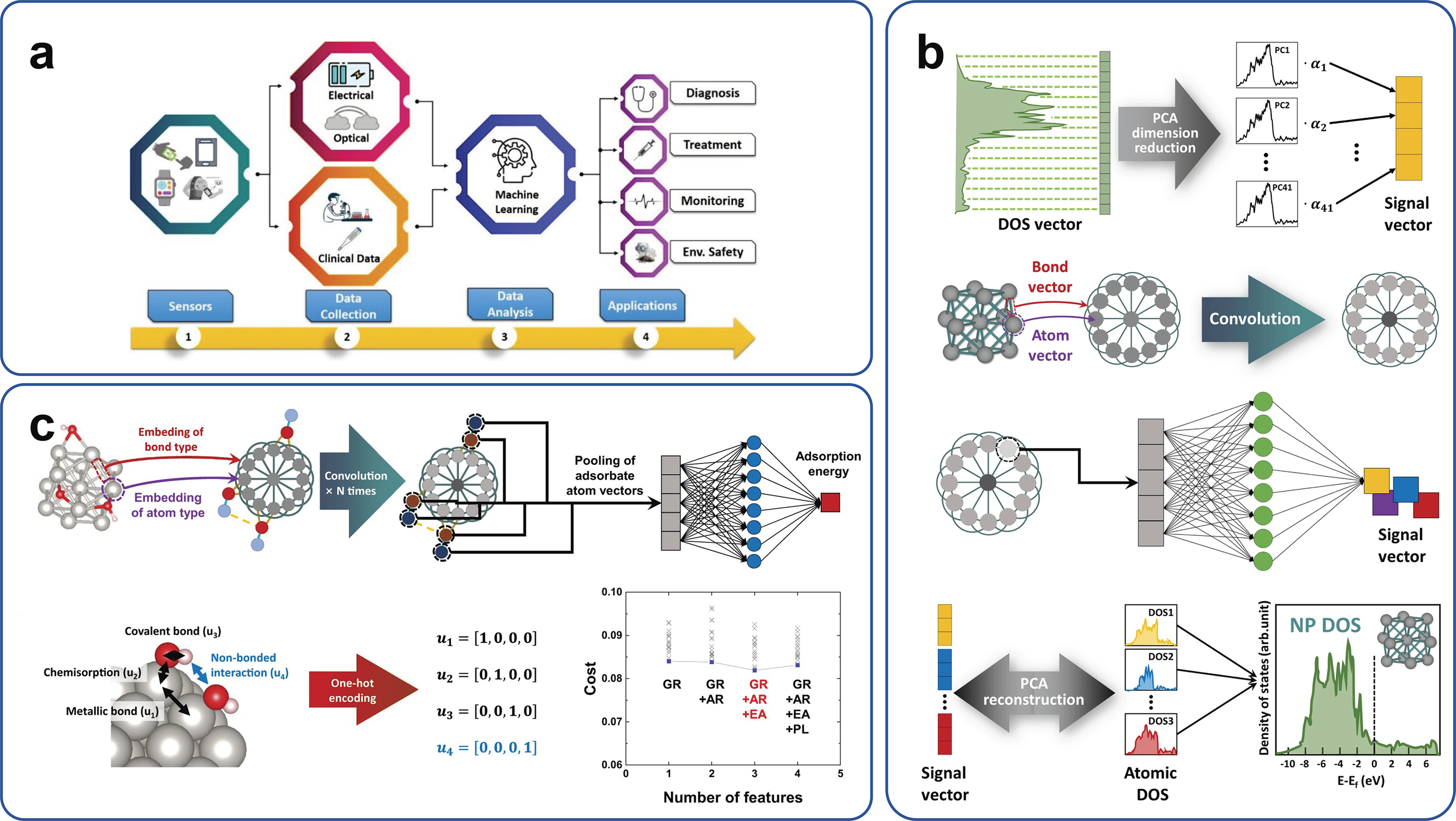
In vivo and in situ, chemical monitoring urgently demands electrochemical sensors with enhanced sensitivity, selectivity, and resistance to biofouling. However, the development of suitable interface materials is constrained by the complexity of their interactions with biological substrates and the high cost of empirical trial-and-error approaches. ML offers data-driven predictions shortening the time span of discovering new electrocatalytic materials and optimizing interface design on electrodes, which is more effective compared to the traditional way. When combined with conventional physics-based approaches, the data-driven ML offers complementary information, linking structure to function while improving generalizability and interpretability (Figure 5a) (Bhaiyya et al., Reference Bhaiyya, Panigrahi, Rewatkar and Haick2024). For example, Bang et al. developed a hybrid principal component analysis–crystal graph convolutional neural network (PCA-CGCNN) ML architecture that compresses high-dimensional density-of-states (DOS) images into low-dimensional vectors using PCA, while CGCNN captures the relationship between local atomic environments and DOS patterns via simple periodic table–derived features. Trained on only a few density functional theory (DFT)–computed DOS samples, this versatile model accurately predicted the electronic structures of both pure and bimetallic nanoparticles with a ~ 104-fold speedup over conventional DFT simulations (Figure 5b) (Bang et al., Reference Bang, Yeo, Kim, Han and Lee2021). This model is generalizable across diverse material systems, as it constructs input graphs from atomic coordinates, allowing applicability to a broad range of nanostructures.
ML-driven frameworks for material property prediction and structure–function analysis in nanoscale systems. (a) Schematic overview of the ML process flow, from sensing and data collection to model-based prediction and application. Reprinted from Bhaiyya et al. (Reference Bhaiyya, Panigrahi, Rewatkar and Haick2024) with permission from American Chemical Society. (b) Accelerated mapping of electronic density-of-states patterns of metallic nanoparticles via ML-based modeling. Reprinted from Bhaiyya et al. (Reference Bang, Yeo, Kim, Han and Lee2021) with permission from Springer Nature. (c) BE-CGCNN model accelerates the construction of Pourbaix diagrams, enabling the exploration of electrochemical stability over various NP sizes and shapes. Reprinted from Bang et al. (Reference Bang, Hong, Park, Kim, Han and Lee2023) with permission from Springer Nature.

Building on this framework, the authors proposed a bond-type embedded CGCNN (BE-CGCNN) to construct large-scale nanoparticle surface Pourbaix diagrams, which can predict surface coverages and adsorption energies, enabling high-throughput screening of optimal nanostructured electrode modifiers and accelerating electrode interface design (Figure 5c) (Bang et al., Reference Bang, Hong, Park, Kim, Han and Lee2023).
ML-enhanced signal preprocessing and feature extraction
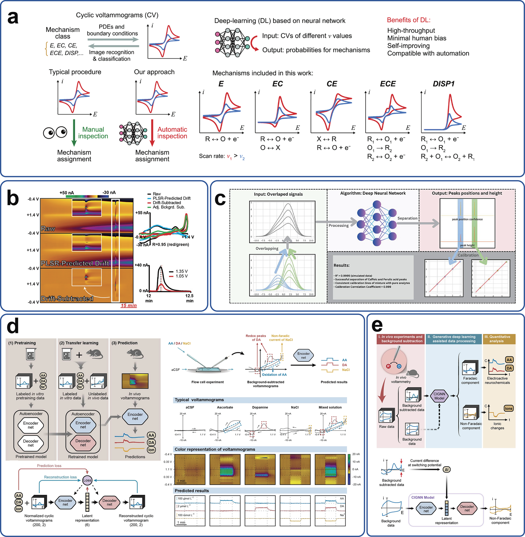
ML techniques such as elastic-net regression, support vector machines (SVMs), and deep neural networks (DNNs) are increasingly integrated into electrochemical signal processing workflows to correct baseline, remove drift, deconvolute overlapping peaks, and extract robust features automatically, thereby boosting both sensitivity and selectivity across multiple voltammetric and impedance-based measurements. For decades, investigations employing CV have required manual inspection of voltammograms. Recently, a deep learning–based algorithm has automated the extraction and multi-output quantification of multiple neurotransmitter signals, avoiding manual peak fittings (Figure 6a) (Hoar et al., Reference Hoar, Zhang, Xu, Deeba, Costentin, Gu and Liu2022). Despite the dominance of FSCV in subsecond monitoring of neurotransmitters in vivo, the high scan rates generate large capacitive charging currents that drift over time and obscure the true faradaic signal (Movassaghi et al., Reference Movassaghi, Alcañiz Fillol, Kishida, McCarty, Sombers, Wassum and Andrews2024). Traditional background-subtraction methods typically are effective only over short time windows, limiting long-term analysis. To overcome this, new ‘background-inclusive’ FSCV techniques are paired with ML algorithms such as elastic-net regression model and subtract the non-Faradaic drift directly from raw voltammograms, greatly improving signal-to-noise ratios and quantitation accuracy (Puthongkham and Venton, Reference Puthongkham and Venton2020; Hoar et al., Reference Hoar, Zhang, Xu, Deeba, Costentin, Gu and Liu2022; Movassaghi et al., Reference Movassaghi, Alcañiz Fillol, Kishida, McCarty, Sombers, Wassum and Andrews2024). To further mitigate background drift, Meunier et al. developed a dual-waveform PLS regression (DW-PLSR) model. A small triangular waveform captures electrochemical drift characteristics, which are then used by PLSR to predict and subtract the background drift across the full potential window, thereby improving the reliability of FSCV recordings beyond 10 minutes (Figure 6b) (Meunier et al., Reference Meunier, McCarty and Sombers2019).
ML enables automated voltammetric classification, drift correction, peak deconvolution, and integrated in vitro–in vivo signal analysis. (a) The bijective relationship between molecular electrochemical mechanisms and cyclic voltammograms. Comparison between manual inspection and automated classification and the function and proposed benefits of a deep learning algorithm trained on five molecular electrochemical mechanisms modeled by partial differential equations. Reprinted from Hoar et al. (Reference Hoar, Zhang, Xu, Deeba, Costentin, Gu and Liu2022) with permission from American Chemical Society. (b) FSCV data preprocessing using dual-waveform partial least squares regression (DW-PLSR). Color plots show raw signals, model-predicted drift, and drift-subtracted signals. FSCVs compare DW-PLSR and conventional background subtraction at the time of adenosine injection and current–time traces at the primary (+1.35 V) and secondary (+1.05 V) adenosine oxidation potentials. Insets show adenosine-induced fluctuations. Reprinted from Meunier et al. (Reference Meunier, McCarty and Sombers2019) with permission from American Chemical Society. (c) One-dimensional convolutional neural network (1D-CNN) trained on simulated and experimental voltammetric data, enabling accurate deconvolution of overlapping electrochemical peaks. Reprinted from Ciepiela et al. (Reference Ciepiela, Wójcik and Jakubowska2025) with permission from Elsevier. (d) Schematic of the deep learning–based voltammetric (DLV) sensing platform and representative deconvolution of in vitro voltammograms using the DLV model. Reprinted from Xue et al. (Reference Xue, Ji, Jiang, Yu and Mao2021) with permission from Wiley. (e) Methodological workflow of the CIGNN-based in vivo voltammetric analysis, comprising in vivo experiments (Step I), generative deep learning–assisted data processing (Step II), and quantitative analysis (Step III) and diagram of the CIGNN model architecture used to predict the non-Faradaic component of background-subtracted data. Reprinted from Li et al. (Reference Li, Xue, Sun, Wei, Wu and Mao2025) with permission from American Chemical Society.

CNNs are especially effective in electrochemical signal processing due to their ability to extract local features automatically and encode them at multiple scales. By applying one- or two-dimensional convolutional filters across potential–current traces, CNNs learn characteristic electrochemical patterns, such as peak shapes, step changes, and inflection points, without manual feature engineering. Their inherent parameter sharing and sparse connectivity reduce model complexity and enhance noise robustness. For example, a one-dimensional deep CNN (1D-DCNN) composed of multiple stacked 1D convolutional layers with rectified linear unit activations and pooling operations can extract features from raw voltammetric signals and predict peak height and position with high precision, achieving automated resolution of overlapping voltammetric signals (Figure 6c) (Ciepiela et al., Reference Ciepiela, Wójcik and Jakubowska2025).
Despite advances in background drift subtraction, selectively measuring multiple neurochemicals in vivo remains a grand challenge owing to their broad concentration ranges, rapid temporal dynamics, and overlapping electrochemical signals. To address this, Mao’s group developed a deep learning–based voltammetric sensing platform, which trains neural networks to deconvolve mixed currents recorded from a single CFME (Figure 6d). This approach enables the spatiotemporal separation of AA, DA, and ionic signals (Xue et al., Reference Xue, Ji, Jiang, Yu and Mao2021). Later, they introduced a chemistry-informed generative neural network (CIGNN) that leverages the distinct electrochemical signatures of Faradaic redox and non-Faradaic charging currents. By extracting instantaneous current changes at switching potentials, CIGNN enhances robustness against complex background interference and identifies variations in differential capacitance (Figure 6e) (Li et al., Reference Li, Xue, Sun, Wei, Wu and Mao2025).
Meanwhile, Internet of Things (IoT)–enabled electrochemical platforms upload raw sensor data via narrowband (NB)-IoT modules to cloud servers, where deep learning models perform multicomponent signal deconvolution and visualization, thus enabling scalable remote monitoring and management. While current implementations demonstrate robust lactate quantification, the modular architecture allows rapid adaptation to other biomarkers (Bill et al., Reference Bill, Jasper, Weltin, Urban, Rupitsch and Kieninger2023). Future iterations could incorporate simultaneous monitoring of multiple targets using multienzyme-functionalized arrays, predictive analysis using long short-term memory (LSTM) networks to anticipate metabolic crises, and edge–cloud hybrid architectures where preliminary feature extraction occurs on-device to reduce latency. These advances are shifting the electrochemical analysis paradigm from isolated single-point measurements toward dynamic, network-based in vivo monitoring, enabling seamless integration of molecular events with digital ecosystems.
ML in electrochemical sensing has progressed beyond purely data-driven feature extraction toward hybrid frameworks that embed domain-specific chemical and physical information. These algorithms can more effectively disentangle true analyte signals from complex background noise, offering superior robustness and interference resistance. Recent deep learning architectures now tackle the complexity of in vivo neurochemical mixtures, revealing dynamic biochemical processes beyond the reach of conventional methods. Future integration of online and federated learning may facilitate adaptive calibration across electrodes and collaborative model refinement across laboratories, ensuring reliable, long-term in vivo monitoring. Meanwhile, IoT-enabled systems are laying the groundwork for the next generation of precision neurochemical diagnostics and biosafety monitoring, bridging chemical information with the digital world. Ultimately, these innovations are transforming neurochemical monitoring, offering unprecedented spatiotemporal precision in decoding brain chemistry.
Prospective
Despite significant advances in in situ and in vivo brain electrochemical sensing, achieving long-term, high-fidelity neurochemical monitoring remains a multifaceted challenge. One major obstacle is electrode fouling caused by protein adsorption and glial encapsulation, which leads to signal drift and loss of sensitivity over time. Addressing this issue will require the development of next-generation biomimetic antifouling coatings, such as fluorinated porous membranes or GO composites, integrated with electrocatalysts that not only boost electrocatalytic activity but also offer robust resistance to chemical and biofouling (Gao et al., Reference Gao, Wei, Ma, Wu, Ji, Mao, Yu and Mao2024).
Another critical hurdle is the coexistence of multiple neurotransmitters with similar redox potential, which complicates sensing selectivity. To address this, integrating electrochemical impedance spectroscopy (EIS) with various voltammetric methods, such as differential pulse voltammetry, in a single microfluidic platform could provide multidimensional electrochemical fingerprints, paving the way for the construction of multiplexed ‘neurochemical maps’ with high spatiotemporal resolution (Buttkewitz et al., Reference Buttkewitz, Heuer and Bahnemann2023).
Chronic implantation of sensors in the brain also presents substantial biological challenges. Foreign-body responses and inflammation can degrade sensor performance within days. Mitigating these effects may involve engineering electrode interfaces with mimic antioxidant enzymes for ROS scavenging, employing flexible and biodegradable substrates (e.g. polyimide and poly(lactic-co-glycolic acid) (PLGA)) to reduce mechanical mismatch, and incorporating self-healing, drug-eluting coatings that respond to inflammatory cues to preserve sensor function over time (Xu and Hsu, Reference Xu and Hsu2023; Sun et al., Reference Sun, Li, Yang, Sun, Hou, Guan, Chen, Liu, Chen, Ma, Huang, Li, Wang, Wang, Chen, Cheng, Xiong, Sheng, Zhang, Peng, Wang, Wang and Yin2024).
In terms of data interpretation, electrochemical modalities such as voltammetry and amperometry often suffer from high background noise and overlapping multicomponent signals. This underscores the need for hybrid modeling frameworks that combine deep learning (e.g. convolutional autoencoders such as DiscrimNet, a network recently shown to accurately predict individual tonic concentrations of DA, norepinephrine, and serotonin from voltammetry data in vitro and in vivo in rats (Goyal et al., Reference Goyal, Yuen, Sinicrope, Winter, Randall, Rusheen, Blaha, Bennet, Lee, Shin and Oh2024) with chemometric techniques such as PLS and PCA. Such approaches could enable online signal deconvolution and real-time quantification, with enhanced generalizability across electrode types, brain regions, and subjects. Currently, ML-based background subtraction still relies on manual identification of interference sources and extraction of relevant features for iterative training. Future deep learning architectures may autonomously detect and suppress latent noise components, thereby improving both efficiency and accuracy in electrochemical signal processing.
Moreover, high-density sensor arrays and the demand for wireless, real-time data transmission impose strict constraints on power consumption and communication bandwidth. Addressing these limitations will likely require the integration of ultra-low-power analog-to-digital converters (ADCs), neuromorphic event-driven sampling hardware, and compact communication modules such as ultrawideband (UWB) (Ando et al., Reference Ando, Takizawa, Yoshida, Matsushita, Hirata and Suzuki2016) or near-field communication (NFC) to create compact, low-power closed-loop neural chemical sensing systems suitable for freely moving animal studies and future clinical applications.
Given the extreme complexity of neurochemical and electrophysiological signaling in the brain, it is imperative to integrate ML technologies to markedly increase data processing throughput and efficiently handle vast amounts of information in real time. Convergent advances across these domains, including material engineering, sensor design, signal processing, and intelligent computation, will ultimately unlock the potential of continuous, high-resolution monitoring of neurochemicals, providing new insights into brain function and dysfunction in both health and disease.
Open peer review
To view the open peer review materials for this article, please visit http://doi.org/10.1017/qrd.2025.10015.
Data availability statement
All relevant data are included in this publication or can be found in the cited literature.
Author contribution
JC and LC contributed to the writing and creation of figures. YL, JZ, XN, CL, and MX provided critical feedback and revised the manuscript. CY and XL supervised this work.
Financial support
This work was supported by the National Key Research and Development Program of China (2022YFA1206100), the Beijing Natural Science Foundation (Z230022 and L251024), the National Natural Science Foundation of China (22374005 and 21974154), and the Recruitment Program of Global Experts (Youth).
Competing interests
The authors declare none.


 Open access
Comments
Dear Professor Phan,
Thanks for your invitation of contributing to the special issue of on Nanoscale Analysis of Brain Chemistry and Structure. We would like to submit the attached perspective “Recent Advances of In-Situ and In-Vivo Electrochemical Analysis of Brain Chemistry at Micro- and Nano-Scale” to Quarterly Review of Biophysics Discovery. This work is original, unpublished, and is not considered for publication elsewhere. I hope you find it interesting for the Quarterly Review of Biophysics Discovery. We think this will be extremely important as there is a large interest in this area now and a lot of arguments!
In this study, we provide a concise review of recent advances in electrochemical analysis of brain chemicals. First, novel electrochemical techniques, including tailored electrode modifications have achieved unprecedented spatiotemporal resolution for in vivo and in situ detection of neurotransmitters and neuromodulators. At the same time, hybrid electrophysiology-electrochemistry approaches now allow simultaneous monitoring of neural activity and chemical signaling. Reflecting these trends, machine learning (ML) methods are increasingly applied to brain chemical analysis: ML not only accelerates electrode interface design but also effectively removes background noise and corrects for interference in high throughput neurochemical recordings, thereby boosting processing speed and quantitative accuracy. Building on this momentum, we propose a series of forward‑looking perspectives that span three key areas: biofouling‑resistant electrode coatings; microfluidic platforms integrating multiple electrochemical methods to generate multidimensional fingerprints; and machine‑learning–driven workflows that accelerate throughput and manage large datasets in real time. We are confident that our perspectives resonate with the Quarterly Review of Biophysics Discovery’s mission to focus on bold, hypothesis driven insights and new findings, advance the frontiers of biophysics, and ignite breakthroughs across disciplines.
Thank you in advance for your consideration and looking forward to hearing from you.
Sincerely,
Xianchan Li, Ph. D.
Assistant Professor
State Key Laboratory of Natural and Biomimetic Drugs
School of Pharmaceutical Sciences
Peking University
E-mail: xcli@pku.edu.cn