

Depression is common in older people, affecting at least 3% of people over 65 years old. Reference Beekman, Copeland and Prince1 It is associated with significant functional impairment and comorbidity, and there is growing evidence that vascular lesions may increase an older person's risk of developing depression. Reference Teodorczuk, Firbank, Pantoni, Poggesi, Erkinjuntti and Wallin2,Reference Vaishnavi and Taylor3 A limited number of functional neuroimaging studies, including single photon emission computed tomography (SPECT) and positron emission tomography (PET), have demonstrated focal or diffuse changes in regional cerebral blood flow (CBF) and cerebral glucose metabolism in older individuals with depression, but there are considerable inconsistencies between studies. In SPECT, regional CBF deficits in frontal and prefrontal areas appear a consistent finding in people with late-life depression, Reference Awata, Ito, Konno, Ono, Kawashima and Fukuda4–Reference Nobler, Roose, Prohovnik, Moeller, Louie and Van Heertum7 although anterior cingulate, temporal and parietal regions as well as caudate have also been shown to be implicated. Reference Awata, Ito, Konno, Ono, Kawashima and Fukuda4,Reference Ishizaki, Yamamoto, Takahashi, Takeda, Yano and Mimura5 Using PET, greater depressive symptoms were found to be associated with frontal lobe activity in late-life depression. Reference Dotson, Beason-Held, Kraut and Resnick8 However, others have found elevated bilateral glucose metabolism in frontal, precuneus and parietal areas in older study participants with depression. Reference Smith, Kramer, Ma, Kingsley, Dhawan and Chaly9
Arterial spin labelling (ASL) magnetic resonance imaging (MRI) is a non-invasive procedure for quantifying CBF. Unlike PET and SPECT techniques, ASL MRI has the advantage of not using radioactive sources or injected contrast agents, allowing for frequent scanning, especially useful in follow-up studies. In addition, anatomical scans can be obtained in the same session, allowing adjustment for effects of atrophy. Evidence from previous functional imaging suggests that frontal, cingulate, parietal and striatal regions may be affected in late-life depression, although there is a major lack of consistency between studies in terms of results and actual brain regions involved. This may be due to differences in samples, depression severity, illness duration or antidepressant medication, as well as in acquisition and analysis methods. Therefore, to help resolve some of the discrepancies in the limited literature in late-life depression and to obtain direct measures of CBF, we applied ASL MRI and region of interest (ROI) methods to investigate grey matter CBF in patients with late-life depression and similarly aged healthy individuals, focusing on specific brain areas including the lateral and medial frontal, cingulate, central and parietal cortices. White matter CBF was also examined, since among the most striking structural features of late-life depression are abnormalities in white matter, with both increased lesions Reference Herrmann, Le Masurier and Ebmeier10–Reference Teodorczuk, O'Brien, Firbank, Pantoni, Peggesi and Erkinjuntti12 and changes on diffusion imaging. Reference Shimony, Sheline, D'Angelo, Epstein, Benzinger and Mintun13,Reference Yang, Huang, Hong and Yu14 The striatum was not investigated owing to insufficient sampling of this region during the ASL acquisition. The decision to include participants with a current or previous episode of major depression was based on previous structural imaging studies indicating that brain changes are not reversible, are independent of current mood state and pre-date future depressive episodes. Reference Teodorczuk, Firbank, Pantoni, Poggesi, Erkinjuntti and Wallin2,Reference Teodorczuk, O'Brien, Firbank, Pantoni, Peggesi and Erkinjuntti12,Reference Godin, Dufouil, Maillard, Delcroix, Mazoyer and Crivello15 In view of previous research we predicted that regional CBF changes would be seen in frontal cingulate grey matter and more generalised changes in white matter in late-life depression compared with healthy individuals.
Method
Participants
We recruited from secondary care services patients over 60 years old with a history of a DSM-IV major depressive episode, current or previous. 16 These individuals were assessed by an old age psychiatrist (A.V.) and excluded from the study if they had dementia or a Mini-Mental State Examination (MMSE) score below 24, Reference Folstein, Folstein and McHugh17 comorbid or previous drug or alcohol misuse, a history of head injury, epilepsy or cerebrovascular accident, or a myocardial infarction in the preceding 3 months. Healthy older individuals with no personal psychiatric history were recruited from the same geographical area. The local research ethics committee approved the study and all participants gave written informed consent. Their assessment included family history of depression, previous psychiatric history, medical history and current medication. Current depression severity was rated using the Montgomery–Åsberg Depression Rating Scale (MADRS) and the 30-item Geriatric Depression Scale (GDS). Reference Montgomery and Åsberg18,Reference Yesavage19 Other measures recorded included age at onset of depression and duration of depression. Age at onset was determined by interviewing patients and reviewing all available psychiatric records. Duration of depression was ascertained by taking a life history and asking about the time of onset of each depressive episode and time taken for remission for each episode. This was based on information given by the patient and where available, clarification was sought from a partner or caregiver. The duration of each episode was then added up to give a lifetime estimate of duration of depression.
Imaging
Imaging was performed using a 3.0 T Achieva magnetic resonance scanner (Philips Medical Systems, Eindhoven, The Netherlands). The ASL MRI data were acquired using a multislice pulsed ASL flow-sensitive alternating inversion recovery (FAIR) sequence with six slices. The bottom slice was aligned to mid corpus callosum (Figs 1 and online DS1), no gaps, in-plane resolution 4 mm, slice thickness 6 mm, echo time (TE) 24 ms, repetition time (TR) 4000 ms and inflow time 1700 ms. In addition to ASL images, T 1-weighted images covering the whole brain were acquired: magnetisation-prepared rapid gradient echo (MP-RAGE), 150 sagittal slices, 1.2×1.15×1.15 mm3; TR 9.6 ms, TE 4.6 ms, flip angle 8°, SENSE factor 2. We used T 1 volumes for image registration, normalisation into standard space, and grey matter and white matter identification.
Data processing and image analysis
Using SPM8 (www.fil.ion.ucl.ac.uk/spm), the T 1-weighted three-dimensional image was segmented into grey and white matter, and coregistered with the mean perfusion image. Perfusion images were transformed to Montreal Neurological Institute space (MNI; www2.bic.mni.mcgill.ca/) using information from their T 1 coregistration/segmentation. Perfusion volume voxels common to all participants were used to define a mean perfusion mask. From their T 1 segmentation binary grey matter and white matter masks were created for each participant. Using the SPM toolbox MarsBaR (MARSeille Boîte à Région d’Intérêt, version 0.42, http://marsbar.sourceforge.net/), ROIs were then generated (Figs 2(a) and online DS2(a)). A white matter ROI (Figs 2(b) and online DS2(b)) was also derived from the white matter binary image described by the WFU PickAtlas version 2.4 (www.nitrc.org/projects/wfu_pickatlas/). Last, to reduce type 1 errors, corresponding ROIs from each hemisphere were combined.
Sagittal view depicting the acquisition volume.

Selected MarsBaR regions of interest overlaid on a magnetic resonance imaging template used for the measurement of regional blood flow in participants with late-life depression and healthy older individuals: (a) lateral frontal, medial frontal, cingulate, central, parietal; (b) white matter (blue).

From the ASL data we calculated an average and a subtraction image from the labelled and non-labelled scans for each participant; FMRIB's software library (FSL, version 4.01, www.fmrib.ox.ac.uk/fsl/) was used for further image analysis. For grey matter CBF measurements, images were multiplied by the mean perfusion, grey matter and ROI masks, whereas for white matter CBF measurement images were multiplied by the mean perfusion, white matter and ROI white matter masks. Specifically, the combined masks were applied to the average and subtraction ASL images to derive mean values in each ROI. The CBF was then calculated for each ROI from the mean values of average and subtraction ASL images by applying the general kinetic model, Reference Buxton, Frank and Wong20–Reference Kim and Tsekos22 using a blood T 1 of 1490 ms and blood–brain partition coefficient (γ) of 0.9. To reduce the possible inter-individual variability in CBF, we normalised all CBF ROI data by dividing each participant's ROI data by their global mean CBF value for grey matter.
Statistical analysis
Where appropriate, differences in demographic and clinical data were assessed using analysis of variance, Mann–Whitney U and chi-squared tests. Group differences in normalised CBF were investigated using analysis of covariance, with imaging parameters as dependent, diagnosis as a fixed factor and age as a covariate. Clinical correlates of normalised CBF were investigated using partial correlation r′ (age-corrected). A P value of <0.05 was considered significant.
Group characteristics

| Control group (n = 30) | Depression group (n = 38) | Test statistic | |
|---|---|---|---|
| Gender, m : f: n | 10 : 20 | 11 : 27 | χ2= 0.2, P = 0.7 |
| Age, years: mean (s.d.) | 74.4 (6.4) | 74.1 (6.1) | F 1,66 = 0.05, P = 0.8 |
| MMSE score, mean (s.d.) | 29.5 (0.8) | 28.8 (1.1) | F 1,66 = 9.02, P = 0.004** |
| TIV, cm3: mean (s.d.) | 1534.9 (165.9) | 1519.4 (162.8) | F 1,66 = 0.2, P = 0.7 |
| MADRS score, mean (s.d.) | NA | 12.9 (11.2) | |
| GDS score, mean (s.d.) | NA | 11.6 (7.9) | |
| Age at depression onset, years: mean (s.d.) | NA | 51.8 (22.3) | |
| Duration of depression, years: mean (s.d.) | NA | 23.0 (1.5) | |
| Medication, n | |||
| Antidepressants | 0 | 32 | |
| SSRIs | 0 | 9 | |
| Venlafaxine | 0 | 14 | |
| Duloxetine | 0 | 3 | |
| Mirtazapine | 0 | 19 | |
| Tricyclics | 0 | 0 | |
| Antihypertensives | 14 | 18 | |
| Diuretics | 6 | 10 | |
| Beta blocker | 7 | 11 | |
| ACE inhibitor | 5 | 10 | |
| ARB | 2 | 2 | |
| Calcium channel blocker | 7 | 5 | |
| Alpha blocker | 0 | 0 | |
| Statins | 10 | 14 | |
| Antiplatelets | 9 | 13 | |
| Warfarin | 3 | 1 |
ACE, angiotensin converting enzyme; ARB, angiotensin II receptor blocker; GDS, Geriatric Depression Scale; MADRS, Montgomery-Åsberg Depression Rating Scale; MMSE, Mini-Mental State Examination; NA, not applicable; TIV, total intracranial volume; SSRI, selective serotonin reuptake inhibitor.
* <P<0.05
** P<0.01.
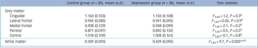
Regional cerebral blood flow volumes in the two study groups, normalised to mean global grey matter flow
| Control group (n = 30), mean (s.d.) | Depression group (n = 38), mean (s.d.) | Test statistic | |
|---|---|---|---|
| Grey matter | |||
| Cingulate | 1.163 (0.103) | 1.134 (0.108) | F 1,65 = 1.2, P=0.3Footnote a |
| Lateral frontal | 0.944 (0.085) | 0.941 (0.092) | F 1,65 = 0.04, P = 0.9Footnote a |
| Medial frontal | 0.908 (0.129) | 0.948 (0.099) | F 1,65 = 2.1, P=0.2Footnote a |
| Parietal | 0.871 (0.097) | 0.892 (0.132) | F 1,65 = 0.5, P=0.5Footnote a |
| Central | 1.018 (0.109) | 1.008 (0.161) | F 1,65 = 0.1, P=0.8Footnote a |
| White matter | 0.359 (0.093) | 0.429 (0.095) | F 1,65 = 9.7, P = 0.003Footnote ** Footnote a |
a. Statistical evaluations performed on age-corrected means.
* P<0.05
** P<0.01.
Results
Demographic and clinical characteristics of the sample are given in Table 1. The depression and control groups were comparable for gender, age and total intracranial volume. Thirty-two patients with late-life depression were receiving antidepressants (selective serotonin reuptake inhibitors, venlafaxine, duloxetine or mirtazapine). No patient was taking a tricyclic antidepressant. A third of the currently depressed group (n = 8) had had no previous episode of depression; just over half (54%, n = 13) had experienced one to five lifetime episodes, 4% (n = 1) had six to ten episodes and 8% (n = 2) had experienced more than ten episodes. Using age at onset <60 years to define early-onset depression, the early-onset (n = 19) and late-onset (n = 19) groups were similar in age, in MMSE, GDS and MADRS scores (F 1,36⩽1.6, P⩾0.2) and in gender (χ2 = 3.2, P = 0.07).
Box plot of normalised white matter cerebral blood flow in the control group and in participants with late-life depression.

Significant increases in normalised CBF (Table 2) were observed in white matter in patients with late-life depression relative to the control group (F 1,65 = 9.7, P = 0.003, 19% increase). Figure 3 shows the distribution of normalised CBF values in white matter. Grey matter CBF in lateral frontal, medial frontal, cingulate, central and parietal regions did not significantly differ between groups (F 1,65⩽2.1, P⩾0.2). Antidepressant effects on normalised CBF measures were also investigated in the depression group, yielding non-significant results (F 1,36⩽1.1, P⩾0.3). Grey matter and white matter CBF were also indistinguishable between the early-onset and late-onset groups (F 1,36⩽2.4, P⩾0.13).
Characteristics of depression group participants categorised by remission

| RemissionFootnote a (n = 21) | Non-remission (n = 17) | Test statistic | |
|---|---|---|---|
| Gender, m : f : n | 6 : 15 | 5 : 12 | χ2 = 0.003, P = 0.9 |
| Age, years: mean (s.d.) | 72.9 (5.1) | 75.5 (7.0) | F 1,36 = 1.8, P=0.2 |
| MMSE score, mean (s.d.) | 28.7 (1.2) | 28.9 (1.0) | F 1,36 = 0.4, P=0.5 |
| TIV, cm3: mean (s.d.) | 1521.6 (74.6) | 1516.6 (152.3) | F 1,36 = 0.009, P = 0.9 |
| MADRS score, mean (s.d.) | 5.2 (3.3) | 22.3 (10.2) | F 1,36 = 52.1, P<0.001Footnote *** |
| GDS score, mean (s.d.) | 6.3 (4.9) | 18.1 (5.9) | F 1,36 = 45.1, P<0.001Footnote *** |
| Age at depression onset, years: mean (s.d.) | 46.1 (22.9) | 57.2 (20.6) | F 1,36 = 2.4, P=0.1 |
| Duration of depression, years: mean (s.d.) | 26.8 (22.0) | 18.3 (20.6) | F 1,36 = 1.5, P=0.2 |
| Medication, n | |||
| Antidepressants | 16 | 16 | |
| SSRIs | 3 | 6 | |
| Venlafaxine | 6 | 8 | |
| Duloxetine | 3 | 0 | |
| Mirtazapine | 8 | 11 | |
| Tricyclics | 0 | 0 | |
| Antihypertensives | 9 | 9 | |
| Diuretics | 5 | 5 | |
| Beta blocker | 8 | 3 | |
| ACE inhibitor | 5 | 5 | |
| ARBs | 0 | 2 | |
| Calcium channel blocker | 1 | 4 | |
| Alpha blocker | 0 | 0 | |
| Statins | 7 | 7 | |
| Antiplatelets | 6 | 7 | |
| Warfarin | 1 | 0 |
ACE, angiotensin converting enzyme; ARB, angiotensin II receptor blocker; GDS, Geriatric Depression Scale; MADRS, Montgomery-Åsberg Depression Rating Scale; MMSE, Mini-Mental State Examination; TIV, total intracranial volume; SSRI, selective serotonin reuptake inhibitor.
a. Remission defined as an MADRS score of 10 or below.
* P<0.05
** P<0.01
*** P<0.001.
Associations between normalised CBF values and key depression variables (MADRS and GDS scores, age at onset of depression and duration of depression) in late-life depression were examined. A significant relationship was found between white matter CBF and MADRS score (r′ = –0.42, P = 0.005) as well as GDS score (r′ = –0.32, P = 0.03), but not for age at onset of depression (r′ = 0.06, P = 0.4) or duration of depression (r′ = –0.06, P = 0.4). Correlations between grey matter CBF and these variables were all non-significant (0.006⩽r′⩽0.37, P⩾0.18). Age and gender were also investigated but did not correlate with white matter or grey matter CBF (0.003⩽r⩾0.28, P = 0.09).
Participants with depression were then split into groups: remission (n = 21, MADRS⩽10) and non-remission (n = 17, MADRS⩾11). Reference Zimmerman, Posternak and Chelminski23 These subgroups were comparable in age, gender, MMSE score, age at depression onset and duration of depression (Table 3). Relative proportions taking antidepressant medication were also similar. A significant difference in white matter CBF was observed among depression subgroups (Table 4) and controls (F 2,64 = 8.0, P = 0.001), but not for grey matter CBF (F 2,64⩽1.1, P⩾0.3). Post hoc tests revealed white matter CBF was significantly increased in the remission group compared with controls (P = 0.001), suggestive relative to the non-remission group (P = 0.06) but similar between non-remission and control groups (P = 0.80).
Discussion
Using the ASL imaging technique we found significant elevation of normalised white matter CBF in patients with late-life depression compared with healthy older individuals. However, normalised grey matter flow in lateral frontal, medial frontal, cingulate, central and parietal regions did not significantly differ between groups. To our knowledge this is the first report demonstrating increased white matter CBF associated with late-life depression. Younger adults with depression have shown increased CBF in frontal, central, temporal and parietal regions compared with healthy adults when ROI methods are used, although such procedures cannot determine separate flow in grey matter or white matter. Reference Pagani, Gardner, Salmaso, Sánchez Crespo, Jonsson and Jacobsson24 Others have reported elevated cerebral glucose metabolism in frontal, precuneal and parietal areas in older patients with depression, Reference Smith, Kramer, Ma, Kingsley, Dhawan and Chaly9 although this was associated with atrophy in the same regions. As well as increased white matter lesions, impairments to white matter connectivity have also been described in late-life depression using diffusion tensor imaging. Reference Shimony, Sheline, D'Angelo, Epstein, Benzinger and Mintun13,Reference Yang, Huang, Hong and Yu14 Further investigations are required in order to understand the importance of such white matter changes and their influence on depressive symptoms in late-life depression. Our study included participants with depression and those who had recovered, with the mean MADRS score of 12.9 reflecting this.
Regional cerebral blood flow volumes, normalised to mean global grey matter flow, in depression group participants categorised by remission state

| Remission group (n = 21) Mean (s.d.) |
Non-remission group (n = 17) Mean (s.d.) |
|
|---|---|---|
| Grey matter | ||
| Cingulate | 1.135 (0.126) | 1.134 (0.084) |
| Lateral frontal | 0.926 (0.099) | 0.960 (0.080) |
| Medial frontal | 0.939 (0.082) | 0.959 (0.119) |
| Parietal | 0.901 (0.147) | 0.881 (0.114) |
| Central | 1.032 (0.180) | 0.978 (0.135) |
| White matter | 0.456 (0.093) | 0.395 (0.090) |
In the late-life depression group a highly significant correlation was found between white matter CBF and MADRS score. Therefore CBF results may have been influenced by the current mood state of people in this group. In an attempt to investigate this issue further, the cohort was dichotomised into those with remitted and non-remitted depression. Elevation of white matter CBF was apparent in the remission group relative to the non-remission and control groups. Clinical characteristics were broadly similar in the two depression subgroups. Results may suggest that increased white matter CBF could be either a response to, or a predictor of, successful antidepressant treatment, perhaps by facilitating neurotransmission in specific circuits and so reducing depressive symptoms.
Late-life depression research has mainly focused on studying structures implicated within the limbic–cortical–striatal–pallidal–thalamic circuit. White matter hyperintensities and abnormalities of white matter tracts may indicate disturbances to specific neural circuits or fibre tracts involved in mood regulation. Increased white matter CBF could represent a response to white matter pathological change or be a marker of those who respond to treatment. However, the latter theory would need to be tested in prospective studies.
We acknowledge that results specifically in grey matter CBF may have been underpowered owing to the heterogeneity of our depression cohort, almost two-thirds of whom were in a currently depressed state. Other limitations of our study include depressive symptoms in controls and striatal CBF not being measured. In conclusion, our findings combined with previous evidence showing increased white matter lesions and both connectivity and diffusion changes, further suggest that white matter changes may be more relevant than those in grey matter for explaining the underlying neurobiology of late-life depression. Future studies investigating functional imaging changes in the depressed and recovered state of late-life depression are required.
Funding
This work was supported by the UK National Institute for Health Research Biomedical Research Centre for Ageing and Age-related Disease award to the Newcastle upon Tyne Hospitals National Health Service Foundation Trust.
Acknowledgements
We thank all participants for their invaluable contribution.


eLetters
No eLetters have been published for this article.