

Introduction
The planning target volume (PTV) as per the International Commission of Radiation Units and Measurements (ICRU 50 & 62) state that a geometrical margin should be used around the clinical target volume (CTV) to ensure adequate coverage.1 In local prostate radiotherapy the CTV mostly consists of the prostate, any extension with/out some/all of the seminal vesicles (SVs). In order to calculate the PTV margin for a CTV, the systematic and random errors for individual patient/s and for a whole population sample must be calculated.2 Most centres have different values for their systematic and random errors because of different imaging techniques, verification methods, immobilisation, set up procedures, bladder/rectal filling protocols, delineation variability and uncertainties in equipment. Without the use of cone beam computer tomography (CBCT) and tracking of intra-fraction motion, calculation methods are often employed to help guide PTV margin selection based on coverage probability. It is therefore justified to investigate these errors and incorporate them into local PTV margins. Adaptive radiotherapy with patient-specific margins adjusting of the PTV may be ideal, but where this is not feasible, we must use as close a sample that is representative of our patient population.
Our institutions current conformal radiation therapy (CRT) verification uses a 3 mm tolerance offline bony anatomy matching method. MV electronic portal images (EPIs) are taken days 1–3 and weekly. Days 1–3 EPIs are averaged to get the mean and corrected for if ≥4 mm. Any image after week 1, that is >3 mm will be repeated the following day and if both when averaged are >3 mm, the move is implemented. This means the first three images are without any corrections (unless a gross error occurs). These patients are treated with two phases delivering 56 Gy in 28 fractions, 2 Gy per fraction and then 18 Gy in nine fractions. Our phase 1 PTV56 Gy margins are 9 mm RL, SI and anterior with 6 mm posteriorly. Phase 2 PTV74 Gy consists of 6 mm RL, SI, anterior and 3–6 mm posteriorly. These displacements only give combined information regarding set-up errors and phantom transfer errors.2
Arc-modulated radiotherapy (AMRT) patients have daily online kilovoltage (kV) image-guided radiotherapy (IGRT). Patients have 74 Gy in 37 fractions, 2 Gy per fraction and undergo fiducial marker insertion before the planning computed tomography (CT). The full displacements in the RL, SI and AP direction between isocentres of the CT and EPIs are applied using the automatic couch. This allows the measure not only of the set up/phantom transfer errors, but also systematic and random inter-fraction motion of the prostate, which is related to bladder/bowel changes. The current PTV71 Gy used for these patients is 1 cm RL, SI and ant with a 5 mm posteriorly. PTV74 Gy consists of 5 mm in all directions. Patients undergoing CT and treatment were supine with knee and ankle immobilisation. A comfortably full bladder and empty rectum was required. Any patients with a rectal diameter ≥5 cm would undergo a repeat CT with laxatives. This would continue throughout treatment unless contraindicated.
This study aims to:
• Address the uncertainties to be considered when deciding on calculating PTV margins.
• Calculate PTV margins with the aid of published geometric uncertainties for four different verification methods including offline 3 mm tolerance bony anatomy for our CRT patients, daily online bony anatomy, skin alignment only and full online prostate marker matching for AMRT patients.
• Compare our current margins with those calculated and the literature.
Methods and materials
Verification techniques
Seventy patients were assessed that had CRT and offline bony imaging. This consisted of orthogonal megavoltage (MV) images that were assessed manually by two trained therapeutic radiographers using Royal College of Radiologists (RCR) guidelines.2 Displacements in the RL, SI and AP were recorded. Only phase 1 images were included in this study which consisted of the majority of images. The total pairs of images assessed were 490. The CRT sample verification was based on a 3 mm tolerance. No corrections were applied until after three fractions were averaged. Therefore, analysis of the first three fractions alone was assessed to demonstrate the likely errors if no correction were applied (no CS, first 3 #s). Then the entire sample displacements were assessed, thus representing the ‘current 3 mm tolerance’ sample.
Twenty-three separate AMRT patients underwent daily online orthogonal kV EPIs. These images were manually matched by the same staff group overlaying the fiducial markers from the EPI and the CT. The full shift in the RL, SI and AP was applied using the automatic couch. No rotational, pitch or yaw errors were corrected. The total pairs of images assessed were 851. All the displacements in the RL, SI and AP directions from both CRT and AMRT patient's data were exported from ARIA to Microsoft Excel 2003 for statistical analysis. All SI errors were averaged over both lateral and anterior digitally reconstructed radiographs (DRRs).
Measuring systematic (∑) and random errors (∂)
Systematic errors are deviations occurring in the same direction and magnitude over the treatment course shifting the dose from the CTV.Reference Van Herk3 These are pre-treatment as opposed to treatment errors and are more important than random errors. According to the RCR and British Institute of Radiology (BIR),2, 4 the combined systematic error consists of four sources combined in quadrature. This ensures margins are not excessively large1, Reference Stroom and Heijmen5:
 $${ \mathop{\sum}{^{{\rm{2}}} } \: = \:\mathop{\sum}{^{{\rm{2}}} } \,{\rm{delineation}}\:\cr\quad + \:\mathop{\sum}{^{{\rm{2}}} } \,{\rm{motion}}\: \ (\rm {inter\hbox{-}fraction})\cr\quad+ \:\mathop{\sum}{^{{\rm{2}}} } \,{\rm{phantom}} \ {\rm{transfer}} \ {\rm{error}}\:\cr\quad + \:\mathop{\sum}{^{{\rm{2}}} } \,{\rm{patient}} \ {\rm{set}} \ {\rm{up}} \eqno\rm$$
Similarly, the combined random error consists of three sources combined in quadrature:
 $${ \partial^2 {\rm{}}\, = \,\partial^2 {\rm{}}\,{\rm{patient}}\,{\rm{set}}\,{\rm{up}}\, \cr\quad+ \,\partial^2 {\rm{}}\,{\rm{motion}}\, (\rm {inter\hbox{-}fraction})\cr\quad+ \,\partial^2 {\rm{}}\,{\rm{intra }}- fraction \ {\rm{motion}} \eqno\rm$$
The intra-fraction motion is displayed as a separate entity to demonstrate that this was not corrected for in any of the verification methods and published data was used. For the CRT patients, bony anatomy EPIs shows the combined effect of ∑2 phantom transfer + ∑2 patients set up error, as well as the random ∂2 patient set up. It gives no information regarding delineation or any motion.4, Reference McGarry, Cosgrove, Fleming, O'Sullivan and Hounsell6 Therefore, ∑2 = ∑2 delineation + ∑2 motion + ∑2 patient set up (includes ∑2 phantom transfer error). Combined treatment execution consists of ∂2 = ∂2 patient set up + ∂2 motion + ∂2 intra-fraction motion. The assessment of fiducial marker displacements gives information of the combined effect of ∑2 motion + ∑2 phantom transfer + ∑2 patient set up, but not delineation. It also gives a combined ∂2 patient set up + ∂2 inter-fraction motion, but no intra-fraction motion information. Therefore, ∑2 = ∑2 delineation + ∑2 patient set up (includes ∑2 motion + ∑2 phantom transfer error). Combined treatment execution will be ∂2 = ∂2 patient set up (includes ∂2 inter-fraction motion) + ∂2 intra-fraction motion. The population systematic errors for the CRT and AMRT patients were calculated according to the RCR.2
Prostate PTV margin calculation
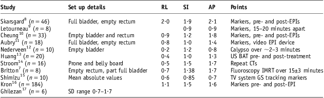
The full displacements measured for the prostate marker sample represent the required PTV margins for skin alignment only. As a result of full online fiducial marker correction for the AMRT patients, the only errors not corrected for are ∑2 delineation and ∂2 intra-fraction motion. The intra-fraction motion and delineation parameters were taken from the literature. Table 1 shows the intra-fraction motion values from literature. The parameters used in this study were that of Britton et al.Reference Britton, Yoshihiro, Masatoshi, Kenji, Yoshihiro and Shogo7 This appears to be a reasonable average estimation for this uncertainty among several studies.
Literature-based prostate intra-fraction motion SDs (mm)

Abbreviations: SD, standard deviation; RL, right–left; SI, superior–inferior; AP, anterior–posterior; EPI, electronic portal image; US, ultrasound; BAT, B-mode acquisition and targeting; CT, computed tomography; GS, Gold Seed.
Table 24 shows geometric uncertainties used in the margin calculations. The ∑2 motion (inter-fraction) and ∂2 motion values below were used for the CRT patients but were measured within the overall error in the prostate marker patients. MRI has been shown to be more accurate with reduced intra and inter-observer variability in prostate delineation.Reference Rasch, Barillot, Remeijer, Touw, van Herk and Lebesque18 It has also been shown to result in smaller prostate volumes (∼40%) versus CT implying that what we treat/contour may indeed be larger.Reference Rasch, Barillot, Remeijer, Touw, van Herk and Lebesque18, Reference Aubin, Leverdiere and Gingras19 Thus, some do not consider this error because of CT overestimation, but our calculated margins considers it and may therefore be somewhat generous. BIR4 recommends a ∑2 delineation of 2·0 mm for MRI, but as we use CT, those of Rasch et al.Reference Rasch, Barillot, Remeijer, Touw, van Herk and Lebesque18 were used.
Geometric uncertainties and calculation sheet used for prostate PTV margins

Abbreviations: PTV, planning target volume; RL, right–left; SI, superior–inferior; AP, anterior–posterior; FM, fiducial marker.
PTV margin calculations
The commonly used Van Herk et al.Reference Van Herk, Remeijer, Rasch and Lebesque20 PTV margin formula 2·5∑ + 0·7∂ was used. This ensures a minimum dose to the CTV of 95% for 90% of patients. This margin takes into account a beam penumbra of ∼3·2 mm and planning parameter of 1·64.Reference Van Herk, Remeijer, Rasch and Lebesque20
Results
Systematic and random errors
Table 3 shows the combined population systematic ∑ and random errors ∂ for the bony anatomy matching CRT and AMRT prostate marker patients. Here for CRT patient calculation purposes total ∑2 = ∑2 delineation + ∑2 motion + ∑2 patient set up (includes ∑2 phantom transfer). Similarly, ∂2 = ∂2 patient set up + ∂2 motion + ∂2 intra-fraction motion. Daily online bony anatomy only eliminates the set up/phantom transfer errors.
Combined population systematic and random errors for MV offline bony anatomy in CRT patients and daily kV online marker correction in AMRT patients

Abbreviations: MV, megavoltage; CRT, conformal radiotherapy; kV, kilovoltage; AMRT, Arc-modulated radiotherapy; RL, right–left; SI, superior–inferior; AP, anterior–posterior; CS, correction strategy.
For the fiducial marker patients, the total ∑2 = ∑2 delineation + ∑2 patient set up (includes ∑2 motion + ∑2 phantom transfer). Also, ∂2 = ∂2 patient set up (includes ∂2 motion) + ∂2 intra-fraction motion.
CTV-PTV margins
Table 4 shows the calculated PTV margins for the different verification methods. It can be seen that the margins required are greatly reduced when preforming daily marker matching. Tattoo alignment only and daily bony anatomy margins do not differ greatly however.
Prostate PTV margins using the Van Herk et al.Reference Van Herk, Remeijer, Rasch and Lebesque20 method 2·5∑ + 0·7∂

Abbreviations: PTV, planning target volume; CRT, conformal radiation therapy; CTV, clinical target volume, RL, right–left; SI, superior–inferior; AP, anterior–posterior.
Discussion
Systematic and random errors
As showed by studies,Reference Van Herk3, 4, Reference Van Herk, Remeijer, Rasch and Lebesque20 the systematic error is the main contributor to PTV margins. Table 3 shows that when comparing the ∑ and ∂ for no CS patients and our current 3 mm tolerance, there is not much difference. This demonstrates most patient set ups are within 3 mm bony tolerance with no action required. By introducing full online bony anatomy correction, this does reduce ∑ and ∂, but not by a significant amount. Using online prostate marker correction, the corresponding ∑ and ∂ is greatly reduced and in turn the PTV margin and volume as per formula: Volume = 4/3 × pi × r 3.Reference Langenhuijsen, Smeenk and Louwe21
Intra-fraction motion and marker matching
Intra-fraction motion is a limiting factor in PTV margin reduction.Reference Beltran, Herman and Davis22Table 1 shows literature results for intra-fraction motion. This source of uncertainty is not corrected for in online marker matching. Therefore, we must recognise it, quantify it and account for it in the PTV.Reference Boda-Heggemann, Köhler, Wertz, Ehmann, Hermann and Riesenacker23 The values used were that of Britton et al.Reference Britton, Yoshihiro, Masatoshi, Kenji, Yoshihiro and Shogo7 where intra-fraction motion was measured on 40 patients with an empty rectum and partially full bladder. It was felt this was a close match for our currently used bladder/rectal protocol and overall a good average estimation. Kron et al.Reference Kron, Thomas and Fox16 also found similar results of 1·1 mm RL, 1·5 mm SI and 1·6 mm AP on 184 patients with pre- and post-treatment marker displacements. Similar values have also been found using real time tracking. These intra-fraction values were only used within the random uncertainties and felt not to have a systematic element. Ideally extracting other department's figures may be potentially dangerous, but if the figure used represents an average/over approximation, this will give a worse case scenario representing a good estimate of the safety margin.
The current study was on both CRT and AMRT patients with different amounts of time per fraction. Van den Heuvel et al.Reference Van den Heuvel, Slagmolen and Budiharto24 found that there is a significant increase in probability of prostate motion as time elapses. Our CRT patients have appointments of 10 minutes with AMRT delivery time ∼2 minutes.Reference Kjaer-Kristoffersen, Ohlhues, Medin and Korreman25 Therefore, as suggested by others,Reference Kron, Thomas and Fox16, Reference Boda-Heggemann, Köhler, Wertz, Ehmann, Hermann and Riesenacker23, Reference Otto26 reducing time may result in reduced intra-fraction motion. The intra-fraction motion standard deviations (SDs) used in this study may therefore be abit generous for the AMRT group. Options to reduce the time include quicker delivery and verification. Manual matching may introduce observer variability versus automatic and may take longer. Another approach would be to correct for intra-fraction motion by online fiducial tracking as suggested by Shimizu et al.Reference Shimizu, Shirato and Kitamura15
Prostate markers migration has been looked at and Nederveen et al.Reference Nederveen27 found a systematic error of 0 and SD of 0·6 mm. Although the study highlights the difficulty in distinguishing between migration and prostate deformation, it is of the author's opinion that it would not be unreasonable to disregard this minute uncertainty of migration.
PTV margins and current margins
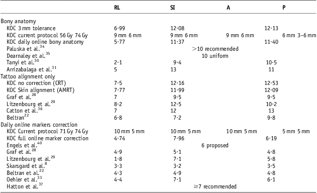
Table 5 shows the calculated margins and margins from other centres. Based on the calculations, our CRT bony anatomy patient's margins may be reduced for phase 1 laterally, whereas the current phase 2 margins are likely too small by ∼6 mm SI and AP.
Prostate PTV margins (mm)
Abbreviations: PTV, planning target volume; KOC, Kent Oncology Centre; RL, right–left; SI, superior–inferior; AP, anterior–posterior; CRT, conformal radiotherapy; AMRT, Arc-modulated radiotherapy.
Aligning to skin tattoos only requires larger PTV margins. Graf et al.Reference Graf, Wust, Budach and Boehmer28 found slightly smaller results for supine patients whereas Beltran et al.Reference Beltran, Herman and Davis22 found margins (mm) of 6·8 RL, 7·2 SI and 9·8 AP. Litzenbourg et al.Reference Litzenberg, Balter and Hadley29 who tracked implanted markers providing intra-fraction motion found similar margins to ours. If daily online bony anatomy verification was introduced at our centre, this would only reduce the PTV margins (mm) by 1 RL, 0·7 SI and 0·7 AP. This would, however, increase time and possible intra-fraction motion and therefore not justified given the small set up error. Online bony anatomy matching margins found in this study would be (mm) 6 RL, 11 SI and 11 AP. Tanyi et al.Reference Tanyi, He and Summers30 found PTV margins for bony anatomy alignment of 2·1 RL, 9·4 SI and 10·5 AP whereas those by Arrizabalaga.Reference Alonso-Arrizabalaga, Brualla González, V Roselló Ferrando, Pastor Peidro and Torrecilla31 which included intra-fraction motion consisted of 5 RL, 13 SI and 11 AP.
When undertaking full online marker correction versus tattoos, margins (mm) are reduced to 5 RL, 8 SI and 6 AP. The current AMRT margins imply the 71 Gy margins (mm) as being excessive by 5 RL, 2 SI and 4 anterior. Skarsgard et al.Reference Skarsgard, Cadman and El-Gayed8 stated that correcting for all pre-treatment displacements using markers that the margin required for intra-fraction motion would be (mm) 3·3 RL, 3·2 SI and 3·5 AP. These small margins, however, do not consider ∑2 delineation and use a recipe by Antolak et al.Reference Antolak, Rosen, Childress, Zagars and Pollack32 (SD × 1·65). Litzenberg et al.Reference Litzenberg, Balter and Hadley29 included intra-fraction motion only for markers patients and found a similar PTV margin to ours using Van Herks recipe.Reference Van Herk, Remeijer, Rasch and Lebesque20 BeltranReference Beltran, Herman and Davis22 and GrafReference Graf, Wust, Budach and Boehmer28 also using this found margins of ∼5 mm. Oehler et al.Reference Oehler, Lang and Dimmerling33 found a similar margin which also included delineation and intra-fraction prostate motion.
Clinical issues
The effect of small changes of the PTV margins on control and late toxicity is not clear. A balance must be maintained between intent and minimising toxicity and at our institution a smaller post margin to reduce rectal toxicity similar to elsewhere is used.Reference Barrett, Dobbs, Morris and Roques38 Langenhuijsen showed the dosimetric advantage of reducing the PTV margins (mm) from 10 to 7 and 7 (5 post). This resulted in a reduction in the PTV mean volume by 27% and dose to the bladder, rectal wall and anal canal was reduced by a mean of 17%, 12% and 19%, respectively. A further reduction was evident when a 5 mm post margin was used.Reference Langenhuijsen, Smeenk and Louwe21 Safe margin reduction was clinically supported by Dearnaley in a randomised controlled trial comparing 1 with 1·5 cm PTV margins using EPI verification.Reference Dearnaley, Hall and Lawrence35 The larger margin resulted in increased acute bowel/bladder side effects and late bowel toxicity. This was despite both margins resulted giving similar control in terms of PSA at 5 years. It was therefore not justified to use the larger margin and using the smaller 1 cm margin may allow further safe dose escalation that has been shown to increase freedom from biochemical failure.
This reduction in toxicity was not supported elsewhere, however. A recent study by Crehange et al.Reference Crehange, Mirjolet, Gauthier, Martin, Truc and Peignaux-Casasnovas39 compared PTV margins of 10 mm (5 mm post) to 5 mm for IGRT prostate IMRT. No differences were found in terms of late genitourinary and gastrointestinal toxicity (GI), as well as biochemical progression free survival at 3 years. However, the follow-up time is not long enough and a 5 mm post margin was used in both groups, which may have resulted in similar GI toxicity. There is a potential danger in margin reduction when using numerical information and doing such should be done carefully ideally supported with volumetric analysis. PaluskaReference Paluska, Hanus and Sefrova34 demonstrated how margin reduction is unsafe without the presence of daily prostate alignment. Crevoisier et al.Reference de Crevoisier, Tucker and Dong40 found that found that large rectal distension at CT was associated with a worse PSA failure due to the highly likelihood of geographical displacement of the prostate during treatment and missing the target. This would not be expected to occur in the presence of daily prostate marker matching. Despite this, Engels et al.Reference Engels, Soete, Verellen and Storme41 showed that rectal distension at CT had a negative impact on PSA failure at 5 years, but that patients with prostate markers had a worse effect than those undergoing bony anatomy verification. This appears to be difficult to understand, but the margins used for the marker patients were possibly too small at (mm) (3 RL, 4 SI and 5 AP) whereas bony anatomy patients had 6 mm RL and 1 cm AP/SI. They therefore recommended a 6 mm margin when considering intra-fraction motion.
AMRT is used more now in prostate radiotherapy because of its greater ability to reduce normal tissue doses and allow for dose escalation.Reference Kjaer-Kristoffersen, Ohlhues, Medin and Korreman42, Reference Palma, Vollans and James43 The physical properties of steeper gradient isodoses in AMRT provide a greater benefit for surrounding tissues when margins are reduced compared with CRT. Jensen et al.Reference Jensen, Carl, Lund, Larsen and Nielsen44 found a marked decrease in rectal normal tissue complication probability with narrower margins in IMRT and being more pronounced than CRT.
Limitations of study
Although the total numbers of images were quite large, the sample size used was not. Including a greater number of patients would allow a better variety and thus more accurate representation of a given population. Inter-fraction motion was not measured directly by differences between bony anatomy and fiducial markers. The values from literature was used but closely matched ours upon further analysis. This study does not address rotations, pitch or yaw. Prostate deformation, conformity indexes or assessing volume reductions because of hormonal therapies were not included either. Currently at our institution, EPI/MV images and fiducial markers/kV images are manually matched. This may introduce small observer variability, but staff are trained to use common stable anatomical points and any disagreements are averaged and would not be generally >1 mm. A small error of ± 1 mm may be accepted when performing daily QA comparing a known displacement value on a phantom to that matched with the IGRT software. Isotropic prostate margins only were used. The inter-observer variability in prostate delineation was not measured locally, so this may be slightly larger or smaller than that used from literature. Despite this, it is felt the calculated margins are generous given the CT likely overestimation of prostate volume. The same can be said for inter-fraction motion in the CRT patients and intra-fraction motion for the prostate marker patients. Therefore, locally analysis of delineation variability is warranted to assess prostate contours with volumetric verification to confirm our margin calculation method. Currently intra-fraction motion is being measured through the use of the AMRT beam during treatment and hopefully this can provide a more accurate estimation of the calculation for our patient population and technique.
Avoiding unnecessary large margins should be considered to avoid normal tissue toxicity and improve the therapeutic ratio. It may be difficult to justify PTV margin reductions based on numerical recipes/information, but they can serve as a good estimate of the appropriate margin in certain scenarios. They can therefore prevent the use of non-realistic margins. This study has shown the calculation of the margins and what geometric uncertainties need to be considered. The true clinical assessment should ideally be evaluated through the use of CT at the time of treatment with DVH analysis based on accurate dose calculations. Further clinical randomised studies are needed evaluating control and toxicity, but may be difficult because of confounding variables.
Conclusion
• Our current PTV margins for the 3 mm tolerance CRT patients undergoing offline MV imaging on bony anatomy appear to be too small, mostly in the SI and AP directions by ∼3 mm. The RL margins could be kept at 7 mm throughout both phases, as opposed to 9 mm and 6 mm in phase 2. Incorporation of IGRT is required across all patients to ensure coverage, without the use of excessive margins, which can increase toxicity.
• The PTV margins for the AMRT patients seem to be excessive for the prostate. Full online daily marker correction showed PTV71 Gy margins as being excessive by 5 mm RL, 2 mm SI and 4 mm anterior. Prostate only margins of 5 mm RL, 8 mm SI and 6 mm AP appear to be adequate and may reduce toxicity.
• The current margins of 10 mm for PTV71 Gy are adequate, but perhaps not posteriorly or the PTV74 Gy margins of 5 mm. However, these calculations do use delineation errors, which are a large component of the total systematic error.
Acknowledgements
The author thank Jennifer Sebastian Pillai for all her help and support.
Conflicts of interest
There is no conflict of interest to report for this article.