



Eating disorders are disabling, critical and expensive mental disorders, causing 10 200 deaths per year, equating to 1 death every 52 min.1 As reported in the Global Burden of Disease Study, around 41.9 million cases were uncounted in the number of global eating disorders in 2019.Reference Santomauro, Melen, Mitchison, Vos, Whiteford and Ferrari2 Treatment and recovery from eating disorders is a long and difficult process, increasing economic costs and service utilisation (e.g. US$64.7 billion in health-related expenses in the USA yearly).1 In China, the prevalence of binge eating disorder (BED) in adult women is the highest (3.53%) among all subtypes of eating disorders.Reference Tong, Miao, Wang, Yang, Lai and Zhang3 Crucially, subclinical binge eating is characterised by overeating, accompanied by a sense of loss of control while eating. Its prevalence is particularly high (15.5–22.2%) in children and adolescents, possibly increasing the risk for future development of full-syndrome eating disorders.Reference Goldschmidt, Wall, Loth and Neumark-Sztainer4–Reference Tanofsky-Kraff, Schvey and Grilo6 Accumulating evidence has suggested that binge eating is strongly associated with adverse health consequences and several psychological difficulties, such as depression, anxiety, poor quality of life and suicidal ideation, which can have long-term effects. Thus, probing the neural underpinnings of subclinical binge eating behaviour would be key to identifying vulnerable individuals who are at heightened risk for dysregulated eating and developing more targeted and effective prevention strategies.
The role of single system in binge eating
Previously, theorists and researchers have largely focused on the role of a single psychological function or neural system (e.g. inhibitory control, reward sensitivity) in the origin and persistence of binge eating.Reference Stice and Yokum7–Reference Murray, Zhang, Duval, Nagata and Jann18 These studies have confirmed that neurofunctional alterations involved in executive functioning (e.g. inhibitory control and response inhibition) and reward processing (e.g. reward sensitivity, reward reactivity and reward-based eating drive) are a robust and clinically relevant neurobiological feature of binge eating symptomatology. However, how the brain's reward and inhibition control systems potentially interact to contribute to susceptibility to problematic eating remains poorly understood. As early symptomatic behaviours are predictive of the later development of clinical disordersReference Bartholdy, O'Daly, Campbell, Banaschewski, Barker and Bokde11 and identifying the full range of adaptive to maladaptive phenomena is essential for a comprehensive understanding of the full dynamic range and means for the promotion of well-being in as many individuals as possible, it is of critical importance to investigate the precise neural interaction mechanisms of subclinical binge eating in the general population, which will advance our understanding of symptomatologic variability associated with clinical BED.Reference Chen, Dong, Zhou, Gao, Liu and Wang19–Reference Li, Chen, Luo, Luo and Chen21
The role of reward-inhibition dual-system in binge eating
Theoretically, the underlying interaction of the brain's reward and inhibitory control systems plays a key role in excessive food intake and overeating.Reference Tanofsky-Kraff, Schvey and Grilo6,Reference Lowe, Morton and Reichelt22 The bipartite interaction model of dietary decision-makingReference Lowe, Morton and Reichelt22 proposes that the developing adolescent prefrontal cortex (PFC) does not have full top-down control over behaviour, and the time difference in maturation creates an imbalance between reward-driven behaviours (limbic system) and top-down cognitive regulation (from the PFC), which is manifested as enhanced sensitivity to rewards and diminished behavioural regulation. This imbalance between top-down regulatory regions and subcortical regions may drive excessive consumptive behaviours, motivated by food rewards and binge eating behaviour. Empirical evidence also suggested that abnormal information communication, as reflected by alterations in brain connectivity between the executive control network (ECN; e.g. superior frontal gyrus [SFG], middle frontal gyrus [MFG] and inferior parietal gyrus) and the reward network (e.g. caudate [CAU], putamen [PUT] and medial orbitofrontal cortex [mOFC]), may relate to the decreased ability to maintain balance between dietary self-control and reward sensitivity, thus promoting more binge-type eating in the general population.Reference Chen, Li, Liu, Xiao and Chen20,Reference Li, Chen, Luo, Luo and Chen21,Reference Oliva, Morys, Horstmann, Castiello and Begliomini23–Reference Li, Chen, Luo, Xiao, Liu and Chen25
Key questions unanswered
Despite increasing knowledge on the reward-inhibition dual-system interaction patterns of binge eating symptomatology, most studies have relied on correlation-based indices of functional connectivity. The functional connectivity allows researchers to identify shared variation in activations between brain regions, but it does not allow conclusions to be drawn about the direction of information flow. Moreover, the specific focus on resting-state functional magnetic resonance imaging (fMRI) has also precluded knowledge of impairments in dynamic engagement of functional circuits during dual-system conflict processing. This leaves key questions unanswered. Is abnormal reward-inhibition processing in individuals with recurrent binge eating episodes caused by abnormalities in top-down processes that stem from executive functioning deficits, bottom-up processes such as aberrant reward sensitivity, or both? Here we address this gap by examining the regional activation, undirectional synchronisations and directional pathways involved in food reward-based response inhibition in people with binge eating episodes, especially the intrinsic effective connectivity between prefrontal control (top-down) and subcortical reward (bottom-up) circuits.
Dynamic causal modelling
Dynamic causal modelling (DCM) is a powerful tool for describing the effective connectivities of fMRI blood oxygen level-dependent (BOLD) activity, allowing for testing the directional influence exerted by one brain region on another (i.e. the direction of influences).Reference Friston, Harrison and Penny26 DCM estimates underlying neural dynamics in terms of excitatory and inhibitory connections (i.e. the valence of influences) between regions, self-inhibition parameters that account for natural decay over time within regions and changes in connections as a function of experimental contexts, such as task conditions.Reference Friston, Harrison and Penny26–Reference Friston, Preller, Mathys, Cagnan, Heinzle and Razi28 In classical DCM, a few models are specified, and these models differ in the presence or absence of the influence of experimental manipulations on certain connections. Notably, the current study applied a novel method of DCM with a parametric empirical Bayes (PEB) framework to examine the bidirectional modulatory changes in intrinsic and extrinsic effective connectivity to model context-sensitive changes in information flow.Reference Friston, Litvak, Oswal, Razi, Stephan and Van Wijk27,Reference Zeidman, Jafarian, Corbin, Seghier, Razi and Price29,Reference Zeidman, Jafarian, Seghier, Litvak, Cagnan and Price30 In contrast to classical DCM analyses involving model comparison, the key advantage of the DCM-PEB approach is that it removes the need to contend with the multiple-comparison problem. This technique provides both the posterior distribution of the connection strengths at the group level and the marginal likelihood or Bayesian model evidence of the PEB model itself for Bayesian model comparison of alternative hypotheses.Reference Friston, Harrison and Penny26,Reference Friston, Litvak, Oswal, Razi, Stephan and Van Wijk27,Reference Zeidman, Jafarian, Corbin, Seghier, Razi and Price29–Reference Friston and Penny31 Using fMRI data acquired during neurocognitive task performance, the recently developed DCM-PEB approach has been applied to map directed links underlying force control, repetition suppression, memory performance, body weight, perceived criticism and clinical outcomes (schizophrenia, autism and major depression). However, this technique remains to be used in the identification of directional interaction patterns of binge eating in subclinical samples. Thus, it is still unclear whether and how the ECN and reward network show aberrant task-evoked modulation during inhibition of the food reward stimuli in adults with binge eating episodes.
The current study
By focusing on the directional architecture between reward and inhibition control systems through more advanced brain connectivity techniques (i.e. DCM-PEB), the present research sought to characterise the directionality of abnormal influences of one region on another during dual-system conflict processing in subclinical binge eaters. Notably, we constructed a food reward picture library specific to individuals with binge eating episodes for the food reward Go/NoGo (GNG) paradigm given the cultural and geographical aspects of food preferences.Reference Labonté and Nielsen32 Based on the altered brain responses identified in a previous task-evoked fMRI study,Reference Oliva, Morys, Horstmann, Castiello and Begliomini12 we hypothesised that the binge eating group would exhibit abnormal activation and functional connectivity involved in cognitive control and/or reward sensitivity. In the absence of any prior knowledge of impairments in dynamic engagement of functional circuits, we expected to observe an imbalance between reward and control regions (i.e. effective connectivity) during the food reward_nogo condition in the binge eating group, compared to controls. Finally, we explored the proximal risk factors for binge eating behaviour and its neural basis using mediating model analysis (cross-sectional level), and further probed the predictive ability of brain indices at baseline on disordered eating and body weight 6 months later (longitudinal level). The current study represents an important first step in addressing the essential role of directional interaction patterns in subclinical binge eating behaviour by using a recently developed DCM-PEB approach, which offers novel insights into the neural hierarchical mechanisms of this highly prevalent problematic eating.
Method
Transparency and openness
The codes, research materials and coded data that support the findings of this study are available upon reasonable request from the corresponding author.
Participants
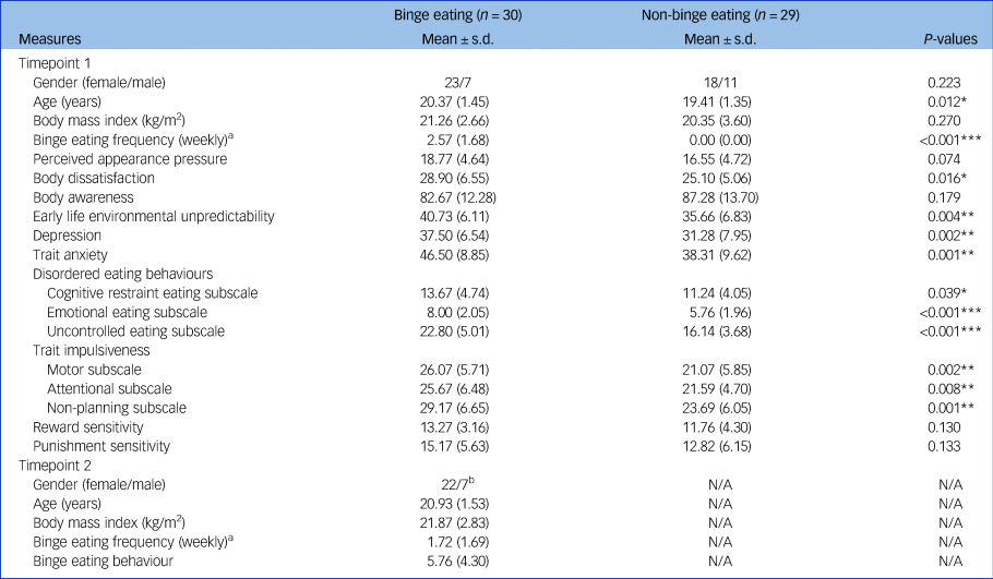
All participants were recruited from the Southwest University in Chongqing, China (via advertisement online). A total of 1451 young adults completed the behavioural assessment (i.e. the Eating Disorder Diagnosis Scale [EDDS] and Binge Eating Scale [BES]). Participants who fulfilled the following criteria constituted the binge eating group: (a) the presence of binge eating episodes marked by a perceived loss of control and the consumption of a large amount of food as indexed by a response of greater than or equal to 1 on EDDS Item 8 (i.e. ‘How many TIMES per week on average over the past 3 MONTHS have you eaten an unusually large amount of food and experienced a loss of control?’); and (b) the absence of any compensatory behaviours (i.e. purging via self-induced vomiting or laxative use, restrictive eating or compulsive exercise) as reflected by a 0 response to EDDS Items 15–18. A total of 30 participants constituted the binge eating group (Mean[number of binge eating episodes per week] = 2.51; s.d. = 1.67; range, 1–8). All of the episodes classified as binge eating were considered subclinical owing to the lack of structured clinical interviews. Meanwhile, non-binge eating control participants were included if they fulfilled the following criteria: (a) the absence of binge eating episodes as indexed by a response of 0 on EDDS Item 8; and (b) the absence of any compensatory behaviours as reflected by a 0 response to EDDS Items 15–18. The final sample involved 30 participants for the binge eating group (23 females) and 29 participants for the non-binge eating group (18 females) (Table 1). Furthermore, we used the BESReference Gormally, Black, Daston and Rardin33 to confirm the surmised binge eating status: participants who reported no binge eating episodes were expected to score lower than 8 in the BES to be included in the non-binge eating group.Reference Oliva, Morys, Horstmann, Castiello and Begliomini23 In our study, the mean BES score of the non-binge eating group was 4.06 (s.d. = 2.25; range, 0−7), suggesting the effectiveness of the grouping.
Descriptive characteristics and between-group comparisons for self-reported measures

N/A, data not available.
a. The Eating Disorder Diagnosis Scale was used to assess binge eating frequency (see the ‘Method’ section for details).
b. One participant was excluded because of model parameter cloning failure (see the ‘Dynamic causal modelling analysis’ section for details).
Significance is indicated by the asterisks (*P < 0.05, **P < 0.01, ***P < 0.001).
Before the brain scanning and self-reported measurement, all participants signed a written informed consent form containing self-report medical and psychiatric disorder information. Only those participants who reported no psychiatric or neurological disorders, no use of psychoactive medications and no other chronic diseases were included in the present research. Participants met the safety requirement for MRI scanning and the exclusion criteria, including claustrophobia, metallic implants, pregnancy and a history of head trauma and fainting. Participants were paid 70 Yuan as compensation for their time. The authors assert that all procedures contributing to this work comply with the ethical standards of the relevant national and institutional committees on human experimentation and with the Helsinki Declaration of 1975, as revised in 2008. All procedures involving human participants were approved by the Ethical Committee for Scientific Research of the Faculty of Psychology, Southwest University (IRB No. H22096).
Procedure
Preliminary screening and rating of picture materials
The food reward pictures were selected from the Chinese Food Image Database for Eating and Appetite Studies,Reference Li, Pan, Han, Liang, Yang and Meng34 given the cultural and geographical aspects of food preferences.Reference Labonté and Nielsen32 A total of 508 food pictures were visually screened by two doctoral students, with the inclusion criteria being high image clarity and generalisability (in line with the Chinese cultural context). After two rounds of preliminary screening, 154 food pictures were included in the first round of the food picture, named Set 1. Meanwhile, 80 pictures of daily necessities (such as scissors and cups) were selected from the standardised picture library of daily necessities compiled by our team. Through randomly repeating 20 pictures, we obtained the first round of neutral pictures, named Set 2, totalling 100 pictures. An additional 53 participants were recruited for this study and asked to rate the arousal, pleasantness and familiarity of 154 food pictures and 100 neutral pictures on a 7-point scale ranging from 1 (e.g. ‘I am very unfamiliar with this food/daily necessity’) to 7 (e.g. ‘I am very familiar with this food/daily necessity’) (for further details, see the Supplementary Results).
Re-rating and determination of picture materials
Before the scanning, a total of 59 participants (n binge eating = 30, n non-binge eating = 29) further rated 110 food pictures and 100 neutral pictures on a 9-point scale ranging from 1 (very unpalatable) to 9 (very palatable) in terms of arousal, pleasantness, familiarity, palatability (food pictures only) and favouritism (food pictures only) (Supplementary Table 2 available at https://doi.org/10.1192/bjp.2024.212).
Formal experiment
For standardisation, participants were required to avoid eating for 2 h before arriving at the laboratory. Participants arrived at the laboratory about 40 min early. Upon arrival at the laboratory, each participant completed the informed consent form related to the experiment. Subsequently, participants provided sociodemographic information and completed a series of self-report questionnaires. The experimenters explained the rules of the GNG task to the participants, and the participants undertook a practice session of the task (for details, see ‘Food reward Go/NoGo task’ section below). Before undergoing magnetic resonance imaging (MRI), participants were required to wear specialised clothing, remove any metal artifacts such as earrings and wear earplugs. Participants with myopia were equipped with the corresponding degree of scanning glasses in advance (for use during scanning). After the experimenters explained the scanning procedure to the participants, they completed the food reward GNG task while undergoing MRI (see Fig. 1 for the experimental procedure).
Schematic illustration of experimental procedure and data analysis strategy. Schematic flow of the experimental procedure, whole-brain activation analysis, seed-based functional connectivity analysis and dynamic causal modelling analysis. Hypothetical model 1 corresponds to the reward-inhibition dual-system model. Hypothetical model 2 represents the reward-inhibition-vision triple-system model.
GNG, Go/NoGo; fMRI, functional magnetic resonance imaging; FWE, family-wise error; FDR, false discovery rate; mOFC, medial orbitofrontal cortex; THA, thalamus; CAU, caudate; PUT, putamen; NAc, accumbens; IFG, inferior frontal gyrus; dmPFC, dorsal medial prefrontal cortex; aPFC, anterior prefrontal cortex; sPar, superior parietal lobule; SPG, superior parietal gyrus; LOC, lateral occipital cortex.

Demographics
Information was solicited about participants’ age, gender, ethnicity and self-reported handedness. Body mass index (BMI) was calculated from subjectively reported height and weight [BMI = weight (kg)/height2 (m2)].
Self-report measures
All participants completed the EDDS, BES, Food Preference Scale, Satisfaction and Dissatisfaction with Body Parts Scale, Three-Factor Eating Questionnaire-R18, The Center for Epidemiologic Studies Depression Scale, State-Trait Anxiety Inventory, Barratt Impulsiveness Scale, Visual Analog Scale, Body Awareness Questionnaire, Perceived Sociocultural Pressure Scale, Early Life Environmental Unpredictability Scale and Sensitivity to Punishment and Sensitivity to Reward Questionnaire. Details of measures can be found in the Supplementary Methods.
Food reward Go/NoGo task
The GNG is a measure of response inhibition that requires individuals to perform speeded responses on Go trials and to inhibit responding on No-go trials. In our study, the food reward GNG paradigm was designed to examine inhibition of prepotent responses to food reward stimuli compared to neutral stimuli (Supplementary Fig. 1). The task consisted of two runs, in which pictures of food reward (e.g. hamburgers and cherries) or neutral objects (e.g. scissors and cups) were presented. After two rounds of material rating, we finally included 43 food picture stimuli and 37 daily necessities as neutral picture stimuli for formal scanning. These picture stimuli not only maximised the reward value of the food stimulus for the binge eating groupReference Labonté and Nielsen32 but also ensured, to the largest extent possible, that the neural response differences were driven by the grouping conditions (for details, see the Supplementary Results). Participants had to either press a button with their right hand or inhibit their response to each picture, according to the instructions at the beginning of each run. The role of food and neutral pictures was different according to the run: in the ‘GO FOOD’ run, food pictures served as target stimuli, and therefore participants were asked to press the button with the right-hand index finger to food pictures (Go) and withhold their response to neutral pictures (No-go). In the ‘GO NEUTRAL’ run, neutral pictures served as target stimuli, and therefore participants were asked to press the button with neutral stimuli (Go) and withhold their response to food stimuli (No-go). Participants were instructed to respond as quickly and accurately as possible. Each run consisted of 129 Go trials (75%) and 43 No-go trials (25%).Reference Oliva, Morys, Horstmann, Castiello and Begliomini12 Before the formal scanning, participants received a practice session of the task (30 trials in total). At the beginning of each run, an instruction slide was presented as a reminder. During each run, the trials began with a fixation cross (1000 ms), followed by a neutral or a food stimulus presented for 1000 ms. The time window to respond lasted 1000 ms. Within a given run, trials were separated by a random inter-trial interval ranging from 1000 to 3000 ms. The stimuli were presented in a pseudorandomised order and the order of the runs was counterbalanced across participants to optimise the efficiency of the design. Total duration of the task was approximately 20 min. The GNG task was programmed and administered using E-Prime 2.0 presentation software (Psychology Software Tools, Inc. Pittsburgh, PA, USA; see https://pstnet.com/products/e-prime/).
Neuroimaging data acquisition and preprocessing
All images were collected using a 3-T Trio scanner (Siemens Prisma, Erlangen, Germany) at the Brain Imaging Center, Southwest University. Foam pads and earplugs were used to minimise head movement and scanning noise. The functional images were collected using a gradient echo planar imaging sequence with the following parameters: repetition time, 2000 ms; echo time, 30 ms; number of slices, 62; slice thickness, 2 mm; field of view, 224 × 224 mm2; flip angle, 90°; voxel size, 2 × 2 × 2 mm3. The GNG task was conducted in two sessions consisting of 522 volumes (261 volumes per session). High-resolution T1-weighted anatomical images were acquired using a magnetisation-prepared rapid gradient echo sequence (parameters: repetition time, 2530 ms; echo time, 2.98 ms; field of view, 256 × 256 mm2; flip angle, 7°; slice per slab, 192; slice oversampling, 33.3%; voxel size, 0.5 × 0.5 × 1 mm3).
A standard preprocessing procedure was implemented using Statistical Parametric Mapping (SPM12, Wellcome Department of Cognitive Neurology, London, UK; see https://www.fil.ion.ucl.ac.uk/spm/) working in MATLAB (R2017b, MathWorks, Inc., Natick, MA, USA; see https://www.mathworks.com). The DICOM data were first converted to Neuroimaging Informatics Technology Initiative (NIfTI) format. The functional images were corrected for temporal shifts between slices and realigned to the middle volume. The anatomical T1 images were then coregistered to the realigned mean functional image, then images were transformed into standard Montreal Neurological Institute (MNI) space using segmentation-based normalisation parameters, resliced to a final voxel size of 2 × 2 × 2 mm3 (as acquired). The resulting functional images were spatially smoothed using an 8-mm full-width at half-maximum Gaussian kernel.
Head movements were generally small in the current sample, with the majority of participants moving less than 1 mm/degree across the task, and two participants with a maximum of still less than 2 mm/degree. Therefore, a total of 59 participants were included in subsequent analyses.
Whole-brain activation analysis
First-level analysis
The fMRI data were modelled using a general linear model (GLM) by convolving the onsets of each condition with the canonical hemodynamic response function (HRF). This study defined four conditions: (a) food reward_nogo; (b) neutral_go; (c) neutral_nogo; and (d) food reward_go. To account for head movement, the six movement parameters of the rigid body transformation applied by the realignment procedure were also included as regressors in the first-level analysis. The resulting GLM for each participant included 16 regressors representing each experimental condition (four conditions in total) and movement parameters (six parameters per run, 12 parameters in total for two runs). Before model estimation, a high-pass filter of 128 s was applied to remove low-frequency noise and slow drifts in the signal. Notably, the present research focused on the brain activation involved in inhibition of prepotent responses to food reward stimuli (i.e. food reward_nogo conflict condition).Reference Gholamipourbarogh, Ghin, Mückschel, Frings, Stock and Beste35 Therefore, the beta image for the food reward_nogo condition and two types of beta contrast images (i.e. food reward_nogo minus neutral_nogo, food reward_nogo minus food reward_go) were output as a result of this analysis.
Second-level analysis
We used the two-sample t-test to examine between-group differences in brain activation. Age and gender were included as covariates.Reference Tozzi, Goldstein-Piekarski, Korgaonkar and Williams36 Analysis was conducted on the whole brain and statistical inference was performed by using cluster-wise control of family-wise error (FWE). Statistical images were first assessed for cluster-wise significance with a primary cluster-defining threshold of P = 0.001, then the thresholded cluster was considered significant at a FWE rate of 0.05.Reference Oliva, Morys, Horstmann, Castiello and Begliomini12,Reference Tozzi, Goldstein-Piekarski, Korgaonkar and Williams36 Coordinates of significant local maxima are reported in a standard stereotaxic reference space of the MNI system. Anatomical labels are based on Automated Anatomical Labeling (AAL; Neurodegeneratives Diseases Institute, University of Bordeaux, Bordeaux, France; see https://www.gin.cnrs.fr/en/tools/aal/) implemented in SPM12.
Seed-based functional connectivity analysis
Regions of interest definition and time series extraction
The five regions of interest (ROIs) of the ECN (dorsal medial PFC [dmPFC], left-hand anterior PFC [aPFC], right-hand aPFC, left-hand superior parietal lobule [sPar] and right-hand sPar) were defined based on previous studies of this network.Reference Li, Liu, Zheng, Li, Liang and Chen37 Two cortical ROIs for the reward network (left- and right-hand mOFC) were derived using AAL3, and eight subcortical ROIs of the reward network (bilateral thalamus [THA], CAU, PUT and accumbens [NAc]) were obtained from the HarvardOxford Atlas. See Supplementary Table 1 for the detailed ROI definition. We also included the regions with significant group differences in the activation analysis (i.e. one region; see ‘Group comparison of whole-brain activation’ section below). This study limited the network scale to 16 ROIs as they are representative nodes in the networks.Reference Chen, Li, Liu, Xiao and Chen20,Reference Li, Chen, Luo, Luo and Chen21,Reference Chen, Gao, Qin, Wang, Xiao and Tian24,Reference Li, Liu, Zheng, Li, Liang and Chen37 We defined each ROI as a 10-mm radius centred on the peak coordinates and extracted their time series by performing a principal components analysis across voxels and retaining the principal eigenvariate.
Psychophysiological interaction analysis
This study used psychophysiological interaction (PPI) analysis to examine connectivity differences between the binge eating and non-binge eating groups during three conditions of interest, respectively, namely the food reward_nogo condition, food reward_nogo minus neutral_nogo condition and food reward_nogo minus food reward_go condition. The PPI model consisted of the physiological terms (the time series of a seed), psychological terms (HRF convolved main effect of condition of interest) and PPI terms (deconvolved raw time series of the seed multiplied by the main effect of condition of interest, and then convolved with the HRF). These three regressors were included in the GLM with six movement parameters included as covariates. In the group-level two-sample t-test we conducted multiple PPI analyses and, in each analysis, one of the 16 ROIs was used as a seed and the remaining ROIs were used as targets for each condition of interest (ROI-to-ROI analysis). Statistical significance was set at P < 0.05 with false discovery rate (FDR) correction. In addition, we also tested the group differences in correlations between a seed ROI and each voxel in the rest of the brain for each condition of interest (seed-to-voxel analysis). Statistical significance was set at voxel-wise threshold P < 0.001 (uncorrected) threshold and cluster-size FDR corrected significance of P < 0.05. The task-based connectivity analyses were performed using the Functional Connectivity Toolbox (CONN 19.c; Computational Neuroscience Research Lab, Boston University, Boston, MA, USA; see https://www.nitrc.org/projects/conn).
Multiple-comparison correction
Since a total of 16 seed regions were tested in the current study, a Bonferroni correction was applied to account for multiple testing, with the threshold for significance set to P FDR < 0.05/16 = 0.003. The between-group difference of functional connectivity is considered statistically significant if the P FDR value of the connectivity is less than 0.003.
Dynamic causal modelling analysis
The DCM uses an input-state-output model based on a bilinear state equation:Reference Friston, Harrison and Penny26

where ż is the temporal derivative of the state variable z, which describes neuronal activity resulting from intrinsic effective connectivity (A), changes in connectivity caused by the contextual modulations (B) and the direct influence of the driving input u (C). Here, ż, z and u are the observed parameters, and A, B and C are the estimated parameters. The thus defined neuronal model is coupled to a biologically plausible neurovascular model of the BOLD response, and the coupled models are used to predict the BOLD time series in a priori defined volumes of interest. The present study aimed to investigate the directional connectivity pattern during food reward-inhibition conflict processing. We implemented the PEB framework for DCM in SPM12 (revision 7771), a recently developed technique.Reference Zeidman, Jafarian, Corbin, Seghier, Razi and Price29,Reference Zeidman, Jafarian, Seghier, Litvak, Cagnan and Price30 We briefly explain each step as follows.
DCM-specific design matrix construction
Since our study focused on altered effective coupling during the food reward_nogo condition,Reference Gholamipourbarogh, Ghin, Mückschel, Frings, Stock and Beste35 we constructed a DCM-specific regression design matrix for subsequent analyses by including the food reward_nogo condition as a condition of interest in the DCM-specific model and re-estimated the GLM.
Region of interest definition and time series extractionFootnote a
We defined a total of six ROIs (i.e. significant activation and connectivity regions), namely the right-hand inferior frontal gyrus (IFG; x = −50, y = 14, z = 18), right-hand mOFC (x = 17, y = 37, z = −22), right-hand lateral occipital cortex (LOC; x = 34, y = −96, z = −10), right-hand superior parietal gyrus (SPG; x = 32, y = −42, z = 70), right-hand CAU (x = 13, y = 10, z = 11) and left-hand lingual gyrus (x = 0, y = −74, z = 4). The time series BOLD activity for each participant and each node was extracted by computing the first eigenvector of all voxels within the 8-mm radius sphere.32,33
Model specification,Footnote b estimation and diagnosis (first level)
Time series extracted from individual ROIs were carried into DCM analysis for the first level, in which a fully connected model was estimated for each participant.Reference Koussis, Burgher, Jeganathan, Scott, Cocchi and Breakspear38 The DCM consisted of three different sets of parameters: (a) matrix A: the ‘intrinsic’ connectivity representing the latent connectivity between regions irrespective of experimental conditions; (b) matrix B: the ‘modulatory’ connectivity representing the influence of experimental conditions on the intrinsic connectivity; and (c) matrix C: ‘input’ representing the driving influence on brain regions by the experimental conditions. The inversion (estimation) of the model uses the variational Laplace estimation scheme, which allows finding the predicted time series that matches the observed time series as much as possible, minimising movement of the parameters from their prior values. By doing so, the score of the quality of the mode (i.e. the negative variational free energy) may be maximised, finding the neural parameters that offer the best trade-off between model accuracy and complexity. Before group-level analyses, we checked that for each participant the variance explained by the model was at least of 10%, as an index of the success of model inversion.Reference Zeidman, Jafarian, Corbin, Seghier, Razi and Price29 No subject data was excluded during the model diagnosis.
Notably, the present study focused on two aspects when we specified the model space comprising certain models. First, we were interested in the intrinsic connectivity irrespective of experimental conditions and how the interplay between inhibitory control and reward nodes might be modulated by conflict condition (here, food reward_nogo condition). Thus, we only defined and tested the parameters (differences) in matrices A and B. Second, our current activation and connectivity results suggested that the sensitive neural responses during dual-system conflict processing also involved visual areas (see ‘Group comparison of functional connectivity’ section below). To provide more comprehensive evidence on the potential links between key regions underlying problematic eating, we constructed two hypothetical models: the reward-inhibition dual-system model (four ROIs: CAU, mOFC, IFG, SPG) and the reward-inhibition-vision triple-system model (six ROIs: CAU, mOFC, IFG, SPG, LOC, lingual gyrus). The reward-inhibition dual-system hypothetical model is shown in Supplementary Fig. 2.
Model search, comparison and reduction (second level)
To examine between-group differences in connectivity, this study used a hierarchical model over parameters implemented by the PEB framework, which accounts for variability in individual connection strengths and reduces the weight of subjects with noisy data.Reference Friston, Litvak, Oswal, Razi, Stephan and Van Wijk27,Reference Zeidman, Jafarian, Corbin, Seghier, Razi and Price29 Specifically, we constructed a PEB model to test between-group differences, with the dependent variable being the parameters of each participant (such as 16 A parameters and 16 B parameters). The PEB model includes two main regressors: group mean effect and group difference (contrast) effect. After estimating the model parameters for each participant, the PEB approach requires one to perform Bayesian model reduction (BMR) and Bayesian model average (BMA) analysis. Briefly, BMR is a particularly efficient form of Bayesian model selection that, using a greedy search, automatically compares the full model with 256 models where one or more connections, which have the least evidence, are pruned out and thus switched off, whereas the parameters with the most evidence are kept stable.Reference Friston, Litvak, Oswal, Razi, Stephan and Van Wijk27 Each reduced model has a probability density over the possible values of parameters (connection strengths) that maximises the score for the quality of the model.Reference Zeidman, Jafarian, Corbin, Seghier, Razi and Price29 We performed BMA analysis to average the parameters across models, weighted by the evidence of each model. The current study sought to determine the between-group differences in effective coupling, and we thus modelled the difference of the binge eating group (1) versus the non-binge eating group (−1). Finally, we used a threshold based on free energy, taking into account the covariance of parameters, to evaluate whether a parameter contributed to the model evidence. Based on the PEB framework, posterior probability values >0.95 were considered ‘strong evidence’ for significant effects.Reference Kass and Raftery39 Here, the effective connectivity with posterior probability values greater than 0.95 was considered to best describe the between-group differences (see Fig. 1 for data analysis strategy).
Mediation analysis
To explore the proximal predictors of binge eating behaviour and its neural basis, we examined whether neural alterations mediated the association between psychological factors and binge eating behaviour across groups. Spearman's correlation coefficients were first calculated to test the relationships of significant brain signal values (extracted from activation, functional connectivity and effective connectivity analyses) and binge eating behaviour with potential psychological factors (e.g. body awareness, body dissatisfaction, depression, trait anxiety and impulsivity). The mediation analysis was then performed using Mplus 8.0 software (Muthén & Muthén, Los Angeles, CA, USA; see http://www.statmodel.com). A bootstrapping method with 5000 iterations was used to assess the significance of the mediation model. If the 95% percentile bootstrap confidence interval for the indirect effect does not include zero, it is considered significant at the P < 0.05 level.
Longitudinal prediction analysis
This study further investigated the predictive ability of brain activation and connectivity indices on future disordered eating and body weight in the binge eating group. We used linear regression analysis to test whether significant neural indices during the GNG task (baseline) could effectively predict binge eating behaviour, binge eating frequency (times/week), body weight and BMI (6 months later). Age, gender and the corresponding prediction variable at baseline were controlled as covariates. Data analyses were performed using SPSS 21 software (International Business Machines Corporation, New York, NY, USA; see https://www.ibm.com/spss).
Results
Sample characteristics
Table 1 shows the descriptive characteristics and results of self-reported measures. Compared with the non-binge eating group, the binge eating group had significantly higher binge eating frequency (t = 8.39, P < 0.001), body dissatisfaction (t = 2.49, P < 0.05), early life environmental unpredictability (t = 3.01, P < 0.01), depression (t = 3.29, P < 0.01) and anxiety (t = 3.40, P < 0.01). Individuals with binge eating episodes also reported more cognitive restraint eating (t = 2.11, P < 0.05), emotional eating (t = 4.30, P < 0.001) and uncontrolled eating (t = 4.29, P < 0.001), as well as higher levels of impulsivity (motor subscale: t = 3.23, P < 0.01; attentional subscale: t = 2.76, P < 0.01; non-planning subscale: t = 3.31, P < 0.01) than individuals in the non-binge eating group.
Food preferences
Given that an individual's specific diet may influence their reaction to the stimuli,Reference Labonté and Nielsen32 this study evaluated participant preferences for sweet, salty, spicy, sour and bitter foods. The binge eating group reported the highest and closest preference for sweet and spicy foods (means: sweet [5.33] > spicy [5.10] > sour [4.30] > salty [4.23] > bitter [1.53]; range, 1−7), suggesting that individuals with binge eating episodes do not prefer a specific type of food. Thus, it is necessary and important to construct a food reward picture library specific to binge eaters, so as to reduce the potential influence of individual differences in food preference on neural responses (especially rewarding-related response). In addition, there were no significant differences in food preferences between the two groups (Supplementary Fig. 3), demonstrating that food preferences may not be a confounding factor influencing the neural results.
Behavioural performance
There were no significant between-group differences in the reaction times during correct Go trials, the percentage of commission errors in No-go trials or the percentage of response omissions in Go trials (Supplementary Table 3).
Group comparison of whole-brain activation
There were no significant between-group differences in the three conditions of interest: food reward_nogo, food reward_nogo minus neutral_nogo and food reward_nogo minus food reward_go (all P FWE > 0.05). To explore the potential activation during food reward-inhibition processing, a whole-brain uncorrected cluster-forming threshold of P < 0.005 was set, followed by FWE cluster-level correction at P < 0.05. In the food reward_nogo condition, we found that the binge eating group displayed weaker activation in the left-hand IFG (peak MNI: x = −50, y = 14, z = 18; voxel size = 323; P FWE = 0.029) than controls (Fig. 2(a)). Analysis of covariance further showed that the between-group difference in IFG activation remained significant after controlling for age, gender and BMI (F (1, 54) = 14.14, P = 0.0004).
Group comparison of neural substrates of food reward-based response inhibition. (a) Weaker IFG activation in the binge eating group compared to the non-binge eating group. (b) Compared to non-binge eating group, the binge eating group displayed weaker mOFC–LOC connectivity and mOFC–SPG connectivity in the food reward_nogo condition, as well as weaker CAU–lingual gyrus connectivity in the food reward_nogo−neutral_nogo condition. (c) In the reward-inhibition dual-system model, the binge eating group exhibited weaker mOFC → SPG excitatory connectivity in the food reward_nogo condition (corresponding to matrix B; *posterior probability > 0.95). (d) In the reward-inhibition-vision triple-system model, the binge eating group displayed stronger inhibitory self-connection of the SPG, weaker IFG → SPG inhibitory connectivity, weaker IFG → lingual gyrus inhibitory connectivity and weaker LOC → SPG excitatory connectivity (corresponding to matrix A; *posterior probability > 0.95).
IFG, inferior frontal gyrus; mOFC, medial orbitofrontal cortex; LOC, lateral occipital cortex; SPG, superior parietal gyrus; CAU, caudate; FWE, family-wise error; FDR, false discovery rate.
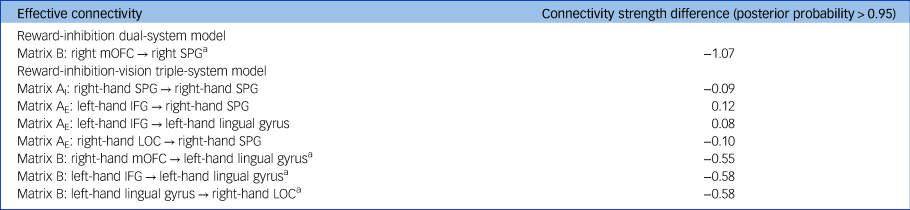
In the food reward_nogo condition, the binge eating group exhibited stronger mOFC → lingual gyrus inhibitory connectivity, weaker IFG → lingual gyrus excitatory connectivity and weaker lingual gyrus → LOC excitatory connectivity (corresponding to matrix B; *posterior probability > 0.95). See Table 2 for the group-difference effect values. For visualisation, we separated the excitatory connectivity (grey) from the inhibitory connectivity (blue). The plus (+) and minus (–) signs indicate the stronger and weaker directed connectivity in the binge eating group compared to non-binge eating group, respectively.

Group comparison of effective connectivity strength (binge eating > non-binge eating)

mOFC, medial orbitofrontal cortex; SPG, superior parietal gyrus; IFG, inferior frontal gyrus; LOC, lateral occipital cortex.
a. Connections modulated by the food reward_nogo condition (conflict condition).
In dynamic causal modelling, matrix A contains the parameters independent of experimental conditions, including matrices AI and AE. Matrix AI represents the intrinsic coupling of the brain region to itself. Matrix AE represents the intrinsic connections between brain regions. Matrix B represents the modulatory effect exerted by specific inputs (i.e. experimental conditions) on the connectivity between regions.
Group comparison of functional connectivity
ROI-to-ROI connectivityFootnote c
In the food reward_nogo condition, the binge eating group showed stronger connectivity within the reward network (left-hand mOFC–right-hand mOFC connection) and ECN (right-hand sPar–left-hand aPFC connection, right-hand sPar–IFG connection) than controls (Supplementary Table 4).
Seed-to-voxel connectivity
The between-group differences in the three conditions of interest are shown in Supplementary Table 4.
Multiple-comparison correction
The right-hand mOFC–LOC connectivity and right-hand mOFC–SPG connectivity in the food reward_nogo condition, as well as the right-hand CAU–left-hand lingual gyrus connectivity in the food reward_nogo−neutral_nogo condition, survived a stringent Bonferroni correction for multiple comparisons (P FDR < 0.05/16 = 0.003) (Supplementary Table 4; Fig. 2(b)). See Supplementary Table 5 for correlations between behavioural performance and connections.
Group comparison of effective connectivity
Reward-inhibition dual-system model
There was no significant between-group difference in the intrinsic connectivity (matrix A). In the food reward_nogo condition, the binge eating group exhibited weaker excitatory connectivity from the right-hand mOFC to SPG (matrix B) (averaged connectivity values: binge eating = −1.222, controls = 0.414, posterior probability = 1.00). See Fig. 2(c) and Table 2 for the group-difference effects.
Reward-inhibition-vision triple-system model
In the A (‘intrinsic’) matrix, compared with controls, the binge eating group showed the following: (a) stronger inhibitory self-connection of the right-hand SPG (averaged connectivity values: binge eating = −0.442, controls = −0.237, posterior probability = 0.96); (b) weaker inhibitory connection from the left-hand IFG to right-hand SPG (averaged connectivity values: binge eating = 0.254, controls = −2.01E–06, posterior probability = 1.00); (c) weaker inhibitory connection from the left-hand IFG to lingual gyrus (averaged connectivity values: binge eating = 0.196, controls = −2.09E–06, posterior probability = 0.97); and (d) weaker excitatory connection from the right-hand LOC to SPG (averaged connectivity values: binge eating = 0.039, controls = 0.256, posterior probability = 0.99). Regarding the B (‘modulatory’) matrix, in the food reward_nogo condition, compared with controls, the binge eating group displayed the following: (a) stronger inhibitory connection from the right-hand mOFC to left-hand lingual gyrus (averaged connectivity values: binge eating = −1.352, controls = −1.74E–06, posterior probability = 0.97); (b) weaker excitatory connection from the left-hand IFG to lingual gyrus (averaged connectivity values: binge eating = −9.39E–09, controls = 1.094, posterior probability = 0.99); and (c) weaker excitatory connection from the left-hand lingual gyrus to right-hand LOC (averaged connectivity values: binge eating = −0.368, controls = 0.282, posterior probability = 1.00). The group-difference effects are shown in Fig. 2(d) and Table 2.
Proximal risk factors for binge eating and its neural basis
The mediation analyses indicated the following: (a) right-hand mOFC–SPG connectivity mediated the link between trait impulsivity and binge eating behaviour (indirect effect = 0.03, s.e. = 0.01, 95% CI [0.005, 0.084], 13.4% of the total effect size); (b) right-hand mOFC–LOC connectivity mediated the relationship between depression and binge eating behaviour (indirect effect = 0.06, s.e. = 0.03, 95% CI [0.011, 0.128], 18.8% of the total effect size); (c) right-hand mOFC–LOC connectivity mediated the link between trait anxiety and binge eating behaviour (indirect effect = 0.04, s.e. = 0.02, 95% CI [0.006, 0.100], 20.0% of the total effect size); and (d) left-hand lingual gyrus → right-hand LOC connectivity mediated the link between trait anxiety and binge eating behaviour (indirect effect = 0.06, s.e. = 0.04, 95% CI [0.001, 0.152], 28.9% of the total effect size). Significant mediation models are shown in Fig. 3(a) (see Supplementary Table 6 for correlation results).
The proximal risk factors for binge eating and its neural basis, and the prediction of future binge eating from the baseline task-based connectivity. (a) The mediation models depict the indirect pathway of key psychological factors (trait impulsivity, depression and trait anxiety) on binge eating via the functional and effective connections. Standardised coefficients are depicted. The dark/light lines represent statistically significant positive/negative effects. (b) Prediction of binge eating (6 months later) from the functional and effective connections (baseline).
mOFC, medial orbitofrontal cortex; LOC, lateral occipital cortex; SPG, superior parietal gyrus.

Task-based connectivity predicts future binge eating
In the binge eating group, baseline binge eating significantly predicted future binge eating after controlling for age and gender (baseline) (β [binge eating frequency] = 0.365, P < 0.05; β [binge eating behaviour] = 0.483, P < 0.01). The baseline right-hand mOFC–SPG connectivity (during the food reward_nogo condition) predicted binge eating frequency 6 months later after controlling for age and gender (baseline) (β = 0.371, P = 0.050). When binge eating frequency (β = 0.413, P < 0.05) and mOFC–SPG connectivity (β = 0.427, P < 0.05) at baseline were included in a regression model, both significantly predicted future binge eating frequency after adjusting for age and gender (baseline). After adjusting for age, gender and binge eating behaviour (baseline), the baseline left-hand lingual gyrus → right-hand LOC connectivity (during the food reward_nogo condition) significantly predicted binge eating behaviour 6 months later (β = 0.374, P < 0.05) (Fig. 3(b)).
Discussion
The present research applied DCM-PEB, a recently developed brain connectivity technique, to identify the directionality of abnormal influences between key regions during food reward-based response inhibition in adults with recurrent binge eating episodes. These novel findings point to a particularly important role of the underlying interactions between cortical reward (mOFC) and frontoparietal control (SPG) circuits in subclinical binge eating, which deepens our understanding of the neural hierarchical mechanisms of maladaptive eating, and may have implications for the early identification of individuals suffering from strong binge eating-associated symptomatology in the general population.
The finding that the binge eating group exhibited weaker IFG activation than controls during the food reward_nogo condition is consistent with the results of a recent task-evoked study that reported that binge eaters from the general population showed lower activation of the right-hand MFG during the Go/No-Go task than the control group.Reference Oliva, Morys, Horstmann, Castiello and Begliomini12 The inhibitory control deficit theory of overeating proposes that individuals with inhibitory control deficits, and lower responsivity of brain regions implicated in behavioural control, are more sensitive to food cues and more vulnerable to the pervasive temptation of appetising foods in our environment, which increases overeating.Reference Stice and Burger13 It is highly plausible that the diminished engagement of cognitive control regions may relate to decreased dietary self-control in individuals with highly disordered eating habits.
At the undirectional synchrony level, we observed that the right-hand mOFC–SPG connectivity and right-hand mOFC–LOC connectivity in the food reward_nogo condition, as well as the right-hand CAU–left-hand lingual gyrus connectivity in the food reward_nogo−neutral_nogo condition, were significantly weakened in the binge eating group. Recent researches have suggested that the involvement of reward and frontoparietal control regions (e.g. OFC–inferior parietal gyrus connectivity, OFC–MFG connectivity and CAU–IFG connectivity) represents the crucial neural substrates that could explain the potential interactions of aberrant reward sensitivity with dysfunctional inhibitory control in binge eating behaviours in the general population.Reference Li, Chen, Luo, Luo and Chen21,Reference Oliva, Morys, Horstmann, Castiello and Begliomini23,Reference Chen, Gao, Qin, Wang, Xiao and Tian24 Theoretical and review studies also demonstrated that dysregulated eating behaviours (such as overeating, binge eating and loss of control eating) are linked to an altered balance of reward and inhibitory processing.Reference Lowe, Morton and Reichelt22 Using the perspective of dynamic engagement of functional circuits, this novel finding that diminished reward-inhibition undirectional synchrony in people with binge eating episodes supports the stable and vital role of the information communication between reward and inhibitory control circuits in both subclinical binge eating behaviours and clinical BED.Reference Chen, Gao, Qin, Wang, Xiao and Tian24,Reference Haynos, Camchong, Pearson, Lavender, Mueller and Peterson40,Reference Murray, Alba, Duval, Nagata, Cabeen and Lee41 In addition, we observed lesser connectivity between reward and visual areas (e.g. right-hand mOFC–LOC connectivity) in the binge eating group. Although no study has yet explored the dynamic integration of neurocircuitry during dual-system conflict processing in subclinical samples, a previous task-dependent study reported altered activation in visual regions (e.g. superior occipital gyrus and inferior occipital gyrus) during response inhibition tasks in binge eaters.Reference Oliva, Morys, Horstmann, Castiello and Begliomini12 Presumably, decreased functional synergy between reward and visual regions might reflect abnormal food-cue processing in adults with problematic eating. Since comparative research on the link between binge eating-associated symptomatology and dual-system functional organisation (modulated by cognitive tasks) is lacking, this result should be interpreted with caution.
The most prominent finding in the present research was the lesser excitatory connectivity from the right-hand mOFC to SPG in the binge eating group compared to the control group during the food reward_nogo condition. Empirical studies have shown that extremes of eating behaviours are strongly linked to an altered balance of reward reactivity and behavioural control in both clinical and non-clinical populations.Reference Li, Chen, Luo, Luo and Chen21,Reference Oliva, Morys, Horstmann, Castiello and Begliomini23,Reference Chen, Gao, Qin, Wang, Xiao and Tian24,Reference Murray, Alba, Duval, Nagata, Cabeen and Lee41 Our study further revealed a directional influence between key nodes in the binge eating group; in general, the excitatory effect means that the brain activity of the right-hand mOFC could increase the rate of change in activity in the right-hand SPG.Reference Zhang, Bo, Li, Zhao, Wang and Liu42 The excitatory effect of the reward region (mOFC)→inhibitory region (SPG) may reflect the potential information exchange during inhibition of the food reward stimuli, in which reward reactivity information in the mOFC is converted into response inhibition information in the SPG, successfully exerting executive control. However, the reduced mOFC → SPG connectivity strength of individuals with recurrent binge eating episodes may suggest a decrease in this bottom-up information exchange ability. This finding offers unique insights beyond those by existing resting-state fMRI studies involving subclinical binge eating participantsReference Chen, Li, Liu, Xiao and Chen20,Reference Li, Chen, Luo, Luo and Chen21,Reference Oliva, Morys, Horstmann, Castiello and Begliomini23 by revealing the directionality of the dual-system information flow involved in food reward-based response inhibition in people with binge eating episodes (for the ‘reward-inhibition-vision triple-system model’, see the Supplementary Discussion).
At the cross-sectional level, three main mediating results were obtained. First, the right-hand mOFC–SPG connectivity mediated the link between trait impulsivity and binge eating behaviour. This finding is in accordance with the results of a previous resting-state fMRI study that reported that binge eaters exhibited a higher level of trait impulsivity and lower functional connectivity between the right-hand insula and MFG, suggesting that higher impulsivity is at the root of binge eating and pointing to a possible disequilibrium between reward sensitivity and cognitive control processes.Reference Oliva, Morys, Horstmann, Castiello and Begliomini12 Our study contributes to the previous literature by further examining the relationship of impulsivity with reward-inhibition functional synchrony and binge eating, demonstrating that trait impulsivity is a strong proximal factor that could weaken the mOFC–SPG connection and facilitate binge eating behaviour. Second, the right-hand mOFC–LOC connectivity mediated the link between depression/anxiety and binge eating behaviour. Negative emotions such as depression and anxiety have been associated with binge eating behaviours/symptoms.Reference Fitzsimmons-Craft, Bardone-Cone, Brownstone and Harney43,Reference Kenny, Fuller-Tyszkiewicz, Moodie, Brown and Williams44 Recent review studies have also highlighted that negative affectivity (e.g. anxiety, depression and stress) are prominent risk factors for adult BED.Reference Tanofsky-Kraff, Schvey and Grilo6 Therefore, the current result may suggest that functional synergy between reward and visual regions involved in aberrant food-cue processing is of great value for explaining the relationship of negative affect with binge eating behaviour. Third, the left-hand lingual gyrus → right LOC connectivity mediated the association between anxiety and binge eating behaviour. To date, examinations of task-dependent functional networks that support the role of cognitive control and reward sensitivity in binge eating symptomatology have rarely involved subclinical samples. Clinically, neurofunctional alterations in the primary visual cortex (such as greater occipital lobe activation, weaker functional connectivity between the right-hand sPar and bilateral lingual gyrus) have been identified as an aspect of the pathophysiological mechanism of BED and bulimia nervosa.Reference Wang, Wang, Wu, Li and Lv45,Reference Mele, Alfano, Cotugno and Longarzo46 Patients with anxiety disorder also displayed weakened voxel-mirrored homotopic connectivity in the lingual gyrus.Reference Han, Yan, Shan, Li, Liu and Li47 Given the well-established association between anxiety and binge eating,Reference Tanofsky-Kraff, Schvey and Grilo6,Reference Fitzsimmons-Craft, Bardone-Cone, Brownstone and Harney43 this study further identified a potential mediating role of the effective connectivity between primary visual processing areas in the link between anxiety and binge eating behaviour in the general population. More importantly, our findings expand previous cross-sectional binge eating studiesReference Li, Chen, Luo, Luo and Chen21,Reference Oliva, Morys, Horstmann, Castiello and Begliomini23,Reference Chen, Gao, Qin, Wang, Xiao and Tian24 by revealing a significant predictive effect of the key dual-system undirectional synchrony (i.e. right-hand mOFC–SPG connectivity) on binge eating frequency 6 months later, which again supports the crucial role of the underlying interplay between reward and response inhibition regions in the onset and maintenance of binge eating.Reference Chen, Li, Liu, Xiao and Chen20–Reference Lowe, Morton and Reichelt22,Reference Chen, Gao, Qin, Wang, Xiao and Tian24,Reference Haynos, Camchong, Pearson, Lavender, Mueller and Peterson40,Reference Murray, Alba, Duval, Nagata, Cabeen and Lee41
Based on these findings, Fig. 4 presents a theoretical model of the neural mechanisms of binge eating from the perspective of the reward-inhibition dual-system interaction. This model builds on existing evidence of binge eating symptomatology emphasising undirectional functional connectivity between reward and inhibitory control circuits.Reference Oliva, Morys, Horstmann, Castiello and Begliomini23,Reference Chen, Gao, Qin, Wang, Xiao and Tian24,Reference Haynos, Camchong, Pearson, Lavender, Mueller and Peterson40,Reference Murray, Alba, Duval, Nagata, Cabeen and Lee41,Reference Murray, Alba, Duval, Nagata, Ganson and Jann48 Moreover, it incorporates directional effective connectivity between key nodes identified in previous studiesReference Chen, Li, Liu, Xiao and Chen20 and in the current study to provide a theoretical framework for future research. Regarding the dual-system undirectional interaction, we proposed that higher levels of binge eating are linked with less efficient information exchange between networks involved in reward sensitivity and executive control (e.g. mOFC–SPG connection, CAU–IFG connection, CAU–SFG connection, PUT–IFG connection, PUT–SFG connection, NAc–SFG connection and insula–MFG connection). Potential commonalities and differences exist in the dual-system directional interaction pattern at rest and during cognitive tasks. The pattern difference is reflected in the valence of influences (i.e. mOFC → SPG inhibitory or excitatory connection), while the pattern commonality is reflected in the strength (i.e. the mOFC → SPG connection strength decreased in both the resting and task states). These novel findings provide empirical support for the bipartite interaction model of dietary decision-makingReference Lowe, Morton and Reichelt22 through revealing the particularly important role of the diminished integration between cortical reward (mOFC) and frontoparietal control (SPG) circuits (i.e. an imbalance between bottom-up reward sensitivity and top-down behavioural regulation) in subclinical binge eating behaviour.
The neural mechanisms of binge eating from the perspective of the reward-inhibition dual-system interaction. Regarding the dual-system undirectional interaction, higher levels of binge eating are linked with less efficient information exchange between reward and inhibitory control systems (e.g. reduced NAc–ACC connection and insula–MFG connection).
BED, binge eating disorder; ACC, anterior cingulate cortex; SPG, superior parietal gyrus; SFG, superior frontal gyrus; MFG, middle frontal gyrus; IFG, inferior frontal gyrus; mOFC, medial orbitofrontal cortex; NAc, accumbens; CAU, caudate.
The thick lines represent functional connections that are weakened in both subclinical and clinical populations (i.e. CAU–SFG connection and CAU–IFG connection). Regarding the dual-system directional interaction, the mOFC → SPG connection strength decreased in both the resting and task states. The plus (+) and minus (–) signs indicate the excitatory connectivity (task state, during the food reward_nogo condition) and inhibitory connectivity (resting state), respectively. Note that this model is not exhaustive and makes no attempt to assimilate reverse evidence.

Several limitations should be considered in the present research. One limitation of this study was that our sample had a fairly restricted age range and the sample size is relatively small, which may affect the generalisability of the findings. This study used the EDDS to screen binge eaters, and self-report bias among participants may have some potential impact on the results. Additional research should further diagnose individuals with (subthreshold) BED through the structured clinical interview for DSM-5 and verify the robustness of the results. Given the limited information provided by the EDDS in healthy controls, and given that the small sample size of the binge eating group may weaken the ability to establish ideal brain–behaviour relationships, the current study could not provide stable evidence on the direction of the relationship between connectivity and binge eating within each group (see Supplementary Table 7). Future studies could investigate these interesting possibilities by increasing the sample size and including some additional measurements (e.g. food portion choice task). An additional limitation is that the number of ROIs in the ECN and reward network was different in the seed-based functional connectivity analysis, although we used pre-defined seed regions based on previous studies.Reference Chen, Li, Liu, Xiao and Chen20,Reference Li, Liu, Zheng, Li, Liang and Chen37 Future research should improve this issue, for example by employing independent component analysis that can determine subject-specific brain networks.Reference Battistella and Simonyan49 Lastly, this study provides information on task-evoked alterations in the dual-system undirectional and directional interaction in subclinical binge eaters, but may not provide clinical insights regarding an actual eating disorder. Also, the decrease in binge episodes at timepoint 2 among binge eaters could in part be caused by the lack of persistent negative life events and emotions (e.g. social stress and depression)Reference Tanofsky-Kraff, Schvey and Grilo6 that may maintain or exacerbate an individual's level of binge eating. Future prospective cohort studies involving newly disordered individuals, with longitudinal follow-ups to determine who goes on to develop binge eating or BED, will deepen understanding of the full range of neuromarkers from non-eating disorder to eating disorder conditions.
The prevalence of subclinical binge eating behaviours is particularly high in the general population, possibly increasing the risk for future development of full-syndrome eating disorders. The present research establishes an important first step to elucidate whether and how the dynamic information integration between cortical reward and frontoparietal control systems contributes to subclinical binge eating, which advances our understanding of the neural hierarchical mechanisms of binge eating-associated symptomatology, and may have implications for the early identification of individuals who are at heightened risk for problematic eating. This application of the recently developed DCM-PEB technique to the task-dependent fMRI data opens a new avenue for characterising the directional architecture underlying subclinical disordered eating, not only in clinical patients.32
Supplementary material
Supplementary material is available online at https://doi.org/10.1192/bjp.2024.212
Data availability
Coded data that support the findings of this research are available from the corresponding author upon reasonable request.
Acknowledgements
The authors would like to express their gratitude to all research assistants (Shuang Zheng, Anqi Zheng, Sibo Ren and Ziqi Liu) for their help with data collection and thank all participants for their time and cooperation. We would also like to thank Professor Feng Zhou (Southwest University) for additional statistical consultation.
Author contributions
X.C.: conceptualisation, formal analysis, investigation, methodology, validation, visualisation, writing – original draft, writing – review and editing. W.L.: conceptualisation, formal analysis, investigation, methodology, validation, visualisation, writing – review and editing. Y. Luo: conceptualisation, investigation, methodology. Y. Liu: conceptualisation, methodology. X.X.: formal analysis, methodology. X.G.: formal analysis, methodology. H.C.: conceptualisation, funding acquisition, project administration, resources, supervision, writing – review and editing.
Funding
This study was funded by National Social Science Foundation of China (no. 22&ZD184), National Natural Science Foundation of China (no. 32271087), Science and Technology Innovation Project of the Ministry of Science and Technology (no. 2021ZD0200530; 2021ZD0200500), Technology Innovation and Application Demonstration Project of Chongqing Science and Technology Bureau (no. 2023GJJL-ZXX0001), and Innovative Research Project for Postgraduate Student of Chongqing (no. CYB240082).
Declaration of interest
None.
Ethical standards
The research protocol was reviewed for compliance with the standards for the ethical treatment of human participants and approved by the Ethical Committee for Scientific Research at the university with which the authors are affiliated.
Research involving human participants
All ethical guidelines for human participants’ research were followed.
Informed consent
All participants provided written informed consents to participate in this study.






eLetters
No eLetters have been published for this article.