Skip to main content Accessibility help
×
  • Cited by 34
Publisher:
Cambridge University Press
Online publication date:
March 2012
Print publication year:
2011
Online ISBN:
9780511921001

Book description

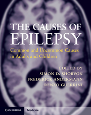
Causation is an aspect of epilepsy neglected in the scientific literature and in the conceptualization of epilepsy at a clinical and experimental level. It was to remedy this deficiency that this book was conceived. The book opens with a draft etiological classification that goes some way to filling the nosological void. The book is divided into four etiological categories: idiopathic, symptomatic, cryptogenic, and provoked epilepsies. Each chapter considers topics in a consistent fashion, dealing with the phenomenon of epilepsy in each etiology, including its epidemiology, clinical features and prognosis, and any specific aspects of treatment. The book is a comprehensive reference work, a catalogue of all important causes of epilepsy, and a clinical tool for all clinicians dealing with patients who have epilepsy. It is aimed at epileptologists and neurologists and provides a distillation of knowledge in a form that is helpful in the clinical setting.

Reviews

'… really very good. I am not aware of anything else that deals with the causes of epilepsy as succinctly and is as well organised and user friendly. This is the sort of book that one would dip into when faced with a particularly interesting or problematic case … should be on the bookshelf of everyone who investigates and manages people with epilepsy.'

Professor Martin J. Brodie - Director, Epilepsy Unit, Western Infirmary, Glasgow

'Most of the authors are world experts on the subjects, and I admire the energy and professional network required to assemble this impressive work … it will have a huge impact.'

Source: The Lancet Neurology

'… extremely comprehensive … fills a significant void in the epilepsy literature … The chapters are well written and address both basic pathogenic mechanisms and clinical diagnosis and management … This book fills a unique niche in the field of epileptology: it is the first to carefully and concisely address the varied etiologies. It is a very readable and comprehensive book that will be valued both by practising clinicians and by trainees and basic neuroscientists. It is an essential resource and should be on the bookshelf of any clinician caring for persons with epilepsy worldwide.'

Source: Epilepsy and Behavior

Refine List

Actions for selected content:

Select all | Deselect all
  • View selected items
  • Export citations
  • Download PDF (zip)
  • Save to Kindle
  • Save to Dropbox
  • Save to Google Drive

Save Search

You can save your searches here and later view and run them again in "My saved searches".

Please provide a title, maximum of 40 characters.
×

Contents


Page 2 of 5


  • Chapter 21 - Sialidosis and Gaucher disease
    pp 164-168
  • View abstract

    Summary

    There were several reports in Japan on cases that had characteristic features of what is now called benign adult familial myoclonic epilepsy (BAFME). Patients with BAFME often had irregular fine dysrhythmic involuntary movement, which became evident on outstretched extended arms. One of the characteristics of BAFME was giant somatosensory evoked potential (G-SEP) with enhanced long-loop cortical reflexes and premovement cortical spikes by the jerk-locked averaging method, suggesting hyperexcitability of cerebral cortex. Differential diagnosis includes a variety of progressive myoclonus epilepsy, hereditary tremor, and idiopathic epilepsies such as juvenile myoclonic epilepsy (JME) and grand mal on awaking. Careful history-taking reveals that the BAFME family history had a dominant hereditary pattern of almost complete penetrance, which is unusual for idiopathic generalized epilepsy (IGE). The presence of involuntary hand tremor with seizure strongly suggests the diagnosis of BAFME.
  • Chapter 22 - Action myoclonus–renal failure syndrome
    pp 169-171
  • View abstract

    Summary

    Idiopathic generalized epilepsies (IGEs) are the commonest group of epilepsies in children and adolescents. IGE syndromes are defined by distinct age at onset, seizure types, and characteristic electroencephalogram (EEG) abnormalities, without structural brain lesions and with normal developmental skills. The EEG shows normal background activity; focal interictal EEG abnormalities, occasionally reported, are not a consistent feature of benign myoclonic epilepsy of infancy (BMEI). Myoclonic-astatic epilepsy epitomizes a spectrum of IGEs with prominent myoclonic seizures, appearing in previously healthy children. Absence epilepsies and more broadly IGEs can sometimes cooccur with paroxysmal movement disorders. The IGEs have a predominant genetic etiology and current data are in favor of a complex model of inheritance with the interaction of two or more genes. Subtle developmental abnormalities of brain architecture are described in patients with IGE. New-generation antiepileptic drugs have been proven to be useful in the treatment of pharmacoresistant IGEs.
  • Chapter 24 - Tuberous sclerosis complex
    pp 177-182
  • View abstract

    Summary

    This chapter considers the etiologies of the benign partial epilepsies recognized in the Classification and Terminology of ILAE published in 2001 that include benign childhood epilepsy with centrotemporal spikes (BCECTS), early-onset benign childhood occipital epilepsy Panayiotopoulos type (PS) and late-onset childhood occipital epilepsy Gastaut type. The authors present clinical and electroencephalographic (EEG) evidence of cases showing the association of other benign focal epilepsies with absence epilepsy, namely, patients with PS and late-onset occipital epilepsy of the Gastaut type, evolving into typical childhood absence epilepsies. Childhood occipital epilepsy (COE) of Gastaut is a rare condition with a probable prevalence of 0.2-0.9% of all epilepsies, and 2-7% of benign childhood focal seizures. Patients with PS, COE of Gastaut, and BCECTS show EEG features in common with idiopathic generalized epilepsies. For example, generalized spike and wave on EEG has been reported in PS, COE of Gastaut, and BCECTS.
  • Chapter 25 - Neurofibromatoses
    pp 183-188
  • View abstract

    Summary

    The developmental/congenital disorders are a gray area between core idiopathic and core acquired epilepsies, and their inclusion under the term symptomatic epilepsy reflects the inevitably artificial nature of all classification schemes. This chapter talks about epilepsy syndromes, temporal characteristics of acquired epilepsy, and provoked epilepsies. The term 'acquired' is used to refer to symptomatic epilepsies excluding the predominately genetic or developmental causes. The main reason for considering epilepsy a symptom is that there are so many different causes, and it is therefore perhaps ironical to note that the current classifications of epilepsy pay no heed to etiology at all, focused as they are on clinical and electrographic semiology. It is clear that the distinctive natures of the underlying pathological and physiological processes underlying symptomatic epilepsy after acute brain insults are very different from those underlying idiopathic epilepsy, and so are the clinical, therapeutic, and prognostic features.
  • Chapter 27 - Other neurocutaneous syndromes
    pp 196-200
  • View abstract

    Summary

    Infantile spasms (IS) are a distinctive form of seizure disorder, mainly observed in infants, in the first year of life, and refractory to conventional antiepileptic drugs. IS are manifested as clusters of increasing plateau-decreasing-intensity brisk flexions or extensions of the neck, with abduction or adduction of the upper limbs. IS must be differentiated from rarer, earlier-onset conditions with ominous prognosis, such as early infantile epileptic encephalopathy and early myoclonic encephalopathy. Differentiating IS from the tonic seizures of Lennox-Gastaut syndrome may be difficult, especially when the attacks are in extension and not repeated in clusters. West syndrome may evolve into Lennox-Gastaut syndrome. The prognosis of IS is heavily influenced by the pathological process underlying the syndrome. Brain MRI has a high prognostic value, which is closely linked to its diagnostic power. Randomized clinical trials in Lennox-Gastaut syndrome have been performed for lamotrigine, topiramate, felbamate, rufinamide, thyrotropin-releasing hormone (TRH) analog, and cinromide.
  • Chapter 29 - Lysosomal disorders and Menkes syndrome
    pp 206-211
  • View abstract

    Summary

    Progressive myoclonus epilepsy of the Unverricht-Lundborg type (EPM1) is an autosomal recessive neurodegenerative disorder that has the highest incidence among the progressive myoclonus epilepsies worldwide. The primary therapeutic approaches for EPM1 patients include rehabilitation and symptomatic pharmacologic management. Pharmacologic intervention includes: valproic acid, clonazepam, high doses of piracetam, levetiracetam and topiramate and zonisamide. Several case reports suggest that treatment of EPM1 patients with the antioxidant N-acetylcysteine (NAC) alleviates key features of the disorder including dysarthria, ataxia, and seizures. Although the role of oxidative stress in EPM1-linked neuronal degeneration is not completely understood, a specific decrease in cerebellar defenses against oxidative stress and a concomitant increase in lipid peroxidation occurs in the mouse model for EPM1. Myoclonic seizures can also be easily misdiagnosed as tonic-clonic seizures or even pseudoepileptic seizures, especially as the majority of the myoclonic movements are not time-locked to electroencephalogram (EEG) discharges.
  • Chapter 31 - Organic acid, amino acids, and peroxisomal disorders
    pp 216-230
  • View abstract

    Summary

    The dentato-rubro-pallido-luysian atrophy (DRPLA) disorder was predominantly seen in the Japanese population. DRPLA is an autosomal dominant neurodegenerative disorder caused by abnormal repeat expansions within the DRPLA gene located on chromosome 12p13.31. Ataxia, choreoathetosis, and/or myoclonus and mental decline are the cardinal signs. Epileptic seizures usually occur in patients with an earlier onset. Unstable expanded CAG repeats in one allele in the DRPLA gene are responsible for this disorder and the size of the CAG expansion is well correlated with age of onset and severity of the disease. There are characteristic degeneration of both the dentato-rubral and pallido-luysian systems in the brain. Diffferential diagnosis includes all types of progressive myoclonic epilepsies, hereditary ataxia, and Huntington chorea. An autosomal dominant hereditary pattern and anticipation from the paternal side make the diagnosis more likely. However, a definitive diagnosis is based on genetic testing.
  • Chapter 32 - Porphyria
    pp 231-236
  • View abstract

    Summary

    Lafora disease typically starts between ages 12 and 17 years, after a period of apparently normal development. Lafora disease-associated mutations are scattered all along the coding regions of the EPM2A and NHLRC1 genes, but also accumulate in discrete spots of high recurrence. The main seizure types in Lafora disease include myoclonic seizures and occipital seizures, although generalized tonic-clonic seizures, atypical absence seizures, and atonic and complex partial seizures may occur. Studies of the combined mutation detection frequency of sequence analysis in EPM2A and NHLRC1 reveal that between 88% and 97% of mutations in these two genes can be detected using sequence analysis alone. Antiepileptic drugs have a major effect against generalized seizures, sometimes controlling seizures for many months. Valproic acid is the traditional antiepileptic treatment for Lafora disease because it is a broad-spectrum drug that controls both the generalized tonic-clonic seizures and myoclonic jerks.
  • Chapter 33 - Pyridoxine-dependent epilepsy
    pp 237-241
  • View abstract

    Summary

    This chapter focuses on disorders due to mitochondrial respiratory chain (MRC) dysfunction and use the collective term mitochondrial cytopathy. It discusses two mtDNA disorders, myoclonus epilepsy with ragged red fibers (MERRF) and mitochondrial myopathy encephalopathy, lactic acidosis, and stroke-like episodes (MELAS). Epilepsy occurs primarily in the group of patients that develop stroke-like lesions (SLL) and seizures are often preceded by or associated with migraine-like headache. Magnetic resonance spectroscopy can demonstrate elevated lactate in regions of the brain, while positron emission tomography (PET) scanning can provide metabolic information suggesting lowered ATP production. Convulsive status epilepticus (CSE) is treated aggressively using traditional protocols. Benzodiazepine infusion is evaluated as a first line together with phosphenytoin and occasionally phenobarbital. The epilepsies in mitochondrial cytopathies often reveal both focal and generalized features. Treatment of mitochondrial cytopathies comprises awareness of the potential complications and early and aggressive control of seizures.
  • Chapter 34 - Rett syndrome and MECP2 and CDKL5 genotypes
    pp 242-245
  • View abstract

    Summary

    The neuronal ceroid lipofuscinoses (NCLs) are a group of inherited neurodegenerative disorders. This chapter summarizes the current classification of NCL disorders and their genetic basis. Seizures are often generalized and initially only occasional, becoming more frequent and with the onset of other seizure types within 1-2 years. Most children subsequently found to have tripeptidyl peptidase 1 (TPP1) deficiency and/or mutations in CLN2 present with seizures between the ages of 2 and 4 years. The EEG may show characteristic occipital discharges in response to slow photic stimulation in CLN2 disease, classic late infantile onset and the other NCLs with onset in the late infantile or early juvenile range. Genes CLN1 and CLN2 code for lysosomal enzymes and there is much interest in the possibilities that gene therapy, stem cell therapy, enzyme replacement therapy, and/or substrate reduction might offer families some hope in the future.
  • Chapter 35 - Urea cycle disorders
    pp 246-248
  • View abstract

    Summary

    This chapter presents the pathophysiology, prognosis, diagnostic tests for sialidosis and Gaucher disease (GD). The epileptic phenotype resulting from sialidoses and from the subtypes of GD presents with myoclonus and generalized seizures and belongs to the category of progressive myoclonus epilepsies (PMEs). Sialidoses are classified in two types presenting with different phenotypes. In both types of sialidosis, electrophysiological studies performed at onset of seizure or myoclonus show paroxysmal electroencephalographic (EEG) activity, which can either present as polyspike-waves or as fast activities. The positive diagnosis can be based on the determination of very low blood glucocerebrosidase enzyme activity. A proposed treatment is substrate reduction therapy (SRT), which is based on chronic oral administration of an inhibitor of the biosynthesis of glucosylceramide and higher glycosphingolipids (miglustat), This inhibitor can cross the blood-brain barrier and might help reducing central nervous system complications in some GD patients.
  • Chapter 36 - Wilson disease
    pp 249-251
  • View abstract

    Summary

    This chapter presents the neurophysiology, pathophysiology, diagnostic testing for action myoclonus-renal failure syndrome (AMRF). AMRF is a form of progressive myoclonus epilepsy first described in four French-Canadian patients belonging to three apparently unrelated sibships living in different regions of Quebec. All patients with AMRF develop action myoclonus and seizures. Most of the patients showed diffuse cerebral and cerebellar atrophy, although some patients had normal computed tomography (CT) and magnetic resonance imaging (MRI) findings. The renal pathology based on renal biopsy specimens showed focal glomerulosclerosis in all patients examined, with features of collapsing glomerulopathy in some. Patients with AMRF have been reported to have normal or low normal β-GC in leukocytes, but very low levels in cultured fibroblasts, and elevated levels in serum. More recently, levetiracetam has been utilized for these patients, and other derivatives are now in clinical trials.
  • Chapter 37 - Disorders of cobalamin and folate metabolism
    pp 252-257
  • View abstract

    Summary

    The progressive myoclonus epilepsies (PMEs) are a rare group of disorders with varied causes manifested by myoclonus, often activated by movement or action, but also occurring at rest. The form of PME is particularly striking in many family members affected by autosomal dominant Alzheimer disease linked to chromosome 14. The presenting features are cognitive decline with prominent seizures and myoclonus. Myoclonus is frequently described as an early feature of patients with amyloid precursor (16) protein gene mutation pedigrees who have limb dyspraxia, brisk reflexes, visual disorientation, and slow gait. Toxic causes of myoclonus include the sialysis syndrome characterized by myoclonus, asterixis, speech disorder, seizures, personality changes, and progressive dementia. The use of serotonin reuptake inhibitors commonly used as antidepressants may lead to severe myoclonus described with the use of citalopram and risperidone. Cephalosporin toxicity, levetiracetam accumulation in renal failure, amlodipine and nifedipine can induce acute myoclonic jerking without convulsive seizures.
  • Chapter 38 - Other single-gene disorders
    pp 258-264
  • View abstract

    Summary

    This chapter first presents the molecular physiology and pathology of tuberous sclerosis complex (TSC), and then discusses diagnostic testing for TSC. In TSC, inadequate suppression of the mTORC1 pathway results in dysgenic lesions and tumor growth in multiple organ systems. Epilepsy is the most common neurological symptom, affecting approximately 85% of TSC patients, and is a significant source of morbidity and mortality. Infantile spasms in TSC frequently develop concurrently with partial epilepsy and poor control of seizures types other than spasms is also correlated with poor outcome. Initial evaluation of a patient with known or suspected TSC should focus on confirmation of the diagnosis by identifying major and minor diagnostic features. Confident diagnosis of TSC in a young child can be difficult, as different organs are preferentially involved at distinct developmental stages. Multicenter clinical trials evaluating rapamycin and other structurally similar chemical analogs are currently underway for use in TSC.
  • Chapter 40 - Fragile X syndrome
    pp 272-276
  • View abstract

    Summary

    Neurofibromatosis 1 (NF1) and neurofibromatosis 2 (NF2) are inherited tumor predisposition syndromes that have a major impact on the nervous system but are clinically and genetically distinct disorders. This chapter discusses the clinical characteristics and genetics of the neurofibromatoses and presents the current knowledge about their relationship with epilepsy. As neurofibromin acts as a tumor suppressor, NF1 individuals are at increased risk of developing benign and malignant tumors, particularly pilocytic astrocytomas, although pilomyxoid astrocytomas and glioblastoma multiforme can occur. The treatment of NF1 is based on assiduous monitoring for disease complications and treatment of the specific disease manifestations. NF2 should be distinguished from schwannomatosis, a rare condition characterized by the development of painful schwannomas involving the cutaneous, peripheral, and spinal nerves. The diagnosis of NF2 is made according a combination of skin, eye, and central and peripheral nervous system manifestations.
  • Chapter 41 - 4p (Wolf–Hirschhorn) syndrome
    pp 277-280
  • View abstract

    Summary

    This chapter presents the clinical features, etiology, and treatment of Sturge-Weber syndrome (SWS). SWS is a rare, sporadic, congenital disorder arising from an early developmental lesion affecting the facial skin, the eye, and the central nervous system. The association of neurological and developmental deterioration and the onset of seizures means that patients with SWS often suffer considerable disability. The effect of choroidal or ciliary body hemangioma in SWS interferes with the angle of the eye, causing elevated episcleral venous pressure or hypersecretion of fluid. Epileptic seizures are the predominant symptom of SWS and occur in about 80% of cases presenting port-wine stain (PWS) and leptomeningeal angioma. Magnetic resonance imaging (MRI) should be performed at a distance of a seizure event to avoid false interpretation of gadolinium enhancement, due to contrast leakage because of alteration of the blood-brain barrier.
  • Chapter 42 - Inverted duplicated chromosome 15 (isodicentric chromosome 15)
    pp 281-284
  • View abstract

    Summary

    This chapter talks about the neurocutaneous syndromes such as hypomelanosis of Ito (HI), incontinentia pigmenti (IP), nevus sebaceous (NS) syndrome and unilateral somatic intracranial hypoplasia. Chromosomal mosaicism is recognized as the pathogenic basis of many cases of HI and related disorders. It can explain the protean clinical manifestations of this condition and their often asymmetrical expression. Cerebral lesions of IP patients commonly extend radially through cortical and subcortical zones, involving cortex, subcortical and deep white matter, ependymal and subependymal zones of one or both cerebral hemispheres. Epilepsy usually appears after a variable period of evolution when the subcortical lesions are apparent in the cerebral hemisphere ipsilateral to the facial hemiatrophy. Unilateral hypoplasia of a polymicrogyric cerebral hemisphere, of the brainstem, cerebellum, and of the intracranial arteries on the same side and a hypoplastic hemibody commonly occur.
  • Chapter 43 - Ring chromosome 20
    pp 285-288
  • View abstract

    Summary

    Angelman syndrome is characterized by developmental delay, absence of speech, motor impairment, epilepsy, and a peculiar behavioral phenotype with happy demeanor. It is caused by lack of expression of the UBE3A gene, which can result from various abnormalities of chromosome 15q11-q13. Interictal high-amplitude rhythmic electroencephalographic (EEG) patterns are distinctive and should be differentiated from epileptic activity. Patterns of seizures, including type, age of onset, other clinical features, and EEG features of patients with Angelman syndrome may show some resemblance to defined epileptic syndromes. Correct characterization of their epilepsy is therefore important for both management and prognosis. Surveys of antiepileptic drugs used in patients with Angelman syndrome have suggested that valproic acid is the most commonly used. Non-pharmacological management is rarely considered, despite the relatively high prevalence of drug resistance. Ketogenic diet has been effective in a few cases.
  • Chapter 45 - Focal cortical dysplasia and related variants
    pp 293-297
  • View abstract

    Summary

    Seizures are a major manifestation of several lysosomal storage diseases. This chapter discusses neurons of lipids or glycoconjugates, which include the neuronal ceroid lipofuscinoses, the gangliosidoses, the sialidoses, and two of the mucopolysaccharidoses, Hunter syndrome and Sanfilippo disease. The galactolipidoses, Krabbe disease, and metachromatic leukodystrophy (MLD), are also discussed, because they secondarily affect neurons and clinically present with seizures. Finally, two transport disorders, one lysosomal, Niemann-Pick disease type C, and the other, Menkes disease, a non-lysosomal X-linked disorder, join these other seizure-provoking hereditary metabolic syndromes because of identifiable metabolic abnormalities within neurons. Progressive neuropsychiatric deterioration appears from the late infantile period manifesting as ataxia, dystonia, dysphagia, dysarthria, seizures, cataplexy, and cognitive deterioration. Seizures are almost constant and include asymmetric myoclonic jerks that can be stiumulus-provoked. Plasma dopamine and dihydroxyphenylacetic acid are elevated due to low dopamine beta-hydroxylase activity.
  • Chapter 46 - Agyria–pachygyria band spectrum
    pp 298-304
  • View abstract

    Summary

    Neuroacanthocytosis syndromes form a genetically heterogeneous group of disorders characterized by the association of neurological abnormalities with acanthocyte. This chapter describes both chorea-acanthocytosis (ChAc) and McLeod syndrome. In the majority of ChAc families, the disease is inherited as an autosomal recessive trait, and is caused by mutations in the VPS13A gene on chromosome 9q21, encoding for chorein. McLeod syndrome is caused by mutations of the XK gene encoding the XK protein which carries the Kx red blood cell antigen. The diagnosis of ChAc is confirmed by the detection of two mutations in the VPS13A gene. The treatment of epilepsy in patients with ChAc or McLeod syndrome represents a challenge, since seizures may at times be intractable and some antiepileptic drugs may worsen the involuntary movements. Cardiovascular events, seizures and aspiration pneumonia are the major causes of death in the older McLeod patients.
  • Chapter 47 - Agenesis of the corpus callosum
    pp 305-310
  • View abstract

    Summary

    Inherited disorders of amino acid, organic acid, and peroxisomal metabolism are individually rare but have a high cumulative frequency. The peroxisomal disorders are subdivided into two groups: the peroxisome biogenesis disorders and the single peroxisome enzyme deficiencies. Impaired mitochondrial glutamate transport has been reported in an autosomal recessive neonatal myoclonic epilepsy. Epileptic seizures may be the inaugural or the main symptom in rare inborn errors of intermediary metabolism. Biochemical analyses in Zellweger spectrum disorders show elevated plasma very long chain fatty acids (VLCFA) and usually deficient erythrocyte membrane plasmalogens. Metabolic acidosis with ketosis is suggestive of organic acidurias such as methylmalonic aciduria, propionic aciduria, and isovaleric acidemia, and may indicate a large number of rarer and less known organic acidurias. Therapy with biotin in biotinidase deficiency and with vitamin B6 in vitamin B6-dependency represent two of the most successful therapies for inborn errors of metabolism.
  • Chapter 48 - Polymicrogyria and schizencephaly
    pp 311-321
  • View abstract

    Summary

    The porphyrias are a group of conditions in which there are deficiencies in one of the eight enzymes of the heme biosynthetic pathway (the porphyrin pathway): four of the enzymes are located in the mitochondria and the other four in the cytosol. There are three acute porphyrias which cause epilepsy and other neurological symptoms: acute intermittent porphyria (AIP), variegate porphyria, and hereditary coproporphyria (HCP). This chapter discusses the clinical features, diagnostic tests and treatment options for porphyria. The diagnosis of acute porphyria depends on demonstrating increased levels of urinary d-aminolevulinic acid (ALA) and porphobilinogen (PBG) in urine. Genetic testing can confirm the disease, but as there are many different mutations in the PBGD gene it is not used widely for screening purposes. Non-enzyme-inducing drugs such as gabapentin, pregabalin, topiramate, or levetiracetam are much safer. For acute therapy, diazepam and clonazepam are relatively safe. Magnesium sulfate has also traditionally been used.
  • Chapter 49 - Periventricular nodular heterotopia
    pp 322-329
  • View abstract

    Summary

    Pyridoxine-dependent epilepsy (PDE) occurs as a result of an inborn error of metabolism where patients are dependent upon regular pharmacologic doses of pyridoxine. While it is possible that unrecognized phenotypes of PDE may present with only encephalopathy and developmental disability, the author's current understanding of this condition indicates that all patients present with lifelong anticonvulsant-resistant epileptic seizures. Patients with PDE may have normal findings on brain imaging studies, a variety of abnormal neuroradiologic features have been described in patients with PDE. It is important for clinicians to realize that a clinical diagnosis of PDE should not be made solely by examining the concurrent effects of pyridoxine administration on the interictal electroencephalographic (EEG). Clinical effectiveness, as well as biochemical and/or genetic confirmation of PDE, are necessary. While the neurodevelopmental outcome of PDE patients is multifactorial, early diagnosis and treatment is clearly important.
  • Chapter 50 - Microcephaly
    pp 330-340
  • View abstract

    Summary

    Rett syndrome (RTT, MIM 312750) is an X-linked neurodevelopmental disorder which occurs in 1. 09/10,000 females. RTT individuals with R168X, R294X, and C-terminal methyl-CpG-binding protein 2 (MECP2) mutations have been observed to be less likely to have seizure onset before 4 years of age. Partial and generalized seizures are reported to occur in RTT. Video-electroencephalogram (EEG) studies have been performed in RTT individuals with a history of seizures. Early onset of seizures is reported to be linked to the combined MECP2 mutations and brain-derived neurotrophic factor (BDNF) polymorphisms. In addition to efficacy in treatment of seizures and potential side effects, antiepileptic drugs (AEDs) may improve non-epileptic behaviors, such as anxiety and breathing dysrhythmia, commonly observed in RTT. A changing epilepsy phenotype has been described in individuals with X-linked cyclin-dependent kinase-like 5 (CDKL5) mutations. The pathogenesis of epilepsy in CDKL5 mutations may be similar to that of MECP2 mutations.

Page 2 of 5


Metrics

Altmetric attention score

Full text views

Total number of HTML views: 0
Total number of PDF views: 0 *
Loading metrics...

Book summary page views

Total views: 0 *
Loading metrics...

* Views captured on Cambridge Core between #date#. This data will be updated every 24 hours.

Usage data cannot currently be displayed.