


Implications
This review summarises key milestones and individuals involved in the evolution of our current knowledge on sperm biology and semen technologies associated with artificial insemination (AI) in cattle.
Introduction
It is fitting that this conference on bull fertility is being held in the west of Ireland; bull fertility is not a new concept in this part of the world. In the Ulster Cycle of Irish mythology, Donn Cúailnge, the Brown Bull of Cooley, a mountainous area in Co. Louth in the east of Ireland, was an extremely fertile bull over whom the Táin Bó Cúailnge (‘Cattle Raid of Cooley’) was fought. It is said that Queen Maeve of Connaught, the most westerly of the four Irish provinces, became jealous of her husband, Ailill, who owned a great white bull, Finnbennach. The only bull in Ireland that was the equal of Finnbennach was the so-called Brown Bull of Cooley. Meave decided that she must have the Brown Bull and left the Royal Palace at Rathcroghan, County Roscommon, for Louth to steal him. According to the myth, the battle for the Bull of Cooley continued for many months. The two bulls eventually met and began their own battle with the Brown Bull finally emerging victorious, but mortally wounded. He made his final journey back across Ireland to his home in Cooley, with the remains of the white bull hanging from his horns, where he succumbed from his wounds.
It is highly unlikely that anyone ever carried out a breeding soundness evaluation on the Brown Bull of Cooley. Although the existence of ‘semen’ has been known throughout recorded history, sperm were only discovered about 350 years ago by Antonie Van Leeuwenhoek (1632–1723). Prior to that discovery, interest in the male’s role in reproduction focused on semen. Indeed, the term ‘spermatozoa’, coined in 1827 by Karl von Baer (1792–1876), who incidentally, was the first to report seeing an oocyte, is derived from the Greek ‘sperein’ meaning ‘to sow’ (Birkhead and Montgomerie, Reference Birkhead and Montgomerie2009). The aim of this review is to highlight some of the main milestones and individuals in the history of sperm biology and the development of technologies associated with AI in cattle (Table 1). For further details beyond the scope (and page limit) of this article, the reader is referred to excellent and very comprehensive reviews by Cobb (Reference Cobb2006 and Reference Cobb2012), Pinto-Correia (Reference Pinto-Correia1997), Birkhead and Montgomerie (Reference Birkhead and Montgomerie2009), Foote (Reference Foote2002), Moore and Hasler (Reference Moore and Hasler2017) and the references therein. In addition, much of the early text draws on a chapter in McGeady et al. (Reference McGeady, Quinn, Fitzpatrick, Ryan, Kilroy and Lonergan2017) written by the author.
Table 1 Major milestones in the discovery of sperm and the development of artificial insemination in cattle. For more details, see text and references cited therein

AI=artificial insemination; IVF=in vitro fertilisation.
History of embryology
In the early history of embryology, two theories of ‘generation’, the term used for reproduction, existed. Preformationism proposed that organisms develop from miniature versions of themselves, already fully formed in the gametes (eggs or sperm) of their parents before conception. The alternative theory, termed epigenesis, contended that each organism was gradually produced, through a series of stages, from an undifferentiated mass. Preformists were split into two camps; ovists believed that the maternal egg was the location of the preformed organism, while spermists believed that offspring developed from a tiny, fully formed, individual contained within the head of the sperm.
The Ancient Greeks
In Europe, up to the second half of the 17th century, beliefs on virtually every question relating to the origins of life, were dominated by the teachings of the Ancient Greeks. Until the Renaissance, the male and female reproductive organs, both referred to as ‘testes’, were considered as homologous anatomical structures. Democritus (460–370 BC) suggested that female offspring originated from the left testis and males from the right. Hippocrates (c. 460–370 BC) argued that generation took place through the joint action of two kinds of semen, one from the male ejaculate, the other from the menstrual blood of the female. In De generatione animalium (The Generation of Animals), published around 350 BC, Aristotle (384–322 BC) outlined the first comprehensive theory of the mechanisms of reproduction in a variety of animals. He made the important observation that the organs develop gradually in the embryo (epigenesis) and are not performed. In contrast to Hippocrates, Aristotle believed that only the male’s semen or ‘seed’ contributed to the ‘form’ of the foetus and that this form was imprinted onto the ‘matter’ which was provided by the menstrual blood of the female, much like a seal stamping hot wax or a seed sown on fertile ground. Aristotle argued that lower animals such as insects generated spontaneously from decay, a theory stimulated by the everyday observation of maggots appearing suddenly on rotting matter, but this concept was ultimately refuted by the experiments of Francesco Redi (1626–98) in the mid-1600s.
Galen (129–c.200) supported the assertion of Hippocrates that the seeds of both the male and female contribute to procreation. This was partly due to his mistaken view that women’s genitalia were identical to those of men but turned inward. His anatomical reports, which were based mainly on dissection of monkeys and pigs, remained uncontested until descriptions and illustrations of human dissections were published in 1543 in the classical work on human anatomy De humani corporis fabrica (On the Fabric of the Human Body) by the Belgian anatomist and physician Andreas Vesalius (1514–64). Although Galen adopted Hippocrates’ view that there were two types of ‘semen’ – one male, the other female – acceptance of this theory was hampered by the fact that it was not possible to identify female semen and therefore Aristotle’s view persisted. It was not until the late Renaissance, with a resurgent interest in science, that these ideas were challenged.
The 1600s: a century of scientific discovery
Hieronymus Fabricius (Girolamo Fabrizio da Aquapendete, 1537–1619), a student of Gabriele Falloppio (Fallopius, 1523–62) after whom the Fallopian tubes were named, could find no trace of semen in the egg or the uterus after copulation and postulated, in De formatione ovi et pulli (1621), that semen was stored in a blind sack near the uterus never coming into contact with the egg. He proposed that fertilisation was elicited by means of an ‘aura seminalis’, thus resurrecting Aristotle’s view that the male semen played only a secondary role in generation.
In the 1630s, William Harvey (1578–1657), a former student of Falloppio and personal physician to King James I and King Charles I, best known for his discovery of blood circulation, carried out a now famous experiment in which he dissected the deer of King Charles I during rutting and mating. He found no trace of semen in the uterus, nor did he find any changes in the female ‘testicles’, the generally accepted term at the time for the ovaries. In addition, he failed to recognise the filamentous conceptus characteristic of ruminants. He ultimately concluded that Aristotle was correct, and that semen acted in some way by shaping menstrual blood. Harvey’s lack of evidence for any direct contribution by the semen is epitomised by his publication in 1651 Exercitationes de generatione animalium (On the Generation of Animals) with the now famous frontispiece illustrating the Greek god, Zeus, liberating all creation from an egg bearing the inscription ‘ex ovo omnia’ (all things come from the egg) (Figure 1). Harvey was convinced that the egg, rather than sperm, was fundamental to generation, apparently challenging Aristotle’s belief that sperm were of greatest importance, although what exactly he meant by ‘egg’ is unclear (Cobb, Reference Cobb2006). He had no understanding of there being equivalent male and female gametes and no idea of what might be contained in semen.
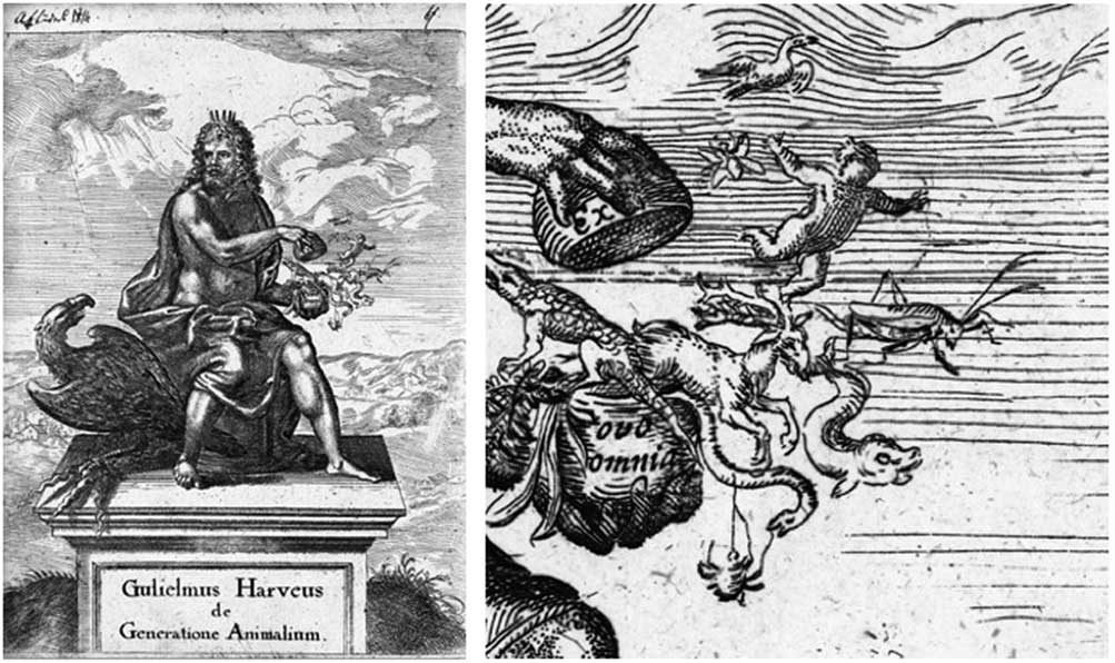
Figure 1 The frontispiece of William Harvey’s Exercitationes de generatione animalium, published in 1651, showing Zeus liberating all living things from an egg bearing the inscription ‘ex ovo omnia’. Courtesy of Wellcome Library, London.
By the mid-1670s, the ‘egg’ theory of generation was widely accepted. This was a remarkable change of direction from the notion of a ‘seed’, but it did not persist for long, following the discovery of microscopic organisms during the period 1665–83 by Robert Hooke (1635–1703) and Antonie van Leeuwenhoek (1632–1723), made possible by the skill of both men in fabricating and using simple microscopes.
The discovery of sperm
van Leeuwenhoek, a Dutch draper from Delft, was entirely self-taught (Corliss, Reference Corliss2002). He did not speak or write Latin, the scientific language of the day. He had been introduced to the Royal Society by Regnier de Graaf (1641–73), after whom the Graafian follicle is named, in 1672, as a maker of exceptional microscopes (Figure 2). The Royal Society subsequently asked him to examine a variety of bodily fluids including semen. Feeling that looking at semen would be inappropriate, he initially did not accede to the request. A few years later, in 1674, a student, Nicolaas Hartsoeker (1656–1725) and van Leeuwenhoek were the first to examine semen microscopically, a situation that would later lead to a dispute between them over the discovery of sperm (cited in Clarke, Reference Clarke2006). Hartsoeker postulated the existence of a preformed individual in the sperm, consistent with his spermist theory of preformation, and produced the now famous drawing of a tiny man or ‘homunculus’ curled up inside the sperm head in his Essai de Dioptrique in 1694 (Figure 3), something which he did not claim to see but rather supposed might be present and which is one of the most illustrative examples of preformationism (Corcos, Reference Corcos1972; Pinto-Correia, Reference Pinto-Correia1997). A contemporary, Dalenpatius (Francois de Plantade, 1670–1740) published drawings in 1699 of homunculi in sperm, a concept later exposed as a hoax (Corcos, Reference Corcos1972).

Figure 2 Antonie van Leeuwenhoek and an example of his hand-lens microscopes. Courtesy of Wellcome Library, London.

Figure 3 Illustration of a homunculus in sperm, drawn by Nicolaas Hartsoeker, published as part of his 1694 French‐language paper entitled Essai de Dioptrique, a semi‐speculative work describing the potential new scientific observations that could be made using magnifying lenses. Courtesy of Wellcome Library, London.
Van Leeuwenhoek was amongst the most prolific contributors to Philosophical Transactions of the Royal Society, the world’s first scientific journal, which published over 100 of his letters to the Society (Glover, Reference Glover2015). These concerned his microscopic observations of the most diverse biological subjects imaginable from sperm to ‘the white matter on the tongues of feverish persons’, the ‘animalcula’ in pepper-water, the structure of the spleen, the proboscis of fleas, the configuration of diamonds and the texture of the skin of elephants.
In 1677, a student from the medical school at Leiden, Johannes Ham (1651–1723), took a specimen of semen to van Leeuwenhoek, ostensibly collected from a man with gonorrhoea, in which Ham had found small living ‘animalcules’ with tails. Van Leeuwenhoek subsequently resumed his own observations and in his own semen, acquired, he stressed, not by sinfully defiling himself, but as a natural consequence of conjugal coitus, he observed a multitude of ‘zaaddiertjes’ less than a millionth the size of a coarse grain of sand and with thin, undulating transparent tails. In the summer of 1677, he reported his findings to Lord Brouncker, president of the Royal Society, urging him not to publish them if he thought it would give offence. The first public mention of sperm, based on van Leeuwenhoek’s observations, was apparently in a letter by Christiaan Huygens (1629–95) in Journal des Scavans. Following further experimentation, van Leeuwenhoek’s findings were eventually published in January 1679, in Latin, presumably due to their delicate nature. The drawing accompanying the article represented sperm of rabbits and dogs (Figure 4). Van Leeuwenhoek’s letter to Brouncker challenged the prevailing ideas about animal generation and represented a return to the ancient Greek view on the origin of life, a sperm‐centric view.

Figure 4 Sperm from rabbits and dogs, drawn by Antonie van Leeuwenhoek. Published in Philosophical Transactions, the journal of the Royal Society, London, 1678. Courtesy of Wellcome Library, London.
The division between ovists and spermists persisted for many years. Although he was originally unaware of their involvement in reproduction, van Leeuwenhoek wrote that sperm were seeds and that the female merely provided the nutrient soil in which the seeds were planted, thus returning to the notion promulgated by Aristotle some 2000 years earlier. Indeed, although sperm was discovered in the 1670s, the detailed events associated with fertilisation were not elucidated until 1876. Thus, for some 200 years, the role of sperm in generation was unclear. This lack of clarity was further compounded in 1744, when the Swiss naturalist Charles Bonnet (1720–93) published Traite d’insectologie, in which he described parthenogenesis in aphids which could apparently breed for numerous generations in the absence of males, providing further support for the ovist theory of generation.
One of the last supporters of ovism was the Italian priest and physiologist Lazzaro Spallanzani (1729–99). More than 100 years after the discovery of sperm, and building on novel experiments by the French scientist, Réne Antoine Ferchault de Réaumur (1683–1757), Spallanzani placed ‘trousers’ made of taffeta on male frogs to prevent sperm from coming into contact with eggs. These experiments provided the first hard evidence of the importance of sperm in reproduction and demonstrated that actual physical contact between the egg and the sperm was necessary for embryo development to occur. In 1784, Spallanzani reported the first successful AI in a dog, resulting in the birth of three pups 62 days later, followed soon after by the first successful AI in humans, around 1790, by the renowned Scottish anatomist and surgeon, John Hunter (1728–93). While many of Spallanzani’s experiments clearly indicate that sperm are necessary for fertilisation, he did not draw this conclusion at the time. Instead, he became further convinced, as suggested in his Experiences pour servir a l’histoire des animaux et des plantes, that the frog egg contained a fully formed tadpole that only needed to be exposed to seminal fluid to begin development (De Felici and Siracusa, Reference De Felici and Siracusa2000).
In the late 18th century, the German embryologist Kaspar Friedrich Wolff (1734–94), in his dissertation Theoria generationis published in 1759, revived and supported the theory of epigenesis, previously proposed by Aristotle and Harvey and discredited that of preformation. Through a detailed study of the development of chick embryos, Wolff demonstrated that the adult bird developed from tissues having no counterpart in the embryo. In Wolff’s De formatione intestinorum, published in 1768 and 1769, he established the principles of formation of organs from foliate layers, through proliferation, folding and wrapping, thus laying the foundations of the theory of germ layers in the embryo. His name remains associated with the Wolffian or mesonephric duct.
In spite of Wolff’s contribution, the preformation theory persisted until the 1820s, by which time a combination of new staining techniques, improved microscopes and the efforts of a talented group of scientists transformed embryology into a defined specialised branch of science. Three friends, Christian Heinrich Pander (1794–1865), Karl Ernst von Baer (1792–1876) and Heinrich Rathke (1793–1860), all of whom came from the Baltic region of Europe, significantly contributed to this advancement of research in embryology (Churchill, Reference Churchill1991). von Baer identified the mammalian oocyte during his studies on the ovary of a bitch, reported in his classic monograph of 1827, ‘De ovi mammalium et homonis genesi’.
Finally … evidence of fertilisation
Around this time, in 1824, Jean‐Louis Prevost (1790–1850) and Jean‐Baptiste Dumas (1800–84) claimed that, rather than being parasites, sperm were the active agents of fertilisation and they proposed that the sperm entered the egg and contributed to the next generation. In Sur les animalcules spermatiques de divers animaux published in 1821, written with Dumas, Prevost made a histological examination of spermatozoa and demonstrated that these cells originate in certain tissues of the male sex glands. His observations were the culmination of a series of experiments, based on those of Spallanzani, which prepared the way for modern discoveries in fertilisation.
In collaboration with Dumas, Prevost published three memoirs in 1824 on generation in the Annales des Sciences Naturelles that are now considered the foundation of experimental embryology. These claims were largely disregarded until the 1840s when the Swiss anatomist and physiologist Rudolph Albert von Köllicker (1817–1905) described the formation of sperm from cells in the adult testes. Advances in staining and microscopy during the 19th century allowed more detailed observations on the initial cleavage stages in the rabbit by German biologist Theodor Ludwig Wilhelm von Bischoff (1807–82) and by von Köllicker in humans and domestic species. von Köllicker published Entwicklungsgeschichte des Menschen und der hoheren Tiere (Development history of humans and higher animals) in 1861, the first textbook on embryology in humans and higher animals. The Swede Gustaf Retzius (1842–1919) described and illustrated spermatozoa from over 400 animal species of a variety of taxa, highlighting the amazing diversity in spermatozoa shape and size (Afzelius, Reference Afzelius1995). George Newport (1803–54), an English biologist, first reported the entry of sperm into the frog egg in 1854. However, it was not until 1876 that the German zoologist Oscar Hertwig (1849–1922) reported sperm and egg pronuclear fusion in the sea urchin, followed 3 years later by the demonstration by the Swiss investigator Hermann Fol (1845–92) that only one sperm is necessary for fertilisation. Thus, after decades of experimentation, fertilisation was finally recognised as the union of the sperm and egg (Briggs and Wessel, Reference Briggs and Wessel2006).
Testes biology
The results of castration demonstrated that the testes exercise control over the characteristics of the male body but provided no clues as to the mechanism(s) involved. It was not until the 17th century that a detailed account of testicular and penile anatomy was presented by de Graaf in his treatise on the male reproductive organs De Virorum Organi Generationi Inservientibus published in 1668 (Ryan, Reference Ryan2015). In an elegant experiment in mice, de Graaf concluded that the testis is simply a collection of minute vessels or tubules which if disentangled would far exceed 20 Dutch ells (one Dutch ell corresponds to ~70 cm) in length (De Felici and Dolci, Reference De Felici and Dolci2013).
The first clear demonstration that the testes have an endocrine role was made by Arnold Berthold (1803–61) in 1849, while studying roosters. He concluded that the regulation of male characteristics was brought about through blood-borne factors. In 1840, von Köllicker discovered that spermatozoa develop from cells located in the seminiferous tubules. This was followed by the German zoologist Franz Leydig’s (1821–1908, Figure 5) description in 1850 of the microscopic characteristics of the interstitial cells, a meshwork of loose connective tissue filling the spaces between the seminiferous tubules and blood vessels, that now bear his name (Christensen, Reference Christensen2007). This discovery in animals was followed by their description in humans by von Köllicker in 1854 (Christensen, Reference Christensen2007). Later, Enrico Sertoli (1842–1910), an Italian scientist, described the columnar cells running from the basement membrane to the lumen of the seminiferous tubules including unique branches of the cell’s cytoplasm that supported germ cell development (Franca et al., Reference Franca, Hess, Dufour, Hofmann and Griswold2016). Anton von Ebner (1842–1925) is credited with introducing the concept of the symbiotic relationship between ‘Sertoli cells’, a term he coined in 1888, and the developing germ cells, which was ultimately confirmed in 1955, when electron microscopy revealed cellular membranes and junctional complexes (Franca et al., Reference Franca, Hess, Dufour, Hofmann and Griswold2016).

Figure 5 Top: Franz Leydig (left), Enrico Sertoli. Bottom: Oscar Hertwig (left), Hermann Fol, see text for details.
For the 50 years after their discovery, Leydig cells were the subject of further studies by light microscopy, and a variety of theories were put forward regarding their possible function. In 1903, Pol Bouin (1870–1962) and Paul Ancel (1873–1961) provided the first substantial, although circumstantial, evidence that Leydig cells had an endocrine function controlling male secondary sexual characteristics. They reported that ligation of the vas deferens in dogs, rabbits and guinea pigs lead to degeneration of the seminiferous tubules, but no castration effects were observed and no degenerative changes of the interstitial cells occurred; on this basis, they concluded that internal secretions of the testes were synthesised by the Leydig cells. Over subsequent decades, additional evidence of an endocrine function was found, but it was not until the 1930s that the male hormone was shown to be testosterone, its endocrine actions were studied extensively, and the role of the pituitary in regulating testicular function was demonstrated. Direct evidence that Leydig cells produced androgen came from histochemistry in 1958 and from biochemistry in 1965 (reviewed in detail by Christensen, Reference Christensen2007).
The seminiferous epithelial cycle
Perhaps two of the most important discoveries in the past century in the field of spermatogenesis were the identification of the seminiferous epithelial cycle in mammals (Leblond and Clermont, Reference Leblond and Clermont1952; Clermont, Reference Clermont1960 and Reference Clermont1972) and the description of the hypothalamic–pituitary–testicular axis that regulates the process (Plant, Reference Plant2015). As noted by Staub and Johnson (Reference Staub and Johnson2018), the notion of a cycle of the seminiferous epithelium was already established at the beginning of the 20th century, thanks in particular to the works of Brown (Reference Brown1885), Von Ebner (Reference Von Ebner1888) and Regaud (Reference Regaud1901). Over a period of more than 50 years, Yves Clermont (1926–2014) and colleagues made a series of contributions that laid the foundation for all modern studies on spermatogenesis (Morales et al., Reference Morales, Hermo and Robaire2014). He described a pattern of organisation of germ cells in seminiferous tubules, and identified the stages of progression of germ cells from spermatogonia to spermatozoa. In addition, he detailed the kinetics of spermatogenesis in many species, including humans and his precise descriptions of most germ cell organelles and their transformation during spermatogenesis remain amongst the standard works of reference. Spermatocytogenesis, meiosis and spermiogenesis, the three major divisions of spermatogenesis, are characterised by division and/or differentiation of spermatogonia, spermatocytes and spermatids, respectively. In the bull, these divisions take 21, 23 and 17 days, respectively, for a total duration of 61 days (Amann, Reference Amann1970; Johnson et al., Reference Johnson, Varner, Roberts, Smith, Keillor and Scrutchfield2000).
Development of artificial insemination in cattle
There are several excellent reviews describing the early days of the establishment of AI in cattle and the reader is referred to those and the references therein for more detail (Herman, Reference Herman1981; Foote, Reference Foote2002; Wilmot, Reference Wilmot2007; Saacke, Reference Saacke2012). Old Arabian documents from the 14th century tell of an Arab chieftain who wanted to mate his prize mare to an outstanding stallion owned by an enemy. By introducing cotton into the mare’s reproductive tract, and then using it to sexually excite the stallion, he collected semen which was subsequently used to impregnate his mare. About four and a half centuries later, Spallanzani, in 1776, in a landmark development, demonstrated that spermatozoa frozen in snow remained viable but were merely temporarily inactivated under cold conditions and brought to life after thawing (Clarke, Reference Clarke2006). This observation predated the development of methods of cryopreserving sperm described below. In 1784, Spallanzani reported the first successful AI in a dog, resulting in the birth of three pups 62 days later.
About 100 years later, in 1897, Walter Heape (1855–1919), working in Cambridge, UK, reported the use of AI in rabbits, dogs and horses. In addition, he carried out ground-breaking experiments on embryo transfer between 1890 and 1897 (Biggers, Reference Biggers1991). In 1899, the first attempts to develop practical methods for AI were described by the Russian Ilya Ivanovich Ivanoff (1870–1932). Ivanoff studied AI in domestic farm animals, dogs, rabbits and poultry. He was the first to successfully inseminate cattle artificially. He was a pioneer in the selection of superior stallions, multiplying their progeny through AI. In 1909, a laboratory was established by the Russian Ministry of Agriculture to train veterinarians in the techniques involved in AI; before 1914, when the Bolshevik Revolution interrupted progress, about 400 technicians had been trained (Herman, Reference Herman1981). Large-scale breeding of cows via AI was first accomplished in Russia, where 19 800 cows were bred in 1931. As a result of improved techniques for the collection, dilution and insemination of semen, the numbers of animals inseminated greatly increased during the 1930s. It is estimated that in 1938 some 40 000 mares, 1.2 million cows and 15 million sheep in Russia were bred by AI (Herman, Reference Herman1981). Ivanoff also became famous, or rather infamous, for attempting to create human-ape hybrids in the mid-1920s, initially with the support of the Russian government – but that’s another story. The work of Ivanoff on AI was continued by Victor Milovanov, another Russian scientist who published a paper on ‘Artificial insemination in Russia’ in 1938, established major projects for cattle breeding and designed the first artificial vaginas suitable for collecting semen from bulls, rams and stallions, similar to those used for semen collection today. This development followed the work of Giuseppe Amantea (1885–1966) in Rome who developed the first artificial vagina, for the dog, in 1914.
In the United States, AI was first used in private herds. Years before the invention of the artificial vagina, some cattlemen were using AI by collecting semen from the vagina of a mated cow and inserting it into the vagina of another cow. In 1907, a Hereford calf was born in the herd of R.L. Hughey of Alva, Oklahoma (Herman, Reference Herman1981). Thomas C. Webster of Fort Steilacoom Washington, began using AI in 1926 and later, after moving to Wisconsin, used AI in herds throughout the state (Herman, Reference Herman1981; Warnick, Reference Warnick1998). Other early attempts at using AI are described by Herman (Reference Herman1981). Closer to home, there is a record of the successful insemination of several Shorthorn cows by the Irish Department of Agriculture’s chief veterinary officer in April 1923, apparently observed by an official of the Shorthorn Society (cited in Wilmot, Reference Wilmot2007). In England, Arthur Walton (1897–1959) working at Cambridge led studies on the properties of semen storage and handling and its long-distance shipping. Cooled rabbit sperm were successfully sent by post from Cambridge to the University of Edinburgh for insemination experiments; from 5 dose inseminated, 46 to 49 h after dispatch, three produced litters of 8, 11 and 2, respectively (Walton, Reference Walton1926). Walton subsequently shipped cooled semen at 10°C in a thermos flask containing chipped ice to Poland in 1936; five ewes were inseminated 2 to 3 days later, two of which became pregnant.
While in the 1920s and early 1930s, it had few supporters, by the 1940s, AI was accepted as a tool for comprehensively regulating cattle breeding (Wilmot, Reference Wilmot2007). The innovating work in Russia inspired Eduard Sørensen (1898–1972) from The Royal Veterinary and Agricultural University in Copenhagen, Denmark, with Jens Gylling-Holm, agricultural adviser of Tranebjerg, to organise the first cooperative dairy AI organisation in Denmark in 1936. The programme enrolled 1070 cows in its first year of which 59% conceived, slightly better than natural service in the same herds. This European success was an important stimulus for the development of AI in dairy cattle in the United States and other Western countries. The Danish veterinarians established the method of rectovaginal fixation of the cervix, allowing semen to be deposited deeply into the cervix or into the body of the uterus; this remains the standard method of AI in cattle. Sørensen is also credited with the invention of the straws for packaging liquid semen (Sørensen, Reference Sørensen1940) which were further developed and modified by Frenchman Robert Cassou (1914–2015, Figure 6), founder of IMV Technologies in 1963, and are now used worldwide for processing and storage of frozen semen (Pickett and Berndtson, Reference Pickett and Berndtson1974). Enos J. Perry (1891–1983) of Rutgers University, New Jersey, visited the AI facilities in Denmark and established the first US farmer-owned AI cooperative, named Cooperative Artificial Breeding Association No.1, Inc., in May 1938 at the New Jersey State College of Agriculture, which was followed by others across the country, including the second such cooperative in Missouri in June of the same year. The organisation started with 102 members and 1050 cows were enrolled in the first year. The first technician, Dr J.A. Henderson, had as his associate and adviser for 2 months Dr K.A.F. Larsen, who had performed the work in Denmark’s first association. Perry’s book ‘The Artificial Insemination of Farm Animals’, first published in 1945 (Perry, Reference Perry1945), was the standard reference on the subject. By 1946, some 84 associations dealing with bull semen existed in the United States and the National Association of Animal (Artificial) Breeders (NAAB) was established (Herman, Reference Herman1981; Derry, Reference Derry2015). By 1955, 30% of registered US dairy cows were inseminated with frozen semen; by 1965, almost all cattle semen in the United States was frozen. The first calf born from commercial AI in the United States in February 1939 was at the Schomp Farm in Stanton, NJ (Figure 7).

Figure 6 Robert Cassou, one of the pioneers of artificial insemination, inventor of the Cassou or ‘French’ straw and founder of IMV Technologies. On his shoulders is the calf ‘Victoire’, son of ‘Uranium’, born on January 8, 1968, 10 years after the death of his father. Courtesy of IMV Technologies.

Figure 7 Left: the first calf born via commercial artificial insemination in the United States in February 1939 at the Schomp Farm in Stanton, NJ. County Agent (holding calf), Dr J. Henderson – Vet/inseminator (centre), Richard Schomp, owner (right). Courtesy Rutgers University Department of Animal Sciences. Right: ‘Frosty’, the first recorded AI calf in the United States born to frozen semen (May 1953 in Janesville, Wisconsin). Melford Hill (left), Berlyn Gruber inseminator (right). Courtesy of Genus ABS.
During the early 1940s, before the development of techniques of cryopreservation, distribution of semen for AI was a challenge, due to the relatively short lifespan of sperm. One ingenious, but impractical, solution in Wisconsin was ‘The Flying Bull’, an airplane that used small parachutes to drop fresh bull semen directly to farms for insemination. Various groups continued to seek a practical extender of semen viability that would reduce the need for immediate deliveries. In 1940, biochemist Paul Phillips and his graduate student Henry Lardy (1917–2010) demonstrated that an egg yolk-buffer medium could preserve the fertility of bull sperm for prolonged periods (Phillips and Lardy, Reference Phillips and Lardy1940). This meant that semen could now be shipped to various parts of the United States and Canada. This technical advance fostered the development of AI as an industry, and gradually lead to the improvement of dairy herds throughout North America. Glenn Salisbury (1910–94) and colleagues improved the media by using egg yolk with sodium citrate, permitting the use of semen at 5°C for up to 3 days (Salisbury et al., Reference Salisbury, Fuller and Willett1941). He also published several classic papers clearly supporting the concept that only a few million sperm per insemination were required. Some likened this dilution to ‘watering the milk’ (Foote, Reference Foote2002) and it lead to the introduction of the term ‘extender’ by Foote and Bratton as the yolk-citrate-antibiotic medium enhanced and extended the usefulness of semen. The net result of these studies was that semen extension could be increased at least 25-fold. Sperm numbers per insemination with liquid semen were reduced from more than 100 million to 4 million sperm per insemination. Salisbury’s subsequent studies with Gunsalus, and later with Robert Foote (1922–2008), on the bacteriology of bull semen and the control of pathogens through the inclusion of antibiotics in the egg yolk-citrate medium led to a major improvement in fertility in dairy herds, and accelerating the adoption of AI.
The yolk-based extender was improved with the so-called Cornell University Extender (Foote and Bratton, Reference Foote and Bratton1950; Foote, Reference Foote2002), containing the antibiotic mixture of penicillin, streptomycin and polymyxim B. Pat Shannon (1928–), a pioneer in the use of liquid semen for AI from New Zealand’s Livestock Improvement Corporation, visited Cornell in the late 1950s and modified the extender for use with liquid semen in the intensive seasonal breeding season in New Zealand. He was instrumental in the development of ‘Caprogen’, an effective extender for preserving bull sperm containing caproic acid and catalase with 5% egg yolk, with as few as 1 to 2 million sperm per insemination (Vishwanath and Shannon, Reference Vishwanath and Shannon2000; Vishwanath, Reference Vishwanath2003).
At Penn State in the late 1940s, John Almquist (1921–2015) discovered that adding antibiotics to diluted bull semen controlled bacterial growth, reducing early embryo mortality and increasing fertility, breakthroughs which were universally adopted by the AI industry. In 1951, Almquist pioneered the use of milk as a medium to extend the life of bull semen. In 1954, he and his staff developed techniques for freezing bull semen in glass ampules (Figure 8), and by the 1960s, breeding associations were exclusively using frozen semen in ampules before they were replaced by more convenient straws. In the early 1970s, he developed new antibiotic combinations that controlled bacterial growth without compromising fertility. He also established that the thawing rate was much more important to sperm survival than the freezing rate.
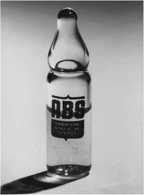
Figure 8 An example of a semen ampule used in the early days of artificial insemination. Courtesy of Genus ABS.
In recognition of their achievements, the Wolf Prize in Agriculture for 1981 was awarded equally to Almquist, Lardy and Salisbury (Figure 9) for their significant contributions to research on storage and preservation of spermatozoa and the application of AI to livestock improvement.
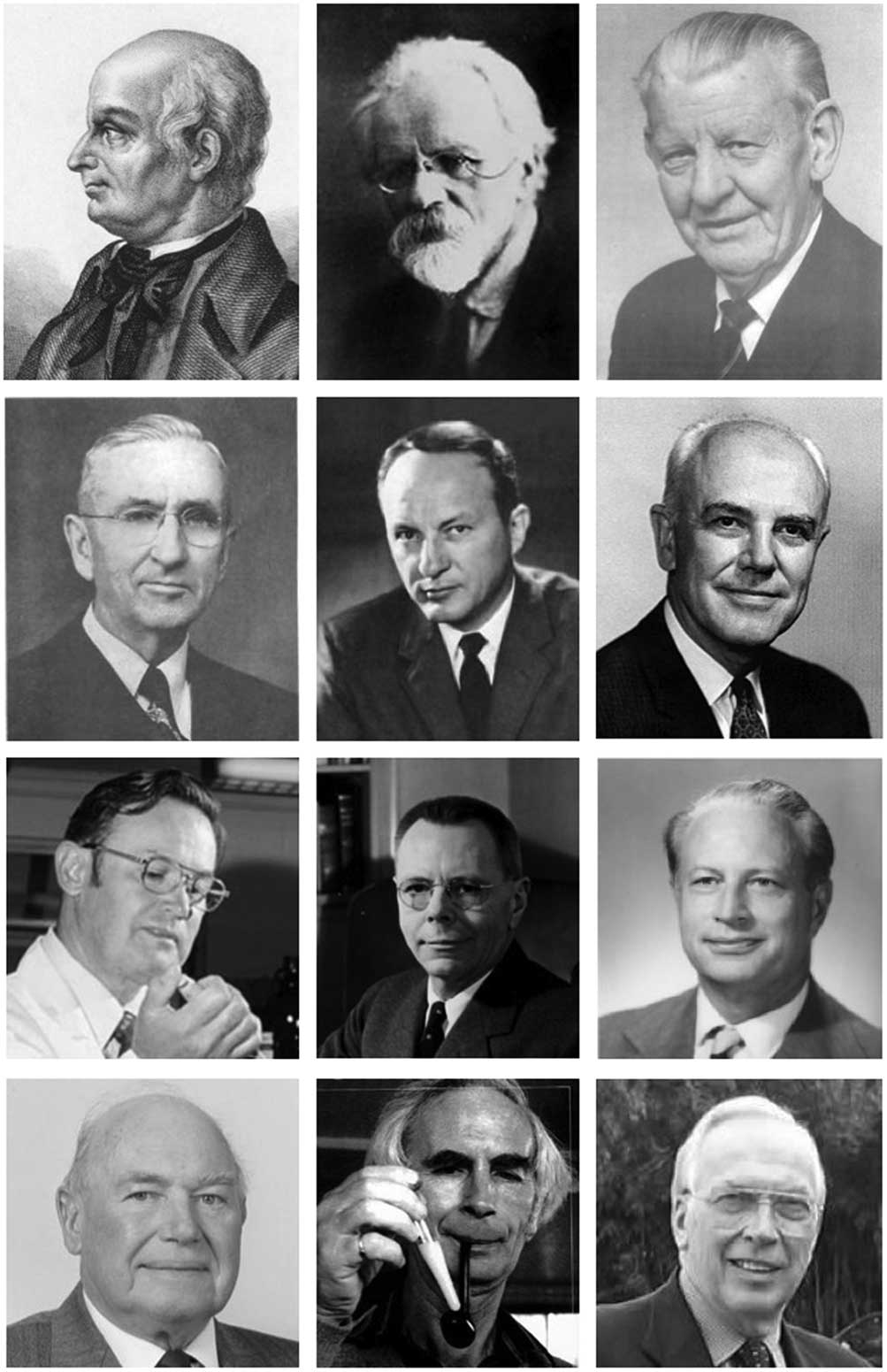
Figure 9 Some of the principal players in the development of artificial insemination and associated techniques. From left to right: top row: Lazzaro Spallanzani, Ilya Ivanoff, Eduard Sørensen; second row: Enos Perry; Henry Lardy, Glenn Salisbury; third row: John Almquist, John Rockefeller Prentice, Robert Foote; Bottom row: Chris Polge, Pat Shannon, Larry Johnston. See text for details.
In a 1980 survey, it was estimated that the total number of inseminations worldwide exceeded 130 million (Vishwanath, Reference Vishwanath2003). An updated survey in 1995 showed that the number of semen doses produced exceeded 200 million, 95% of which was processed as a frozen product (Chupin and Thibier, Reference Chupin and Thibier1995). Five years later world statistics for AI in cattle stood at 232 million doses of frozen semen and 11.6 million as a liquid, with the latter restricted mainly to New Zealand (Thibier and Wagner, Reference Thibier and Wagner2000).
Sperm cryopreservation
In 1776, Spallanzani demonstrated that sperm frozen in snow retained their viability and could be brought back to life on thawing (Clarke, Reference Clarke2006). The modern history of semen cryopreservation dates back ~70 years to the discoveries of the protective agents in egg yolk for cooling and glycerol for freezing fowl and bull sperm by Chris Polge (1926–2006) and colleagues (Polge et al., Reference Polge, Smith and Parkes1949; Polge and Rowson, Reference Polge and Rowson1952) and by the birth of the first calf by AI using frozen/thawed spermatozoa (Stewart, Reference Stewart1951). The cryoprotective properties of glycerol were discovered quite by chance. As a research student in Alan Parkes’ (1900–90) Laboratory at the Medical Research Council, UK, Polge, together with Audrey Smith (1915–81), attempted to cryopreserve fowl spermatozoa using a levulose (sugar) solution but was largely unsuccessful until a solution containing glycerol was inadvertently used and shown to allow sperm survival (Polge, Reference Polge2006). Although Polge et al. have been credited with this breakthrough others have highlighted contemporaneous work, published in Russian, which is not widely acknowledged (Ali et al., Reference Ali, AlHarbi and Ali2017). For example, Bernstein and Petropavlovski in 1937 used 0.5 to 3 M glycerol for freezing of bull, ram, stallion, boar and rabbit spermatozoa to a temperature of −21°C. Smirnov, in 1949, reported successful freezing of rabbit sperm in liquid nitrogen and liquid oxygen using a 0.05-ml aluminium container (cited in Isachenko et al., Reference Isachenko, Isachenko, Katkov, Montag, Dessole, Nawroth and Van Der Ven2004; Ali et al., Reference Ali, AlHarbi and Ali2017). In field trials, Bratton et al. (Reference Bratton, Foote and Cruthers1955) demonstrated that bovine sperm frozen to −79°C and packed on dry ice were still capable of yielding high fertility after AI.
Since then, numerous extenders and cryoprotective compounds have been investigated based on their ability to improve the survivability and post-thaw motility of cryopreserved sperm. That this technology has had a profound effect on the cattle industry has been mainly due to the higher tolerance of bovine sperm to cryoprotectants such as glycerol compared with other species and the relatively few number of sperm required for conception (Holt, Reference Holt2000). Another major change in storage occurred in the 1950s with the shift from solid carbon dioxide storage at −79°C to liquid nitrogen at −196°C following demonstration that some biologic changes occurred with storage at −79°C. John Rockefeller Prentice (1902–72), the owner of American Breeders Service, privately funded the development of liquid nitrogen containers with improved insulation (Foote, Reference Foote2002; Vishwanath, Reference Vishwanath2003).
In vitro fertilisation
Gregory Pincus (1903–67) reported the first successful pregnancy, in the rabbit, following in vitro fertilisation in Reference Pincus and Enzmann1934, although this claim was subsequently questioned as the gametes had been co-incubated in vitro for only a short time before transfer, raising the possibility that fertilisation actually occurred in vivo. This and other studies were later described in Pincus’ seminal work, ‘The Eggs of Mammals’, published in 1936. Developments in the areas of in vitro oocyte maturation, sperm capacitation, fertilisation, and embryo culture during the 1970s and 1980s led to the birth of the first calf from in vitro fertilisation (IVF) in 1981 (Brackett et al., Reference Brackett, Bousquet, Boice, Donawick, Evans and Dressel1982) following the fertilisation in vitro of an ovulated (i.e. in vivo matured) oocyte and the first calf derived from an in vitro produced embryo in 1987 (Lu et al., Reference Lu, Gordon, Chen and McGovern1987; Gordon, Reference Gordon2003). It had long been recognised that to achieve fertilisation, sperm had to undergo ‘capacitation’ in the female reproductive tract (Austin, Reference Austin1951; Chang, Reference Chang1951). The report of IVF of bovine oocytes with frozen-thawed semen and using heparin (Parrish et al., Reference Parrish, Susko-Parrish, Leibfried-Rutledge, Critser, Eyestone and First1986) was central to most subsequent work with bovine IVF (Parrish, Reference Parrish2014). The development of reliable techniques to repeatedly recover oocytes from the ovaries of live cows in the late 1980s (Pieterse et al., Reference Pieterse, Kappen, Kruip and Taverne1988; Kruip et al., Reference Kruip, Pieterse, van Beneden, Vos, Wurth and Taverne1991) opened up major applications for IVF in terms of genetic improvement (Gordon, Reference Gordon2003).
Sexed semen
The next major milestone in AI came with the development of technologies to reliably sort X- and Y-bearing spermatozoa. The history of the development of commercial sexed semen has been the subject of numerous excellent reviews (Garner and Seidel, Reference Garner and Seidel2008; Seidel, Reference Seidel2014; Rath et al., Reference Rath, Barcikowski, de Graaf, Garrels, Grossfeld, Klein, Knabe, Knorr, Kues, Meyer, Michl, Moench-Tegeder, Rehbock, Taylor and Washausen2015). Pinkel et al. (Reference Pinkel, Lake, Gledhill, Van Dilla, Stephenson and Watchmaker1982) were amongst the first to report the flow cytometric measurement of the DNA content of mammalian sperm. The basic technology for sperm sorting was developed in the early 1980s at the United States Department of Energy’s Lawrence-Livermore Laboratory in California using procedures that required demembraning sperm, which resulted in non-viable cells (Garner et al., Reference Garner, Gledhill, Pinkel, Lake, Stephenson, Van Dilla and Johnson1983). Subsequent improvements in procedures maintained sperm viability and fertility post-sorting, led by Larry Johnson at the United States Department of Agriculture in Beltsville, Maryland, led to the birth of the first offspring, in rabbits, with flow cytometrically sex-sorted spermatozoa (Johnson et al., Reference Johnson, Flook and Hawk1989). Procedures were further improved by efforts at Colorado State University by George Seidel et al. and made practical for use for AI. Most of the research in Colorado was funded by XY Inc., which acquired a license from the US Government for the intellectual property regarding the sexing technology developed in Beltsville. The first commercial license for AI was granted by XY Inc. to Cogent in the United Kingdom in 2002. In 2007, XY Inc. was sold to Sexing Technologies in Texas, who have continued to improve semen processing procedures (Vishwanath and Moreno, Reference Vishwanath and Moreno2018). Recently, a laser-based method of cell destruction of the unwanted X- or Y- chromosome bearing sperm was reported (Faust et al., Reference Faust, Betthauser, Storch and Crego2016), followed by the launch, in 2017, by Genus ABS of Sexcel™. Whether this latter technology stands the test of time remains to be seen.
The future
In their 100-year review of reproductive technologies in dairy science, Moore and Hasler (Reference Moore and Hasler2017) highlighted several areas of interest for future research relating to sperm and semen including (i) improved methods of extended sperm storage, particularly for liquid semen, (ii) improved methods for sex-sorting sperm with improved fertility, (iii) better understanding of the role of seminal plasma in eliciting an appropriate environment for early embryo development (Bromfield, Reference Bromfield2018) and (iv) the practical application of powerful new technologies, such as gene editing using CRISPR/Cas9 to animal science (Ruan et al., Reference Ruan, Xu, Chen-Tsai and Li2017).
Computer-aided sperm analysis technology was developed in the late 1980s for analysing sperm movement characteristics and now forms part of the toolbox used routinely to assess sperm motility (Amann and Waberski, Reference Amann and Waberski2014; Lu et al., Reference Lu, Huang and Lu2014; Mortimer et al., Reference Mortimer, van der Horst and Mortimer2015). Advanced computational techniques are facilitating the development of new methods of sperm imaging and tracking that do not rely on standard microscopes. As an example, a holographic on-chip sperm imaging platform, only composed of a light-emitting diode and an opto-electronic image sensor, has been described as a high-throughput, low-cost and portable potential alternative to lens-based traditional sperm imaging and tracking methods (Daloglu and Ozcan, Reference Daloglu and Ozcan2017). This platform has further enabled high-throughput 3D imaging of sperm, revealing various rare locomotion patterns (Su et al., Reference Su, Choi, Feng, Huang and Ozcan2016). It is possible that such computational chip-scale sperm imaging and 3D tracking techniques, perhaps coupled with microfluidic approaches (Nosrati et al., Reference Nosrati, Graham, Zhang, Riordon, Lagunov, Hannam, Escobedo, Jarvi and Sinton2017), will find numerous opportunities in both sperm-related research and commercial applications.
Gene editing has recently allowed the functional significance in terms of fertility of genes expressed uniquely in the male reproductive tract to be tested in a systematic fashion in mice (Miyata et al., Reference Miyata, Castaneda, Fujihara, Yu, Archambeault, Isotani, Kiyozumi, Kriseman, Mashiko, Matsumura, Matzuk, Mori, Noda, Oji, Okabe, Prunskaite-Hyyrylainen, Ramirez-Solis, Satouh, Zhang, Ikawa and Matzuk2016; Abbasi et al., Reference Abbasi, Miyata and Ikawa2018; Fujihara et al., Reference Fujihara, Miyata and Ikawa2018). This data resource may prove to be a very useful starting point to interrogate the phenotypic consequences of alterations in the expression of these genes in the bull. Indeed, as proof of principle, gene editing technology has already been applied to cattle, for example to produced polled animals (Carlson et al., Reference Carlson, Lancto, Zang, Kim, Walton, Oldeschulte, Seabury, Sonstegard and Fahrenkrug2016).
Another emerging area is the application of spermatogonial stem cell (SSC) technologies to applied reproduction. Spermatogonial stem cells are crucial for maintaining spermatogenesis throughout life, and understanding how these cells function has important implications for understanding male infertility. A remarkable feature of SSCs is the capacity to regenerate spermatogenesis following isolation from a donor testis and transplantation into a recipient testis. This capacity has significant potential as a tool for enhancing the reproductive capacity of livestock, thereby improving production efficiency (Oatley et al., Reference Oatley, Kaucher, Yang, Waqas and Oatley2016; Oatley, Reference Oatley2018). Indeed, it has been said that stem cell technology, perhaps coupled with gene editing, will allow us to bypass the bull and cow altogether and produce offspring from oocytes and sperm derived in vitro from embryonic stem cells (Nagamatsu and Hayashi, Reference Nagamatsu and Hayashi2017). If this happens, the Queen Maeve of tomorrow could forego the Brown Bull of Cooley for a good stem cell biologist!
Acknowledgements
The author is supported by funding from Science Foundation Ireland (grant numbers: 13/IA/1983 and 16/IA/4474). Sincere thanks are due to Poul Hyttel, Torben Greve, Henrik Callesen, Dick Saacke, Vish Vishwanath and Maxime Sergent for help in sourcing photos and confirming facts, and to Genus ABS, Rutgers University Department of Animal Sciences, IMV Technologies and the Wellcome Library, London for permission to use photos. Finally, thanks are due to the reviewers of the initial manuscript for suggestions which have significantly improved the text.
Declaration of interest
The author has no conflict of interest to disclose.
Ethics statement
Not applicable to this review paper.
Software and data repository resources
The data in this paper have not been deposited in an official repository.


