Published online by Cambridge University Press: 16 April 2020
Several fMRI and resting-state connectivity studies have demonstrated alterations in the limbic system and frontal areas in social anxiety disorder (SAD).
Here we used high-resolution whole-brain diffusion tensor imaging (DTI) to examine differences in anatomical connectivity between patients and controls in the white matter.
We examined 14 SAD patients (age 26.3 ± 9.0y) and 15 healthy controls (age 25.6 ± 3.3y) using DTI on a 3T Trio MRI scanner (Siemens, Germany). DTI acquisition with 1.6 mm isotropic resolution was performed in 30 directions and a maximum b-value of 800. Fractional anisotropy (FA) maps were obtained using FSL. Group analysis was performed in SPM8 (two sample t-test).
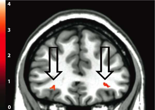
The figure shows a coronal slice through the uncinate fasciculus. Arrows point to areas where SAD patients show decreased FA-values compared to controls (p < 0.05). Note that these areas are limited to the uncinate fasciculus and are found bilaterally.
Reduced FA-values indicate a reduction in anatomical connectivity strength. Our study thus clearly shows reduced connectivity strength in the uncinate fasciculus connecting frontal regions with limbic areas as the amygdalae and hippocampus. This reduced structural connectivity supports functional data demonstrating alterations of brain activation in the amygdala and prefrontal regions in social phobia.

Comments
No Comments have been published for this article.