Hostname: page-component-848d4c4894-wzw2p Total loading time: 0 Render date: 2024-05-01T01:01:30.546Z Has data issue: false hasContentIssue false

Probing the influence of post-processing on microstructure and in situ compression failure with in silico modeling of 3D-printed scaffolds

Published online by Cambridge University Press:  27 July 2018

Sourav Mandal
Affiliation:
Laboratory for Biomaterials, Materials Research Centre, Indian Institute of Science, Bangalore-560012, India
Bikramjit Basu*
Affiliation:
Laboratory for Biomaterials, Materials Research Centre, Indian Institute of Science, Bangalore-560012, India; and Centre for BioSystems Science and Engineering, Indian Institute of Science, Bangalore-560012, India
*
a)Address all correspondence to this author. e-mail: bikram@iisc.ac.in
Get access

Abstract

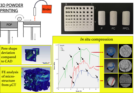
The post-processing treatment plays an important role in tailoring the mechanical and biological properties of the three-dimensional powder-printed porous scaffolds. Depending on scaffold material composition, a combination of post-processing treatments can be used to tailor these properties. This work probes into the impact of post-processing on the microstructure and deformation behavior of 3D-printed scaffolds. In this study, we have chosen CaSO4·xH2O (POP), a system for 3D powder printing and two different post-processing methodologies, namely chemical conversion and polymer infiltration. POP-based scaffolds were fabricated using water-based binder with up to 55% interconnected microporosity and moderate compressive strength of 1.5 MPa. Microcomputed tomography (µCT) is extensively utilized to determine the accuracy and efficacy of the adopted printing and post-processing approach. It was shown that the reproducibility of the fine features depends not only on the size but also on the presence of neighboring features. Crucially, µCT-based microstructure modeling and finite elemental simulation were attempted to computationally capture the compression behavior, in silico. Finally, in situ compression coupled with µCT imaging provided us an insight into fracture behavior of 3D powder-printed scaffolds.

Type
Invited Article
Copyright
Copyright © Materials Research Society 2018 

Access options

Get access to the full version of this content by using one of the access options below. (Log in options will check for institutional or personal access. Content may require purchase if you do not have access.)

References

REFERENCES

Bandyopadhyay, A., Krishna, B., Xue, W., and Bose, S.: Application of laser engineered net shaping (LENS) to manufacture porous and functionally graded structures for load bearing implants. J. Mater. Sci.: Mater. Med. 20, 29 (2009).Google ScholarPubMed
Basu, B.: Biomaterials Science and Tissue Engineering: Principles and Methods (Cambridge University Press, Cambridge, UK, 2017).Google Scholar
Basu, B.: Biomaterials for Musculoskeletal Regeneration: Concepts (Springer, New York, 2016).Google Scholar
Kumar, A., Mandal, S., Barui, S., Vasireddi, R., Gbureck, U., Gelinsky, M., and Basu, B.: Low temperature additive manufacturing of three dimensional scaffolds for bone-tissue engineering applications: Processing related challenges and property assessment. Mater. Sci. Eng., R 103, 1 (2016).CrossRefGoogle Scholar
Barui, S., Chatterjee, S., Mandal, S., Kumar, A., and Basu, B.: Microstructure and compression properties of 3D powder printed Ti–6Al–4V scaffolds with designed porosity: Experimental and computational analysis. Mater. Sci. Eng., C 70, 812 (2017).CrossRefGoogle ScholarPubMed
Barui, S., Mandal, S., and Basu, B.: Thermal inkjet 3D powder printing of metals and alloys: Current status and challenges. Curr. Opin. Biomed. Eng. 2, 116123 (2017).CrossRefGoogle Scholar
Farzadi, A., Waran, V., Solati-Hashjin, M., Rahman, Z.A.A., Asadi, M., and Osman, N.A.A.: Effect of layer printing delay on mechanical properties and dimensional accuracy of 3D printed porous prototypes in bone tissue engineering. Ceram. Int. 41, 83208330 (2015).CrossRefGoogle Scholar
Meininger, S., Mandal, S., Kumar, A., Groll, J., Basu, B., and Gbureck, U.: Strength reliability and in vitro degradation of three-dimensional powder printed strontium-substituted magnesium phosphate scaffolds. Acta Biomater. 31, 401 (2016).CrossRefGoogle ScholarPubMed
Vorndran, E., Klarner, M., Klammert, U., Grover, L.M., Patel, S., Barralet, J.E., and Gbureck, U.: 3D powder printing of β-tricalcium phosphate ceramics using different strategies. Adv. Eng. Mater. 10, B67 (2008).CrossRefGoogle Scholar
Lu, K., Hiser, M., and Wu, W.: Effect of particle size on three dimensional printed mesh structures. Powder Technol. 192, 178 (2009).CrossRefGoogle Scholar
Butscher, A., Bohner, M., Roth, C., Ernstberger, A., Heuberger, R., Doebelin, N., Rudolf von Rohr, P., and Müller, R.: Printability of calcium phosphate powders for three-dimensional printing of tissue engineering scaffolds. Acta Biomater. 8, 373 (2012).CrossRefGoogle ScholarPubMed
Tarafder, S., Balla, V.K., Davies, N.M., Bandyopadhyay, A., and Bose, S.: Microwave-sintered 3D printed tricalcium phosphate scaffolds for bone tissue engineering. J. Tissue Eng. Regener. Med. 7, 631 (2013).CrossRefGoogle ScholarPubMed
Zhou, Z., Cunningham, E., Lennon, A., McCarthy, H.O., Buchanan, F., Clarke, S.A., and Dunne, N.: Effects of poly(ε-caprolactone) coating on the properties of three-dimensional printed porous structures. J. Mech. Behav. Biomed. Mater. 70, 6883 (2017).CrossRefGoogle ScholarPubMed
Gbureck, U., Vorndran, E., Müller, F.A., and Barralet, J.E.: Low temperature direct 3D printed bioceramics and biocomposites as drug release matrices. J. Controlled Release 122, 173 (2007).CrossRefGoogle ScholarPubMed
Asadi-Eydivand, M., Solati-Hashjin, M., Farzad, A., and Osman, N.A.A.: Effect of technical parameters on porous structure and strength of 3D printed calcium sulfate prototypes. Robot. Comput. Integrated Manuf. 37, 57 (2016).CrossRefGoogle Scholar
Suwanprateeb, J., Thammarakcharoen, F., Wasoontararat, K., and Suvannapruk, W.: Influence of printing parameters on the transformation efficiency of 3D-printed plaster of paris to hydroxyapatite and its properties. Rapid Prototyp. J. 18, 490 (2012).CrossRefGoogle Scholar
Dziadek, M., Stodolak-Zych, E., and Cholewa-Kowalska, K.: Biodegradable ceramic-polymer composites for biomedical applications: A review. Mater. Sci. Eng., C 71, 1175 (2017).CrossRefGoogle ScholarPubMed
Lowmunkong, R., Sohmura, T., Takahashi, J., Suzuki, Y., Matsuya, S., and Ishikawa, K.: Transformation of 3DP gypsum model to HA by treating in ammonium phosphate solution. J. Biomed. Mater. Res., Part B 80, 386 (2007).CrossRefGoogle Scholar
Lowmunkong, R., Sohmura, T., Suzuki, Y., Matsuya, S., and Ishikawa, K.: Fabrication of freeform bone-filling calcium phosphate ceramics by gypsum 3D printing method. J. Biomed. Mater. Res., Part B 90B, 531 (2009).CrossRefGoogle Scholar
Asadi-Eydivand, M., Solati-Hashjin, M., Shafiei, S.S., Mohammadi, S., Hafezi, M., and Osman, N.A.A.: Structure, properties, and in vitro behavior of heat-treated calcium sulfate scaffolds fabricated by 3D printing. PLoS One 11, e0151216 (2016).CrossRefGoogle ScholarPubMed
Bouxsein, M.L., Boyd, S.K., Christiansen, B.A., Guldberg, R.E., Jepsen, K.J., and Müller, R.: Guidelines for assessment of bone microstructure in rodents using micro-computed tomography. J. Bone Miner. Res. 25, 1468 (2010).CrossRefGoogle ScholarPubMed
Sarkar, D., Mandal, S., Reddy, B., Bhaskar, N., Sundaresh, D., and Basu, B.: ZrO2-toughened Al2O3-based near-net shaped femoral head: Unique fabrication approach, 3D microstructure, burst strength and muscle cell response. Mater. Sci. Eng., C 77, 1216 (2017).CrossRefGoogle ScholarPubMed
Sarkar, D., Sambi Reddy, B., Mandal, S., RaviSankar, M., and Basu, B.: Uniaxial compaction-based manufacturing strategy and 3D microstructural evaluation of near-net-shaped ZrO2-toughened Al2O3 acetabular socket. Adv. Eng. Mater. 18, 16341644 (2016).CrossRefGoogle Scholar
Hammonds, K. and Baker, I.: Quantifying damage in polycrystalline ice via X-ray computed micro-tomography. Acta Mater. 127, 463470 (2017).CrossRefGoogle Scholar
Réthoré, J., Limodin, N., Buffière, J-Y., Roux, S., and Hild, F.c.: Three-dimensional analysis of fatigue crack propagation using X-ray tomography, digital volume correlation and extended finite element simulations. Procedia IUTAM 4, 151 (2012).CrossRefGoogle Scholar
Limodin, N., Rethore, J., Buffiere, J-Y., Hild, F., Roux, S., Ludwig, W., Rannou, J., and Gravouil, A.: Influence of closure on the 3D propagation of fatigue cracks in a nodular cast iron investigated by X-ray tomography and 3D volume correlation. Acta Mater. 58, 2957 (2010).CrossRefGoogle Scholar
Limodin, N., Salvo, L., Boller, E., Suéry, M., Felberbaum, M., Gailliègue, S., and Madi, K.: In situ and real-time 3-D microtomography investigation of dendritic solidification in an Al–10 wt% Cu alloy. Acta Mater. 57, 2300 (2009).CrossRefGoogle Scholar
Limodin, N., Salvo, L., Suéry, M., and DiMichiel, M.: In situ investigation by X-ray tomography of the overall and local microstructural changes occurring during partial remelting of an Al–15.8 wt% Cu alloy. Acta Mater. 55, 3177 (2007).CrossRefGoogle Scholar
Bale, H.A., Haboub, A., MacDowell, A.A., Nasiatka, J.R., Parkinson, D.Y., Cox, B.N., Marshall, D.B., and Ritchie, R.O.: Real-time quantitative imaging of failure events in materials under load at temperatures above 1600 °C. Nat. Mater. 12, 40 (2013).CrossRefGoogle ScholarPubMed
McDonald, S.A., Dedreuil-Monet, G., Yao, Y.T., Alderson, A., and Withers, P.J.: In situ 3D X-ray microtomography study comparing auxetic and non-auxetic polymeric foams under tension. Phys. Status Solidi B 248, 45 (2011).CrossRefGoogle Scholar
Petit, C., Meille, S., and Maire, E.: Cellular solids studied by X-ray tomography and finite element modeling—A review. J. Mater. Res. 28, 2191 (2013).CrossRefGoogle Scholar
Cao, J-W. and Sakai, M.: Crack-face fiber bridging: Finite element analysis, analytical model, and experimental result. J. Mater. Res. 11, 1537 (1996).CrossRefGoogle Scholar
Zhang, L., Ferreira, J.M., Olhero, S., Courtois, L., Zhang, T., Maire, E., and Rauhe, J.C.: Modeling the mechanical properties of optimally processed cordierite–mullite–alumina ceramic foams by X-ray computed tomography and finite element analysis. Acta Mater. 60, 4235 (2012).CrossRefGoogle Scholar
Michailidis, N., Stergioudi, F., Omar, H., Papadopoulos, D., and Tsipas, D.: Experimental and FEM analysis of the material response of porous metals imposed to mechanical loading. Colloids Surf., A 382, 124 (2011).CrossRefGoogle Scholar
D’Angelo, C., Ortona, A., and Colombo, P.: Finite element analysis of reticulated ceramics under compression. Acta Mater. 60, 6692 (2012).CrossRefGoogle Scholar
Cox, S.C., Thornby, J.A., Gibbons, G.J., Williams, M.A., and Mallick, K.K.: 3D printing of porous hydroxyapatite scaffolds intended for use in bone tissue engineering applications. Mater. Sci. Eng., C 47, 237 (2015).CrossRefGoogle ScholarPubMed
Xue, W., Bandyopadhyay, A., and Bose, S.: Polycaprolactone coated porous tricalcium phosphate scaffolds for controlled release of protein for tissue engineering. J. Biomed. Mater. Res., Part B 91, 831 (2009).CrossRefGoogle ScholarPubMed
Vega, V., Clements, J., Lam, T., Abad, A., Fritz, B., Ula, N., and Es-Said, O.S.: The effect of layer orientation on the mechanical properties and microstructure of a polymer. J. Mater. Eng. Perform. 20, 978 (2011).CrossRefGoogle Scholar
Castilho, M., Moseke, C., Ewald, A., Gbureck, U., Groll, J., Pires, I., Teßmar, J., and Vorndran, E.: Direct 3D powder printing of biphasic calcium phosphate scaffolds for substitution of complex bone defects. Biofabrication 6, 015006 (2014).CrossRefGoogle ScholarPubMed
Castilho, M., Dias, M., Gbureck, U., Groll, J., Fernandes, P., Pires, I., Gouveia, B., Rodrigues, J., and Vorndran, E.: Fabrication of computationally designed scaffolds by low temperature 3D printing. Biofabrication 5, 035012 (2013).CrossRefGoogle ScholarPubMed
Mandal, S., Meininger, S., Gbureck, U., and Basu, B.: 3D powder printed tetracalcium phosphate scaffold with phytic acid binder: Fabrication, microstructure and in situ X-ray tomography analysis of compressive failure. J. Mater. Sci.: Mater. Med. 29, 29 (2018).Google ScholarPubMed
Vincent, L. and Soille, P.: Watersheds in digital spaces: An efficient algorithm based on immersion simulations. IEEE Trans. Pattern Anal. Mach. Intell., 13, 583598 (1991).CrossRefGoogle Scholar
Hege, H-C., Stalling, D., Seebass, M., and Zockler, M.: A Generalized Marching Cubes Algorithm Based on Non-binary; Technical Report No. SC-97-05 (Konrad-Zuse-Zentrum (ZIB), Berlin, 1997).Google Scholar
Zilske, M., Lamecker, H., and Zachow, S.: Adaptive remeshing of non-manifold surfaces. Eurographics 27, 17 (2008). (short papers).Google Scholar
Löhner, R. and Parikh, P.: Generation of three-dimensional unstructured grids by the advancing-front method. Int. J. Numer. Methods Fluids 8, 1135 (1988).CrossRefGoogle Scholar
Jin, H. and Tanner, R.: Generation of unstructured tetrahedral meshes by advancing front technique. Int. J. Numer. Meth. Eng. 36, 1805 (1993).CrossRefGoogle Scholar
Jin, H. and Wiberg, N-E.: Two-dimensional mesh generation, adaptive remeshing and refinement. Int. J. Numer. Meth. Eng. 29, 1501 (1990).CrossRefGoogle Scholar
Castilho, M., Gouveia, B., Pires, I., Rodrigues, J., Pereira, M., Campbell, R.I., and Campbell, R.I.: The role of shell/core saturation level on the accuracy and mechanical characteristics of porous calcium phosphate models produced by 3D printing. Rapid Prototyp. J. 21, 4355 (2015).CrossRefGoogle Scholar
Zhou, Z., Buchanan, F., Mitchell, C., and Dunne, N.: Printability of calcium phosphate: Calcium sulfate powders for the application of tissue engineered bone scaffolds using the 3D printing technique. Mater. Sci. Eng., C 38, 1 (2014).CrossRefGoogle ScholarPubMed
Farzadi, A., Solati-Hashjin, M., Asadi-Eydivand, M., and Osman, N.A.A.: Effect of layer thickness and printing orientation on mechanical properties and dimensional accuracy of 3D printed porous samples for bone tissue engineering. PLoS One 9, e108252 (2014).CrossRefGoogle ScholarPubMed
Vekinis, G., Ashby, M., and Beaumont, P.: Plaster of paris as a model material for brittle porous solids. J. Mater. Sci. 28, 3221 (1993).CrossRefGoogle Scholar
Coquard, P., Boistelle, R., Amathieu, L., and Barriac, P.: Hardness, elasticity modulus and flexion strength of dry set plaster. J. Mater. Sci. 29, 4611 (1994).CrossRefGoogle Scholar
Phani, K.K., Niyogi, S., Maitra, A., and Roychaudhury, M.: Strength and elastic modulus of a porous brittle solid: An acousto-ultrasonic study. J. Mater. Sci. 21, 4335 (1986).CrossRefGoogle Scholar
Supplementary material: File

Mandal and Basu supplementary material

Mandal and Basu supplementary material 1

Download Mandal and Basu supplementary material(File)
File 9.2 MB