
Introduction
Evidence of blunt force cranial trauma incurred through interpersonal violence has been firmly established in recent population studies of the British and European Neolithic (Schulting & Wysocki Reference Schulting and Wysocki2005: 107; Lawrence Reference Lawrence2006: 53; Smith & Brickley Reference Smith and Brickley2007: 25; McKinley Reference McKinley, Mercer and Healy2008: 477; Ahlström & Molnar Reference Ahlström, Molnar, Schulting and Fibiger2012: 17; Schulting Reference Schulting, Schulting and Fibiger2012: 223; Schulting & Fibiger Reference Schulting, Fibiger, Schulting and Fibiger2012; Fibiger et al. Reference Fibiger, Ahlström, Bennike and Schulting2013: 190; Meyer et al. Reference Meyer, Lohr, Gronenborn and Alt2015: 11217). Experimental studies on the means by which these injuries were sustained can greatly aid our understanding of the variable causes and contexts of violence and, more generally, the nature of social interactions in prehistory (Wedel & Galloway Reference Wedel and Galloway2014: 73). Hitherto, however, there has been very little research to identify the specific implements responsible for these types of injury in prehistory.
Experimental studies of blunt force trauma in other time periods have often used human cadavers or animal proxies when attempting to replicate intentional injuries, although both of these mediums are often either susceptible to major faults in accuracy, or raise various questions about their ethicality (Corey et al. Reference Corey, Jones, James, Leadbeatter and Nokes2001: 104; Thali et al. Reference Thali, Kneubuehl and Dirnhofer2002a: 199, Reference Thali, Kneubuehl, Zollinger and Dirnhofer2002b: 178; Byard et al. Reference Byard, Cains and Gilbert2007: 31; Raul et al. Reference Raul, Deck, Willinger and Ludes2008: 359; Wedel & Galloway Reference Wedel and Galloway2014: 140; Smith et al. Reference Smith, James, Pover, Ball, Barnetson, Foster, Guy, Rickman and Walton2015: 427). In recent years, however, new methods using synthetic ‘skin-skull-brain’ models have begun to emerge as an alternative. These polyurethane human skull substitutes are made to uniform specification, meaning that they avoid the inaccuracies and ethical issues of using animal substitutes or human cadavers (Thali et al. Reference Thali, Kneubuehl and Dirnhofer2002a: 195, Reference Thali, Kneubuehl, Zollinger and Dirnhofer2002b: 178; Smith et al. Reference Smith, James, Pover, Ball, Barnetson, Foster, Guy, Rickman and Walton2015: 427).
This article presents the results of the first use of skin-skull-brain models to investigate the causes of blunt force trauma in the Neolithic osteological record. A replica of the ‘Thames Beater’, a Neolithic wooden club, was able to produce fractures in synthetic skulls with remarkable similarities to those found on Neolithic skeletal remains from Asparn/Schletz, a massacre site in Austria (Teschler-Nicola Reference Teschler-Nicola, Schulting and Fibiger2012: 107), thereby demonstrating the suitability of this method for providing appropriate test analogues. The research opens up new and innovative avenues for exploring the mechanisms and context of blunt force trauma in prehistory. This is essential for understanding the meaning of the social and cultural contexts of such events (as varying forms of violence are indicative of different social pressures and interactions), whether considering material from standard funerary contexts or the increasing number of remains from mass graves across Western and Central Europe (e.g. Schulting & Fibiger Reference Schulting, Fibiger, Schulting and Fibiger2012: 2; Teschler-Nicola Reference Teschler-Nicola, Schulting and Fibiger2012: 101; Fibiger et al. Reference Fibiger, Ahlström, Bennike and Schulting2013: 191; Chenal et al. Reference Chenal, Perrin, Barrand-Emam and Boulestin2015: 1329).
Blunt force trauma
Many mechanisms of injury can cause blunt force trauma, and the limited ways in which bone can react to an impact—whether violent or accidental—can complicate diagnosing the intentionality behind an injury (Alcantara et al. Reference Alcantara, Roszler, Guyot and Peterson1994: 521; Lovell Reference Lovell1997: 148; Raul et al. Reference Raul, Deck, Willinger and Ludes2008: 359; Jacobsen et al. Reference Jacobsen, Bech and Lynnerup2009: 2; Sharkey et al. Reference Sharkey, Cassidy, Brady, Gilchrist and NicDaeid2012: 835; Wedel & Galloway Reference Wedel and Galloway2014: 33). Cranial fractures are more often indicative of intentional violence than post-cranial trauma (Lovell Reference Lovell1997: 149; Chattopadhyay & Tripathi Reference Chattopadhyay and Tripathi2010: 102; Schulting Reference Schulting, Schulting and Fibiger2012: 224; Fibiger et al. Reference Fibiger, Ahlström, Bennike and Schulting2013: 191). Certain fractures, however, are often discounted as the result of accidentally incurred trauma (Lovell Reference Lovell1997: 150; Ortner Reference Ortner2003: 121; Freeman et al. Reference Freeman, Eriksson and Leith2014: 64).
Fracture formation from blunt force trauma to the cranium is influenced by the biomechanical properties of the skull (Lovell Reference Lovell1997: 155; Kasrai et al. Reference Kasrai, Hearn, Gur and Forrest1999: 238; Wedel & Galloway Reference Wedel and Galloway2014: 134; Carr et al. Reference Carr, Lindstrom, Jareborg, Champion, Waddell, Miller, Teagle, Horsfall and Kieser2015: 508). Cranial sutures (the joints between the bones of the skull) are able to absorb force, and can stop the progression of fractures across the surface of the cranium (Lovell Reference Lovell1997: 155; Wedel & Galloway Reference Wedel and Galloway2014: 135). The skull is also buttressed with arched areas of thicker bone; fractures follow the path of least resistance and can be influenced by these patterns of strong and weak bone in the skull (Lovell Reference Lovell1997: 155; Kasrai et al. Reference Kasrai, Hearn, Gur and Forrest1999: 238; Wedel & Galloway Reference Wedel and Galloway2014: 141; Carr et al. Reference Carr, Lindstrom, Jareborg, Champion, Waddell, Miller, Teagle, Horsfall and Kieser2015: 508).
Several types of fracture are formed by blunt force trauma (Figure 1). Linear fractures are produced when a low-velocity force is transmitted through a wide surface area; accidental injuries, such as falls, are possible causes, leading linear fractures to be ruled out of many archaeological studies of violence (Lovell Reference Lovell1997: 150; Ortner Reference Ortner2003: 121; Schulting & Wysocki Reference Schulting and Wysocki2005: 110; Freeman et al. Reference Freeman, Eriksson and Leith2014: 64; Wedel & Galloway Reference Wedel and Galloway2014: 137).
Diagram showing three views of blunt force cranial fracture: left) a linear fracture; centre) the exterior of a depression fracture showing the primary impact site along with the secondary and tertiary fractures that may form on the surface of the cranium; right) the internal view of a depression fracture showing the in-bending created at the site of impact (photograph: Meaghan Dyer).
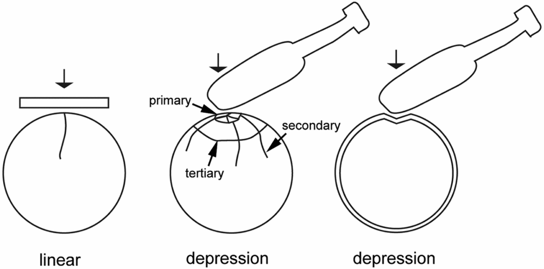
Depressions and penetrating blunt force fractures, formed by higher velocity and more concentrated force patterns, are more closely associated with armed blows (Oh Reference Oh1983: 111; Lovell Reference Lovell1997: 154; Ortner Reference Ortner2003: 121; Schulting & Wysocki Reference Schulting and Wysocki2005: 110; Schulting Reference Schulting, Schulting and Fibiger2012: 225; Wedel & Galloway Reference Wedel and Galloway2014: 62). ‘In-bending’, the process by which the skull table is forced inward towards the brain during a blow, creates bevelled, displaced bone at the impact site, that—along with secondary and tertiary fractures, which form additional fractures radiating out from or circling the impact site—is characteristic of depression fractures (Figure 1) (Oh Reference Oh1983: 116; Lovell Reference Lovell1997: 150; Ortner Reference Ortner2003: 121; Calc & Rogers Reference Calc and Rogers2007: 519; Wedel & Galloway Reference Wedel and Galloway2014: 129; Smith et al. Reference Smith, James, Pover, Ball, Barnetson, Foster, Guy, Rickman and Walton2015: 428).
The Neolithic osteological record: north-west Europe
Skeletal evidence represents the minimum number of injuries that occurred in a past population. Assessments of the skeletal record of Britain and Europe during the Neolithic have clearly established the presence of both healed and peri-mortem examples of intentionally delivered blunt force trauma (Ahlström & Molnar Reference Ahlström, Molnar, Schulting and Fibiger2012: 17; Schulting Reference Schulting, Schulting and Fibiger2012: 223; Fibiger et al. Reference Fibiger, Ahlström, Bennike and Schulting2013: 190; Fibiger Reference Fibiger, Knüsel and Smith2014; Meyer et al. Reference Meyer, Lohr, Gronenborn and Alt2015: 11217). Healed trauma tends to be more prevalent in male skeletons, while peri-mortem trauma is more evenly distributed between the sexes. Overall, there is a higher prevalence of trauma in males, which suggests that they were the principal actors and instigators of violent interaction (Schulting & Wysocki Reference Schulting and Wysocki2005: 123; Ahlström & Molnar Reference Ahlström, Molnar, Schulting and Fibiger2012: 17; Schulting Reference Schulting, Schulting and Fibiger2012; Fibiger et al. Reference Fibiger, Ahlström, Bennike and Schulting2013: 190). The social and cultural context of this violence is still heavily debated, and a better understanding of the implements used for causing these injuries would greatly aid such interpretation.
The identification of weapon typologies can help to establish if certain classes of tool were used opportunistically as weapons, or designed specifically for interpersonal violence, and how this relates to differences in the osteological evidence (Schulting & Wysocki Reference Schulting and Wysocki2005: 107; Ahlström & Molnar Reference Ahlström, Molnar, Schulting and Fibiger2012: 17; Schulting Reference Schulting, Schulting and Fibiger2012: 228; Fibiger et al. Reference Fibiger, Ahlström, Bennike and Schulting2013: 190). Establishing whether implements used at massacre sites differ from those associated with injuries commonly found in standard funerary contexts may demonstrate similarities or discrepancies between the causes.
Neolithic weapons of violence and the Thames Beater
Implements recovered from the material record of Neolithic Britain and north-western Europe are difficult to classify unambiguously as weapons of violence (Christensen Reference Christensen2004: 139; Fowler Reference Fowler2010: 16; Fibiger et al. Reference Fibiger, Ahlström, Bennike and Schulting2013: 191). Instead, potential weapons, including bows and arrows, axes, clubs and possible sling-type tools must all be considered. Current studies have yet to establish which of these implements may have been used as blunt force weapons, and mostly discuss the blunt force mechanism of injury alone, only speculating on the particular implements used (Schulting & Wysocki Reference Schulting and Wysocki2005: 125; Smith & Brickley Reference Smith and Brickley2007: 25; Lorkiewicz Reference Lorkiewicz2011: 432; Ahlström & Molnar Reference Ahlström, Molnar, Schulting and Fibiger2012: 27; Schulting Reference Schulting, Schulting and Fibiger2012: 224; Schulting & Fibiger Reference Schulting, Fibiger, Schulting and Fibiger2012: 2; Fibiger et al. Reference Fibiger, Ahlström, Bennike and Schulting2013: 199; Meyer et al. Reference Meyer, Lohr, Gronenborn and Alt2015: 11220).
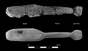
The Thames Beater is an alder club that was found in the Thames River near Chelsea and radiocarbon dated to the Early British Neolithic (Webber & Ganiaris Reference Webber, Ganiaris, Cotton and Field2004: 126). It is one of a very small number of wooden clubs that survive from the Neolithic period in Britain (Webber & Ganiaris Reference Webber, Ganiaris, Cotton and Field2004: 126; Schulting & Wysocki Reference Schulting and Wysocki2005: 125). The artefact is on exhibition at the Museum of London, and a replica was produced by master carpenter David Lewis from Pelynt, Cornwall (Figure 2). Alder was also used for the replica to create an accurate reproduction of the weight, strength and other physical properties of the original artefact. The completed replica (Figure 2) measures 640mm in length (Table 1) and comprises a slightly angled ‘blade’, barrel and pommel (Webber & Ganiaris Reference Webber, Ganiaris, Cotton and Field2004: 124).
The Thames Beater (top) and the replica club used for experimentation (bottom) with the blade, barrel and pommel labelled (photograph: Meaghan Dyer).

Dimensions and weight of the Thames Beater replica.

Wooden clubs have commonly been (and still are) used as weapons in a variety of cultures across time and space (Walker Reference Walker1989: 319; Webber & Ganiaris Reference Webber, Ganiaris, Cotton and Field2004: 124; Dujovny et al. Reference Dujovny, Onyekachi and Perez-Arjona2009: 1005). The low number of examples known from the Neolithic probably reflects the poor preservation of organic materials from the period, rather than their scarcity during this time (Webber & Ganiaris Reference Webber, Ganiaris, Cotton and Field2004: 126; Schulting & Wysocki Reference Schulting and Wysocki2005: 125; Ahlström & Molnar Reference Ahlström, Molnar, Schulting and Fibiger2012). The study of clubs that have survived can greatly aid in furthering our understanding of their potential use as weapons.
Method
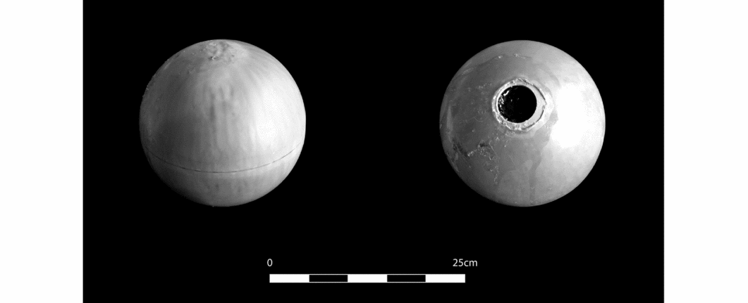
The synthetic bone spheres used for the skin-skull-brain models (Figure 3) were produced by Synbone AG (Switzerland). Two 5mm and two 7mm spheres were selected. The spheres consist of two halves of specialised polyurethane material glued together and coated in an external rubber skin to simulate part of the outer soft tissue of the skull (Synbone AG 2013). The base of the sphere has a central hole, through which it is filled with ballistics gelatin. Four of these spheres were used, two with 5mm-thick walls and two with 7mm-thick walls, to account for variation in skull thickness between individuals (Getz Reference Getz1961: 221; Adeloye et al. Reference Adeloye, Kattan and Silverman1975: 23; Lieberman Reference Lieberman1996: 223; Lynnerup Reference Lynnerup2001: 45).
The assembled synthetic bone sphere: left) showing the join between the two hemispheres; right) showing the aperture at the base of the sphere (photograph: Meaghan Dyer).
Previous studies carried out on synthetic bone have demonstrated the need for the spheres to be filled in order to recreate accurate fracturing (Carr et al. Reference Carr, Lindstrom, Jareborg, Champion, Waddell, Miller, Teagle, Horsfall and Kieser2015: 506; Smith et al. Reference Smith, James, Pover, Ball, Barnetson, Foster, Guy, Rickman and Walton2015: 428). A 10 per cent solution of ordnance level ballistics gelatin, which approximates the density of human soft tissue, was used to fill the spheres (Fackler & Malinowski Reference Fackler and Malinowski1988: 219; Jussila Reference Jussila2004: 91; Carr et al. Reference Carr, Lindstrom, Jareborg, Champion, Waddell, Miller, Teagle, Horsfall and Kieser2015: 506; Smith et al. Reference Smith, James, Pover, Ball, Barnetson, Foster, Guy, Rickman and Walton2015: 428).
Once constructed, the skin-skull-brain spheres were placed on an elevated platform 1.08m high, supported on a cork ring 31mm tall and 138mm in diameter. The hole in the sphere was placed facing down. A right-handed adult male, aged 30, 1.93m tall and weighing 88.5kg, delivered the strikes.
Two types of blow were administered to investigate variable fracture patterns produced by different areas of the club. Figure 4 shows the hand positions for administering the pommel blow and the double-handed blade strike. Each type of strike was delivered once to one 5mm and one 7mm sphere to allow each wall thickness to be observed when struck with the pommel and the blade. For the double-handed strikes, the club was swung through the air and down onto the sphere, contacting at the end of the blade. To deliver the blows with the pommel, the club was drawn up above, aimed and brought down upon the sphere. The strikes with the pommel carried notably less force. Once struck, the resulting fractures were examined, measured and photographed, before and after the rubber skin, and then the gelatin, were removed.
The hand positions used to administer the two types of blow: left) the pommel strike; right) the double-handed strike. Arrows indicate direction of swing (photograph: Meaghan Dyer).

Results
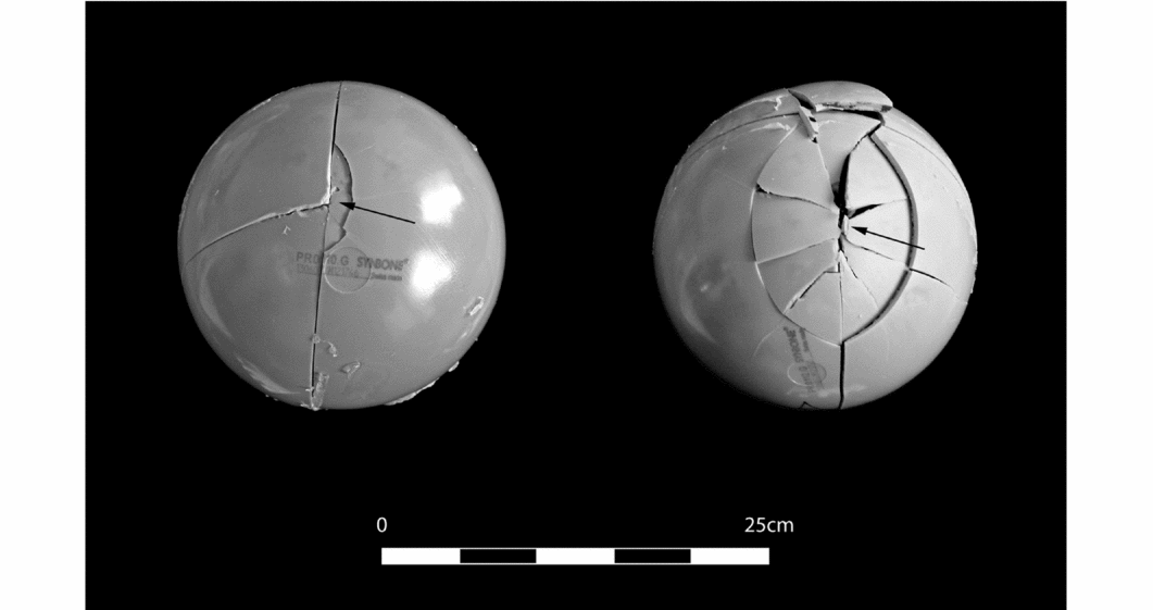
The double-handed blade strikes produced relatively extensive depression fractures in both the 7mm- and 5mm-thick sphere walls. As seen in Table 2 and Figure 5, the impact site on the spheres displaced pieces of bone. Differing numbers of radiating fractures also spread out from the area of impact. This is typical of extensive blunt force trauma (Oh Reference Oh1983: 116; Lovell Reference Lovell1997: 150; Ortner Reference Ortner2003: 121; Calc & Rogers Reference Calc and Rogers2007: 519; Wedel & Galloway Reference Wedel and Galloway2014: 129; Smith et al. Reference Smith, James, Pover, Ball, Barnetson, Foster, Guy, Rickman and Walton2015: 428). In the 5mm-thick sphere, the fractures became linked by circular tertiary fractures caused by further ‘out-bending’, which forms when the force of the impact causes the skull to push out around the area of impact, creating extensively displaced fragments of bone.
Summary of fractures produced with the skin-skull-brain models. (Note that the size of depression fractures relates to the area of depressed bone created at the impact location, and not the bone displaced by intersecting radiating fractures.)
Impact site of the 7mm-thick sphere (left), and 5mm-thick sphere (right), both with central areas of depressed bone surrounded by radiating fracture lines. Arrows indicate the point of impact (photograph: Meaghan Dyer).
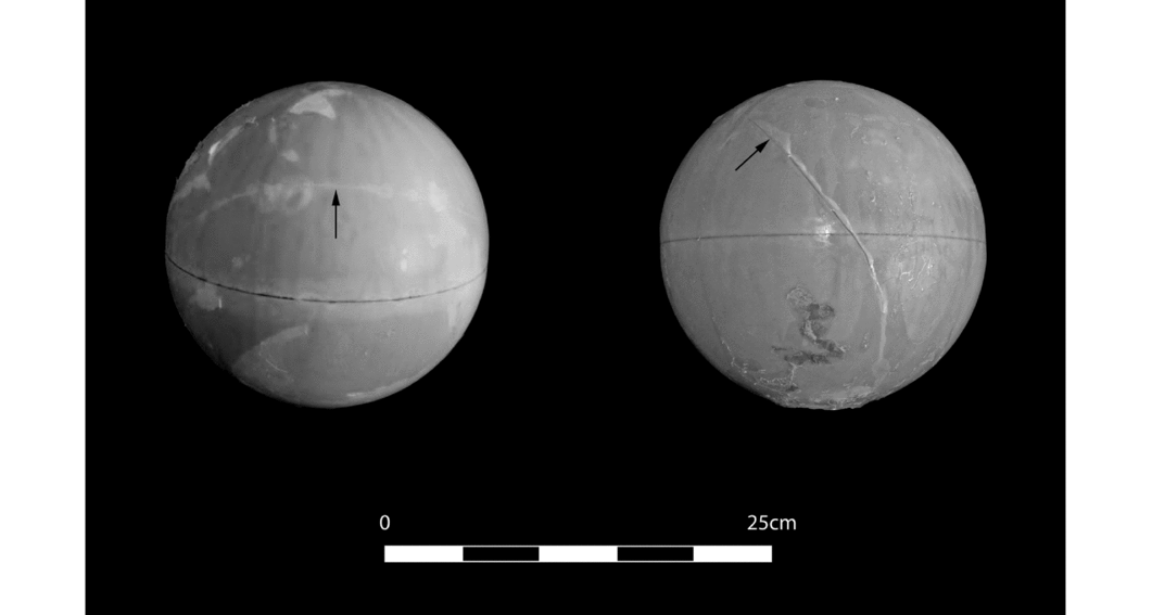
The pommel strikes differed significantly from the predicted results that the blows would produce small, shallow depressed fractures. Both the 5mm- and 7mm-thick sphere walls had long linear fractures extending from the point of impact, as listed in Table 2. These fractures ran in opposite directions from the area of initial impact (Figure 6).
Linear fractures produced by the pommel strikes on the 7mm-thick (left) and 5mm-thick (right) spheres. Arrows indicate the point of impact (photograph: Meaghan Dyer).
Discussion
Synthetic bone as an accurate medium
Despite previous problems in comparing synthetic polyurethane bone with living human tissue at the microscopic level (Smith et al. Reference Smith, James, Pover, Ball, Barnetson, Foster, Guy, Rickman and Walton2015: 427), the present study confirms the ability of a skin-skull-brain model to provide clear results for testing the macroscopic appearance of archaeological blunt force trauma. The depression and linear fractures formed in the skin-skull-brain models display the characteristics of human skull fractures. The internal bevelling of fractured synthetic bone fragments, as seen in Figure 7, along with the formation of radiating secondary and tertiary fractures (Figure 5), are major diagnostic features of blunt force trauma.
Displaced synthetic bone fragments from the double-handed strikes. Bevelled edges are indicated by arrows (photograph: Meaghan Dyer).
Ultimately, synthetic bone adequately represents the biomechanical properties of the frontal and parietal bones of living human crania (Thali et al. Reference Thali, Kneubuehl and Dirnhofer2002a: 199, Reference Thali, Kneubuehl, Zollinger and Dirnhofer2002b: 181; Carr et al. Reference Carr, Lindstrom, Jareborg, Champion, Waddell, Miller, Teagle, Horsfall and Kieser2015: 506; Smith et al. Reference Smith, James, Pover, Ball, Barnetson, Foster, Guy, Rickman and Walton2015: 434). The lack of cranial sutures and buttressing in the test model does, however, influence fracture formation, and the stepped pattern of the bevelled depression fracture edges would tend to have a much smoother appearance in actual human bone.
Most importantly, synthetic bone models remove ethical and legal issues, are cost effective, easily obtainable, able to respond biomechanically in a way that is much closer to real human skull material than can be achieved using animal substitutes, and provide a standardised model for testing (Thali et al. Reference Thali, Kneubuehl, Zollinger and Dirnhofer2002b: 178; Carr et al. Reference Carr, Lindstrom, Jareborg, Champion, Waddell, Miller, Teagle, Horsfall and Kieser2015: 506; Smith et al. Reference Smith, James, Pover, Ball, Barnetson, Foster, Guy, Rickman and Walton2015: 427).
Archaeological comparisons—double-handed strikes
The depression fractures formed by the double-handed blade strikes to the skin-skull-brain models bear a significant resemblance to examples of violence-related blunt force trauma in the Neolithic osteological record. The fracture morphology, shape of displaced fragments and the bevelled fracture edges produced in both spheres very closely match trauma linked to wooden clubs (Teschler-Nicola et al. Reference Teschler-Nicola, Gerold, Kanz, Lindenbauer, Spannagl and Windl1996; Schulting & Wysocki Reference Schulting and Wysocki2005: 125; Teschler-Nicola Reference Teschler-Nicola, Schulting and Fibiger2012: 108).
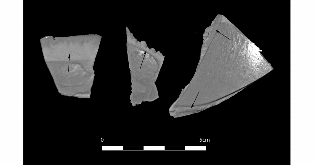
The fractures present on the 7mm-thick sphere wall bear remarkable similarity to injuries recorded on individual 3, a 35–40-year-old male from the Neolithic Austrian site of Asparn/Schletz (Teschler-Nicola et al. Reference Teschler-Nicola, Gerold, Kanz, Lindenbauer, Spannagl and Windl1996; Teschler-Nicola Reference Teschler-Nicola, Schulting and Fibiger2012: 107). As Figure 8 shows, both skulls have a long, thin depression near the top of the skull, with several radiating fractures. The impact sites on both also have one straight and one slightly curved border. This is a remarkable match between the archaeological record and the experimental results.
Comparison between the depression fractures on the 7mm-thick sphere, and the fractures found on individual 3, a 35–40-year-old male, at the site of Asparn/Schultz (skull not to scale) (Synbone sphere photograph on left: Meaghan Dyer; Asparn/Schletz cranium on right adapted from Teschler-Nicola Reference Teschler-Nicola, Schulting and Fibiger2012).

The stark similarities between the experimental models and the archaeological specimens provide a link between many of the cases of cranial trauma noted in the archaeological record and the argument that wooden clubs were used as weapons of violence. Asparn/Schletz represents a single-event massacre site (Teschler-Nicola Reference Teschler-Nicola, Schulting and Fibiger2012: 108; Meyer et al. Reference Meyer, Lohr, Gronenborn and Alt2015: 11217), and the clearly lethal nature of the tested wooden club fits the presumed motivation of intent to kill. It remains to be established whether this type of weapon could also produce some of the healed trauma found in Neolithic population studies. If so, an interpretation would be required that considered why some individuals were able to survive attacks with such a lethal tool. Alternatively, different weapon types might suggest different types of violence. These questions are currently being addressed in an ongoing study.
The anomalous linear fractures
The strikes made with the pommel end of the club deviated greatly from our expectations that the small, rounded surface combined with a more controlled ‘swing’ would produce small, non-lethal depression fractures. Instead, a distinct linear fracture formed, radiating out from the point of contact. Therefore, the lower impact energy that causes linear fractures may indeed result from intentional violence or accidental trauma (Ta'ala et al. Reference Ta'ala, Berg and Haden2006: 996; Sahoo et al. Reference Sahoo, Deck, Yoganandan and Willinger2013; Freeman et al. Reference Freeman, Eriksson and Leith2014: 64; Wedel & Galloway Reference Wedel and Galloway2014: 137). The ambiguity created due to linear fractures forming during accidental and violence-related trauma means that these fractures are commonly excluded in considerations of violence in archaeological trauma studies (Lovell Reference Lovell1997: 154; Ortner Reference Ortner2003: 121; Schulting & Wysocki Reference Schulting and Wysocki2005: 110).
The formation of these fractures may be due to the properties of the synthetic bone. This issue requires further experimental studies, but the presence of documented cases of linear fracture formation from intentional injury lends credence to the idea that blows of a lower impact energy did produce this kind of injury during violent interaction (Ta'ala et al. Reference Ta'ala, Berg and Haden2006: 996; Sahoo et al. Reference Sahoo, Deck, Yoganandan and Willinger2013; Wedel & Galloway Reference Wedel and Galloway2014: 137). The hand placement for the pommel-led strikes greatly limited the swing distance and energy of the attack. The decrease in energy is the most probable reason for linear fracture (Lovell Reference Lovell1997: 150; Ortner Reference Ortner2003: 121).
Wooden clubs as weapons of violence
Although the Thames Beater is a single artefact from England, it provides a good example of the wooden clubs that could have been crafted during the European Neolithic and beyond, and is broadly representative of this general category of implement. The strong correlation between the experimental injuries inflicted by the Thames Beater and the archaeological cases from Asparn/Schletz lends potential support to the theory that wooden clubs were used as short-range weapons of interpersonal violence in Neolithic Europe. With the recent rise in studies of both population and individual sites, information about violence in the Neolithic period has increased, with extensive variation in the interpretation of its social context including highly ritualised individual combat, informal raiding and war (Keeley Reference Keeley1996; Golitko & Keeley Reference Golitko and Keeley2007: 332; Boulestin et al. Reference Boulestin, Zeeb-Lanz, Jeunesse, Haack, Arbogast and Denaire2009; Schulting & Fibiger Reference Schulting, Fibiger, Schulting and Fibiger2012: 2).
Violent events in the Neolithic probably occurred for a variety of reasons, preventing any attempt at a singular contextual explanation (Chenal et al. Reference Chenal, Perrin, Barrand-Emam and Boulestin2015: 1329). A better understanding of the weapons used at massacre sites such as Talheim, Schöneck-Kilianstädten and Asparn/Schletz, and in cases of lower-level endemic violence as documented in larger population studies, could help to improve our understanding of different types of violent behaviour in Neolithic society (Ahlström & Molnar Reference Ahlström, Molnar, Schulting and Fibiger2012: 17; Schulting Reference Schulting, Schulting and Fibiger2012: 223; Teschler-Nicola Reference Teschler-Nicola, Schulting and Fibiger2012: 108; Wahl & Trautmann Reference Wahl, Trautmann, Schulting and Fibiger2012: 85; Fibiger et al. Reference Fibiger, Ahlström, Bennike and Schulting2013: 190).
Conclusion
This study demonstrates a probable connection between what was probably a widespread type of Neolithic weapon and examples of blunt force cranial trauma recorded in the archaeological record. In particular, it matches trauma in at least one individual from Asparn/Schletz. These results are the first to link a particular weapon to blunt force injury recorded from the Neolithic. Further research is currently being undertaken to test other potential weapons, and explore whether they can be differentiated according to the cranial fracture patterns that they produce.
The methodology established here can be applied to studies of other suspected Neolithic weapons to establish variations in the kinds of violence involved. This should provide a better contextualisation of Neolithic violence and the mechanisms by which it was enacted, and thus create a better understanding more generally of social interactions and pressures that led to different forms of violence across Western and Central Europe during this time.
Acknowledgements
The authors kindly thank Maria Teschler-Nicola for her permission to reproduce the images of the Asparn/Schletz individual. Thanks also go to David Lewis, Rebecca Redfern and the Museum of London, Debra Carr and Ian Horsfall of the ‘Impact and Armour Group’ at Cranfield University, Steven Symes, Rick Schulting, Matt Wells, Aleksa Alacia and Jürgen van Wessel. The authors would also like to thank the two anonymous reviewers for their helpful comments.


