




Non-technical Summary
During the early Cambrian (~538–506 million years ago) nearly every major group of animals (e.g., mollusks, arthropods, echinoderms, and so on) independently evolved ways to build a mineral skeleton. Some strategies involved controlling the growth of minerals with an organic scaffolding (called biomineralization) while others utilized the minerals existing in the surrounding sediment to build a protective coating (called agglutination). Most organisms settled on a single method of skeleton building, and that preference has been retained in their lineage to the present day. One unique fossil from the early Cambrian, called Salterella Billings, Reference Billings1865, incorporates both a biomineralized shell and agglutinated sediments, a strategy that has not been observed in almost any other animal group throughout history. This distinctive morphology makes Salterella difficult to place on the tree of life but important for our understanding of how animals evolved skeletons. The slightly older fossil Volborthella Schmidt, Reference Schmidt1888 shares a similar agglutinated skeleton but lacks the biomineralized shell.
In this research, we investigated the morphology of these fossils from multiple localities across North America to characterize their shape, mineral composition, and mineral crystal structure to understand the biology and ecology of these organisms and place them on the tree of life. Through this work, we document that these organisms were selective in the types of minerals they incorporated into their skeletons, suggesting a purpose such as deposit feeding or ballast. We also use the crystal structure of the shell of Salterella to suggest that these fossils are most closely related to the group that includes corals and jellyfish (Cnidaria Hatschek, Reference Hatschek1888). The unique preservation of related taxa, an older form lacking a biomineralized shell (Volborthella) and a younger form with a biomineralized shell (Salterella), may provide crucial insights into the timing and drivers of the evolution of shells in ancient animals.
Introduction
Life has developed numerous ways to build a shell. Many organisms mediate the precipitation of minerals such as calcite, aragonite, silica, or calcium phosphate around an organic scaffolding, a process called biomineralization. Others utilize mineral grains or other materials from the surrounding environment and adhere them to their body, a process called agglutination. The method that any particular organism implements to build its shell is closely linked with its phylogeny (Murdock and Donoghue, Reference Murdock and Donoghue2011). Shell-building behaviors in animals originated in the Ediacaran (e.g., Wood, Reference Wood2011; Yang et al., Reference Yang, Steiner, Schiffbauer, Selly, Wu, Zhang and Liu2020; Leme et al., Reference Leme, Van Iten and Simões2022), but the diversity of shell-building techniques exploded in the early Cambrian with the rapid diversification of animals (e.g., Erwin et al., Reference Erwin, Laflamme, Tweedt, Sperling, Pisani and Peterson2011). At this time, we see independent evolution of mineralized skeletons across multiple animal phyla (e.g., Murdock and Donoghue, Reference Murdock and Donoghue2011), with varying modes employed by different groups. Mineralized skeletons provided many advantages for early animals, most notably including structural support and protection for the body. Hypotheses for what may have driven this synchronized revolution in body development have been numerous, including an evolutionary arms race between predators and prey (Smith and Harper, Reference Smith and Harper2013), genetic repatterning to facilitate diverse body plans (Valentine et al., Reference Valentine, Jablonski and Erwin1999), increased oxygen availability supporting complex food webs (Sperling et al., Reference Sperling, Frieder, Raman, Girguis, Levin and Knoll2013), or oversaturated alkaline environments inducing precipitation of minerals on organic substrates (Grant, Reference Grant1990; Wood et al., Reference Wood, Ivantsov and Zhuravlev2017; Wood, Reference Wood2018). The exploration of different shell-building strategies during this time may provide insights into understanding the mechanism and drivers of this instrumental development in metazoan evolution.
Most early experimenters in shell building settled on a single method—either biomineralizing or agglutinating with a particular mineral. This early “choice” is largely conserved in each lineage to the present day (Porter, Reference Porter2010); that is, organisms have rarely switched back and forth between biomineralization and agglutination over the course of their evolutionary history. While rare, there are some modern organisms that employ a combination of both biomineralization and agglutination to construct their shell, including several species of foraminifera (Borrelli et al., Reference Borrelli, Panieri, Dahl and Neufeld2018), anemones of the genus Palythoa Lamouroux, Reference Lamouroux1816 (Haywick and Mueller, Reference Haywick and Mueller1997), and gastropods of the genus Xenophora Fischer von Waldheim, Reference Fischer von Waldheim1807 (Crippa et al., Reference Crippa, Pasinetti and Dapiaggi2020). One early Cambrian fossil is also known to have integrated both a biomineralized and agglutinated shell—Salterella Billings, Reference Billings1865. Fossils of Salterella are characterized by a conical calcareous shell, which is infilled by layers of agglutinated sediment and a hollow central tube connecting the aperture to the apex of the shell. These fossils occur in shallow-water deposits across the Laurentian paleocontinent during Age 4 of the Cambrian Period (Fig. 1) (Fritz and Yochelson, Reference Fritz and Yochelson1988). Given Salterella may be one of the oldest examples of this shell-building duality, it may provide insights into the initial evolutionary experimentation undertaken by shell-building animals.
Paleogeographic map of Epoch 2 of the Cambrian showing the occurrences of Volborthella and Salterella. Paleogeographic reconstruction modified from Wu et al. (Reference Wu, Pisarevsky, Li, Murphy and Liu2024), Lerosey-Aubril and Ortega-Hernández (Reference Lerosey-Aubril and Ortega-Hernández2024), Golonka (Reference Golonka2007), and Google Earth. References for occurrences of Salterella and Volborthella are provided in Supplementary Figure 1 and Supplementary Table 1.

Since its discovery over a century ago, the phylogenetic affinity of Salterella remains uncertain, limiting its use in inferences about the early evolution of shells. Previous designations include serpulids (Murchison, Reference Murchison1858), cephalopods (Clark, Reference Clark1925), pteropods (Cobbold, Reference Cobbold1920), and hyoliths (Von Zittel, Reference Von Zittel1900), among others. Currently, Salterella resides in its own phylum, the Agmata Yochelson, Reference Yochelson1977, with two other genera (Volborthella Schmidt, Reference Schmidt1888 and Ellisell Peel and Berg-Madsen, Reference Peel and Berg-Madsen1988) and without phylogenetic connection to the rest of the Animalia Linnaeus, Reference Linnaeus1758 (Yochelson, Reference Yochelson1977). The phylum Agmata is characterized by an enclosed cone of agglutinated sediments with a hollow central tube running though the length of the cone. Whereas Salterella shows a calcareous biomineralized shell surrounding the agglutinated layer (Billings, Reference Billings1865), Volborthella fossils are preserved with only agglutinated sediments and no mineralized shell, although it is inferred to have originally possessed an outer organic sheath on the basis of the smooth external surface of the agglutinated layer (Yochelson and Kisselev, Reference Yochelson and Kisselev2003). While placed in the same phylum, the relationship between the two taxa is yet unclear. Volborthella fossils are known from Stage 3 Cambrian strata of Laurentia and Baltica (Fallotapsis Zone and Nevadella Zone) (Glaessner, Reference Glaessner1976; Licari and Licari, Reference Licari, Licari, Licari, Grimer and Licari1999; Hagadorn and Waggoner, Reference Hagadorn, Waggoner and Corsetti2002), while Salterella is known only from Stage 4 Cambrian (former Bonnia–Olenellus Zone) (Fritz and Yochelson, Reference Fritz and Yochelson1988; Hagadorn and Waggoner, Reference Hagadorn, Waggoner and Corsetti2002). The limited stratigraphic range of these taxa and their unique morphology present challenges to both assigning a phylogenetic affinity and resolving their relationship.
Here we explore morphological and ecological comparisons of Salterella and Volborthella with other organisms to place them in a phylogenetic context. This work does not seek to sort out the systematics of the species of Salterella and Volborthella, although this work is needed. Our studied materials were collected from several North American deposits: the Shady Formation in southwestern Virginia, eastern United States; the Harkless and upper Wood Canyon formations in the Great Basin, western United States; and the Illtyd Formation in Yukon, Canada (Fig. 2). Similarities in the divergence angle of the conical fossils and comparison of the ratio of the central tube diameter with the thickness of agglutinated sediments suggest that the taxa are closely related. Both taxa also show similar mineral preference in materials selected for the agglutinated layer, which further suggests a shared evolutionary relationship. We subsequently examine biological constraints, ecological interpretations, and shell microstructures to inform a phylogenetic placement for these taxa. Their body plan includes a body cavity longitudinally restricted by the placement of agglutinated grains and a lack of any evidence of bilateral symmetry, suggesting a simple body form likely with a blind gut. Selectivity in agglutinated grains requires that they had appendages or tentacles to interact with the sediments and suggests that they likely occupied a sessile, benthic life mode on the seafloor. Finally, the shell microstructures consist primarily of homogeneous, blocky prisms with clear growth lamellae, indicating a limited control on biomineralization. Together this evidence points at a cnidarian affinity for the agmatans.
Simplified stratigraphic sections of Cambrian strata at the four localities where Salterella or Volborthella specimens were collected and utilized in this study, showing chronostratigraphy, lithostratigraphy, and fossil occurrences: Shady Formation (Byrd et al., Reference Byrd, Weinberg and Yochelson1973; Pfeil, Reference Pfeil1977; Willoughby, Reference Willoughby1977; Barnaby and Read, Reference Barnaby and Read1990); Illtyd Formation (Fritz, Reference Fritz1991); Harkless Formation (Hagen et al., Reference Hagen, Gill, Vayda and Pruss2024); Wood Canyon Formation (Hagadorn and Waggoner, Reference Hagadorn and Waggoner2000, Reference Hagadorn, Waggoner and Corsetti2002).

Skeleton-building cnidarians are relatively rare in the Cambrian (Leme et al., Reference Leme, Van Iten and Simões2022); from their known fossil record, they do not flourish until the Ordovician (Savarese et al., Reference Savarese, Mount, Sorauf and Bucklin1993; Baars et al., Reference Baars, Pour and Atwoods2013; Elias et al., Reference Elias, Lee and Pratt2020). However, numerous cnidarian taxa are well documented from the early Cambrian, including many conical forms (Chang et al., Reference Chang, Clausen, Zhang, Feng, Steiner, Bottjer, Zhang and Shi2018; Sun et al., Reference Sun, Zhao and Zhu2022; Qu et al., Reference Qu, Li and Ou2023; Zhao et al., Reference Zhao, Parry, Vinther, Dunn, Li, Wei, Hou and Cong2023), many of which are nonbiomineralized. This phylogenetic assignment supports the notion that cnidarians had the capability to both biomineralize and agglutinate a shell since the Cambrian, and thus similar behaviors observed in modern cnidarians may be deeply ancestral. The transition from an organic outer sheath like that of Volborthella to a biomineralized shell like that of Salterella, is potentially coincident with a shift from aragonite seas to calcite seas in the early Cambrian (Wei et al., Reference Wei, Hood, Planavsky, Li, Ling and Tarhan2022; Xiong et al., Reference Xiong, Wood and Pichevin2023), lending support to the hypothesis that this shift in ocean chemistry played a key role in the development of biomineralization in many early animals (Porter, Reference Porter2010).
Geologic background
Wood Canyon Formation
The Wood Canyon Formation is a dominantly clastic unit in the Great Basin region of Nevada (Stewart, Reference Stewart1970; Hagadorn and Waggoner, Reference Hagadorn, Waggoner and Corsetti2002). The studied section is approximately 600 m thick; however, the Wood Canyon Formation can be up to 1,200 m thick (Stewart, Reference Stewart1970). It is informally subdivided into three members (Stewart, Reference Stewart1970) (Fig. 2). The lower member consists of laminated green-brown siltstones with some sandstone beds and three distinct dolomite marker beds (Stewart, Reference Stewart1970; Diehl, Reference Diehl1979; Selly et al., Reference Selly, Schiffbauer, Jacquet, Smith and Nelson2020; Smith et al., Reference Smith, Nelson, O’Connell, Eyster and Lonsdale2023); it contains Ediacaran and Terreneuvian fossils (Grant, Reference Grant1990; Hagadorn and Waggoner, Reference Hagadorn and Waggoner2000; Smith et al., Reference Smith, Nelson, Strange, Eyster, Rowland, Schrag and Macdonald2016, Reference Smith, Nelson, Tweedt, Zeng and Workman2017; Selly et al., Reference Selly, Schiffbauer, Jacquet, Smith and Nelson2020; Evans et al., Reference Evans, Smith, Vayda, Nelson and Xiao2024). The middle member consists of cross-bedded sandstones and conglomerate (Stewart, Reference Stewart1970; Diehl, Reference Diehl1979; Fedo and Prave, Reference Fedo and Prave1991) that are interpreted to represent a terrestrial, braided plain environment (Diehl, Reference Diehl1979; Fedo and Prave, Reference Fedo and Prave1991). The upper member consists of mixed siltstones and fossiliferous grainstones, which have been regionally dolomitized (Stewart, Reference Stewart1970; Diehl, Reference Diehl1979) (Fig. 3.1). The upper member contains abundant early Cambrian fossils, including trilobites, brachiopods, echinoderms, hyoliths, Volborthella, Salterella, archaeocyaths, and complex trace fossils (Stewart, Reference Stewart1970; Nelson, Reference Nelson1978; Langille, Reference Langille1979; Licari and Licari, Reference Licari and Licari1987, Reference Licari, Licari, Licari, Grimer and Licari1999; Hagadorn and Waggoner, Reference Hagadorn, Waggoner and Corsetti2002; Mata et al., Reference Mata, Corsetti, Corsetti, Awramik and Bottjer2012), and it is interpreted to have been deposited in a shallow-marine setting influenced by tides and storms (Stewart, Reference Stewart1970; Diehl, Reference Diehl1979).
Volborthella from the Wood Canyon Formation in southern Salt Spring Hills, California. (1) Field photo showing occurrence of Volborthella in the upper member of the Wood Canyon Formation. Measuring stick marks strata where Volborthella specimens were collected. (2) VMNH 211625, hand sample showing abundance of Volborthella in a fossiliferous bed. (3–7) Thin-section photomicrographs of Volborthella in longitudinal (3, 4, 7) and transverse (5, 6) sections showing agglutinated material (red arrows) and central tube (yellow arrows); (7) size, sorting, and layering of the agglutinated grains. Thin-section numbers: (3, 5) VMNH 211625 (thin section SSSH-1A); (4, 6) VMNH 211625 (thin section SSSH-U2); (7) VMNH 211625 (thin section SSSH-U1).

Harkless Formation
The Harkless Formation is a predominantly clastic unit that outcrops in the White–Inyo Mountains region of Nevada (Nelson, Reference Nelson1978). The studied section is approximately 600 m thick; however, the Harkless Formation can be up to 1,000 m thick (Stewart, Reference Stewart1970). It is informally subdivided into a lower sandy member and an upper silty member (Stewart, Reference Stewart1970; Hicks, Reference Hicks2001) (Fig. 2). The lower sandy member consists of cross-bedded sandstone and dark siltstone (Hicks, Reference Hicks2001; Mata and Bottjer, Reference Mata and Bottjer2013). The upper member is predominately green-gray siltstone with rare fossiliferous grainstone and two distinct biohermal reef deposits near the top (Hicks, Reference Hicks2001; Pruss et al., Reference Pruss, Smith, Leadbetter, Nolan, Hicks and Fike2019). Salterella samples come from fossiliferous beds in the upper member (Fig. 4.1). The Harkless Formation is correlated with the upper member of the Wood Canyon Formation and the Zabriskie Quartzite in the Death Valley region of Nevada (Nelson, Reference Nelson1978). The age of the unit is constrained by the presence of Bonnia–Olenellus Zone trilobites, the occurrence of Salterella (Nelson, Reference Nelson1978; Green, Reference Green1982; Fritz and Yochelson, Reference Fritz and Yochelson1988; Sundberg and Webster, Reference Sundberg and Webster2021), and the mollusk Pelagiella Matthew, Reference Matthew1895 (Savarese and Signor, Reference Savarese and Signor1989). In addition to trilobites and Salterella, the Harkless Formation contains abundant fossils, including sponge spicules, chancelloriids, archaeocyaths, brachiopods, hyoliths, mollusks, echinoderms, bryomorphs, and Volborthella (Lipps and Sylvester, Reference Lipps and Sylvester1968; Yochelson et al., Reference Yochelson, Pierce and Taylor1970; Hicks, Reference Hicks2001; Skovsted and Holmer, Reference Skovsted and Holmer2006; Pruss et al., Reference Pruss, Smith, Leadbetter, Nolan, Hicks and Fike2019, Reference Pruss, Leeser, Smith, Zhuravlev and Taylor2022). The Harkless Formation is interpreted to have been deposited in an offshore environment above storm wave base along the western passive margin of Laurentia (Bond et al., Reference Bond, Christie-Blick, Kominz and Devlin1985; Mata and Bottjer, Reference Mata and Bottjer2013).
Salterella from the Harkless Formation near Gold Point, Nevada. (1) Field photo showing shales of the Harkless Formation with rare limestone beds containing Salterella (white arrows) and archaeocyath reefs at the top of the section. (2) VMNH 211623, hand sample showing abundance of Salterella in a fossiliferous layer. (3–10) Thin-section photomicrographs of Salterella in longitudinal (3–6) and transverse (7–10) sections, showing biomineralized shell (blue arrows), agglutinated material (red arrows), and central tube (yellow arrows); white arrow in (5) marks intergrowth between the biomineralized shell and the agglutinated material. (6) A micritized shell; note the lack of distinction between the layers of the shell. (10) Transverse section showing the grain size and sorting of the agglutinated layer. Thin-section numbers: (3) VMNH 611624 (thin section HS22BF1-2); (4, 6) VMNH 211623 (thin section M2137B-2); (5, 7, 8) VMNH 211623 (thin section M2137B); (9, 10) VMNH 211624 (thin section HS22B-F1(29)).

Illtyd Formation
The Illtyd Formation is a mixed clastic and carbonate unit that is exposed across the Yukon Block of Yukon, Canada (Fritz, Reference Fritz1991). It is up to 1000 m thick and consists of a range of lithologies (Fritz, Reference Fritz1991) (Fig. 2). The lower portion of the Illtyd Formation is dominated by clastic rocks, including cross-bedded sandstones and siltstones, with some interbedded ribbon-bedded limestone (Fig. 5.1), while the upper portion is composed predominately of massive crystalline carbonate (Fritz, Reference Fritz1991; Fritz et al., Reference Fritz, Cecile, Norford, Morrow and Geldsetzer1991). The Illtyd Formation is correlated with the Sekwi Formation of the Mackenzie Mountains and the Misty Creek Embayment of Northwest Canada (Fritz, Reference Fritz1991; Moynihan et al., Reference Moynihan, Strauss, Nelson and Padget2019). The age of the formation is constrained to the upper portion of the Bonnia–Olenellus Zone, which is further supported by the occurrence of Salterella (Fritz and Yochelson, Reference Fritz and Yochelson1988; Fritz, Reference Fritz1991). Archaeocyaths, brachiopods (Paterina sp. Beecher, Reference Beecher1891), and Helcionella sp. Grabau and Shimer, Reference Grabau and Shimer1909 also occur in the unit (Fritz, Reference Fritz1974, Reference Fritz1991, Reference Fritz and Norris1997). The Illtyd Formation was deposited in a shallow marine environment along the evolving passive margin of northwestern Laurentia (Fritz, Reference Fritz and Norris1997; Moynihan et al., Reference Moynihan, Strauss, Nelson and Padget2019).
Salterella from the Illtyd Formation at the type section along the Wind River, Yukon, Canada. (1) Interbedded fine-grained clastic and carbonate strata of the lower Illtyd Formation that locally contain Salterella. (2) YG 836.1, hand sample showing abundance of Salterella in a fossiliferous layer. (3–8) Thin-section photomicrographs of Salterella in longitudinal (3–5) and transverse (6–8) sections, showing biomineralized outer shell (blue arrows), agglutinated material (red arrows), and central tube (yellow arrows). White arrows in (4) show the clear boundary between the agglutinated layer and the shell near the apex, which becomes less defined toward the aperture. (5) Zoomed view of the interfingering of the biomineralized shell and agglutinated layers. (8) Transverse section showing the grain size and sorting of the agglutinated layer. Thin-section numbers: (3, 6–8) YG 836.2 (thin section J1212); (4, 5) YG 836.3 (thin section J1212-2).

Shady Formation
The Shady Formation (also called the Shady Dolomite) is a predominately carbonate unit that outcrops in southwestern Virginia (Barnaby and Read, Reference Barnaby and Read1990). It is subdivided into the Patterson, Austinville, and Ivanhoe members (Fig. 2). The Patterson Member consists of approximately 300 m of nodular- and ribbon-bedded limestone, representing deeper-water ramp facies along the passive margin of eastern Laurentia (Pfeil and Read, Reference Pfeil and Read1980; Barnaby and Read, Reference Barnaby and Read1990) (Fig. 6.1). The Austinville Member is approximately 300 m thick and is composed of cyclic peritidal carbonate mudstone, with outer ramp mud-mound build-ups (Barnaby and Read, Reference Barnaby and Read1990). The Ivanhoe Member does not outcrop widely but consists of up to 40 m of cryptalgal and fenestral laminated mudstone (Barnaby and Read, Reference Barnaby and Read1990). The Shady Formation is interpreted to represent a shallowing-up succession that transitions from carbonate ramp to platform (Barnaby and Read, Reference Barnaby and Read1990). The age of the Shady Formation is constrained by Bonnia–Olenellus Zone trilobites and the occurrence of Salterella (Byrd et al., Reference Byrd, Weinberg and Yochelson1973; Willoughby, Reference Willoughby1977; Fritz and Yochelson, Reference Fritz and Yochelson1988). Other fossils in the unit include echinoderms, calcareous algae (Epiphyton sp. Bornemann, Reference Bornemann1886), rare brachiopods, early mollusks (Helcionella sp.), and archaeocyaths (Pfeil, Reference Pfeil1977; Willoughby, Reference Willoughby1977; Pfeil and Read, Reference Pfeil and Read1980). Except for a small area around Austinville, the Shady Formation has been notably altered by late diagenetic dolomitization that occurred in association with deep burial during Alleghanian deformation (Pfeil and Read, Reference Pfeil and Read1980; Barnaby and Read, Reference Barnaby and Read1992).
Salterella from the Shady Formation at Porters Crossroads near Austinville, Virginia, USA. (1) Field photo showing massive dolomite of the Shady Formation, with white arrow marking fossiliferous bed with Salterella. (2) VMNH 90623, hand sample showing sparse occurrences of Salterella (white arrows). (3–9) Thin-section photomicrographs of Salterella in longitudinal (3–6) and transverse (7–9) sections, showing calcitic biomineralized shell (blue arrows), agglutinated material (red arrows), and central tube (yellow arrows). Note various degrees of recrystallization of the biomineralized shell and agglutinated material (4–6). Thin-section numbers: (3) VMNH 90619 (thin section H8b); (4) VMNH 90621 (thin section H9e); (5, 7) VMNH 90621 (thin section H9b); (6) VMNH 90619 (thin section H8c); (8) VMNH 90621 (thin section H9f); (9) VMNH 90621 (thin section H9c).

Methods
Materials
Salterella specimens from the Harkless Formation were collected from two sections measured ~10 km northeast of the town of Gold Point, Nevada (37°23’30.99"N, 117°15’57.55"W and 37°24’09.48"N, 117°16’50.37"W). Salterella from the Illtyd Formation were collected from the type section in the Wind River area of Yukon, Canada (Fritz, Reference Fritz1991) (65°05’15.4"N, 134°42’09.1"W), which is located within the traditional territory of the First Nation of Na-Cho Nyäk Dun. Salterella from the Shady Formation came from Porters Crossroads near Austinville, Virginia (36°51’53.57"N, 80°59’2.22"W). These materials are supplemented by museum collections, including Salterella specimens from the Shady Formation that are reposited in the Virginia Museum of Natural History (VMNH). The Volborthella specimens were collected in the southern Salt Spring Hills in California (35°36’39.96"N, 116°16’3.36"W).
Thin sections and morphological measurements
Thin sections were prepared by Spectrum Petrographics, Vancouver, Washington. We photographed thin sections using Teledyne Lemenera Infinity 1 cameras on Olympus BX51 and CX41 microscopes in the Department of Geosciences at Virginia Tech. Some thin sections were acid etched to expose the microstructure of the shell, using a dropper of dilute 5% hydrochloric acid for approximately 3 seconds on the thin-section surface, followed by rinsing in water to remove any residual acid.
We made a series of measurements to quantify the morphology of Volborthella and Salterella. Measurements were collected for a combined total of 164 specimens of Salterella (n = 107) and Volborthella (n = 57). Both Volborthella and Salterella are characterized by a conical form and an agglutinated layer with a central tube, so we used measurements of these shared characteristics for morphometric analysis and comparison herein. We measured the divergence angle at the aperture of specimens in thin section (Fig. 7.2). To ensure that we measured a reliable representation of the true divergence angle, only specimens that were sliced longitudinally where the central tube was visible (indicating an axial cut of the specimen) were measured. It is still possible, however, that some measurements could be taken from slightly oblique cuts of specimens and would result in marginally smaller measurements than the true angle. We also measured the diameter of the central tube and the thickness of the agglutinated layer (Fig. 8.2). These measurements were conducted on specimens in thin sections that were cut transversely, and the measurements were taken along the shortest axis of the transverse cut to reduce inaccuracy due to oblique sections.
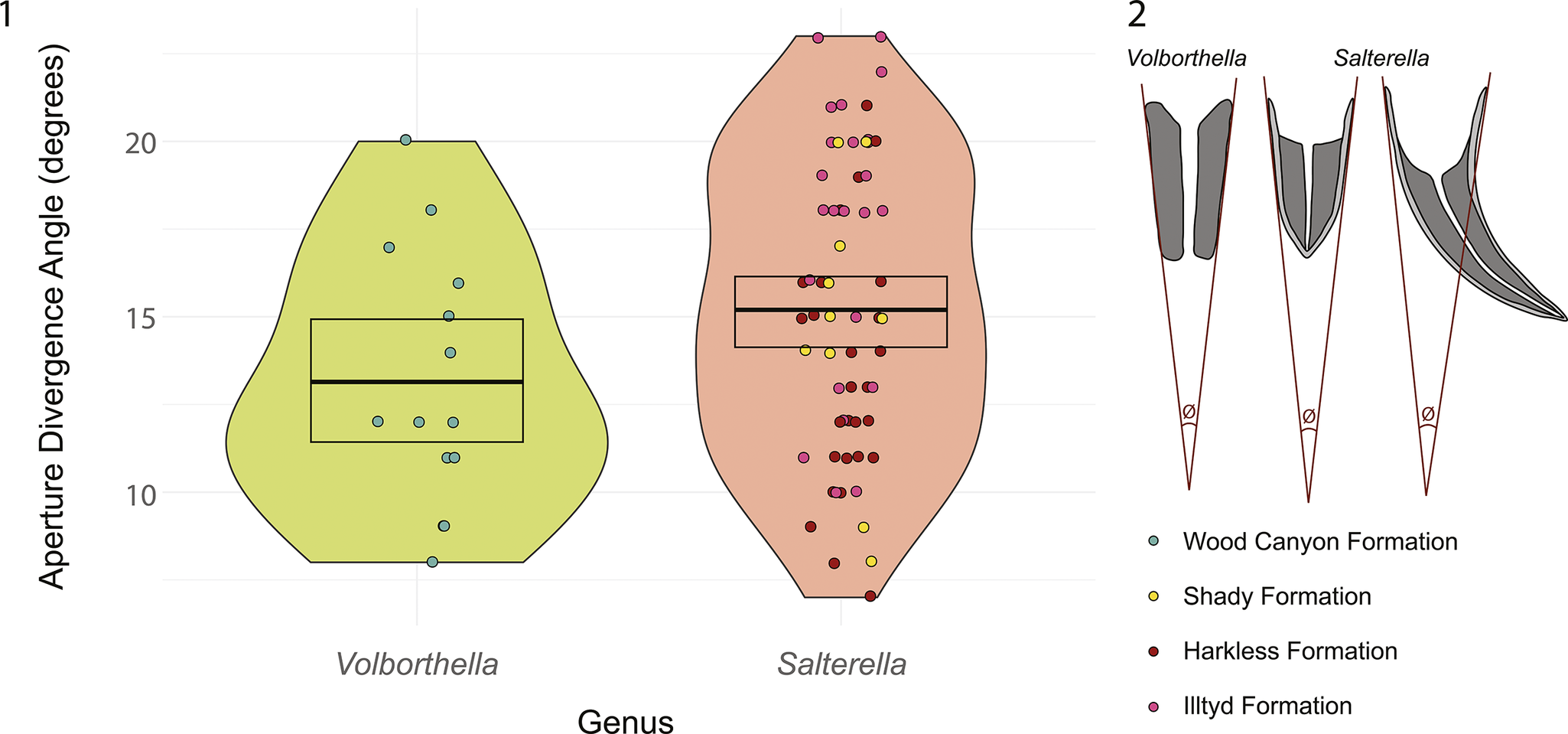
(1) Violin plot showing apertural divergence angle measurements for Volborthella and Salterella. The box marks the 95% confidence interval for the mean, and the horizontal line in the box represents the mean. Data are provided in Supplementary Table 2. (2) Line drawings depicting how divergence angle was measured in specimens of Volborthella and Salterella (angle depicted in dark red, marked by Ø).
(1) Crossplot showing relationship between the central tube diameter and the thickness of the agglutinated layer in Volborthella and Salterella. Linear regression lines with 95% confidence intervals are also shown. Data are provided in Supplementary Table 3. (2) Line drawings showing how central tube diameter and agglutinated layer thickness were measured in specimens of Volborthella and Salterella.

Maceration
Fossils were isolated from rock following a modified method from Jeppsson et al. (Reference Jeppsson, Anehus and Fredholm1999). Samples were broken down to a size that would fit in 1 liter beakers. We then submerged the samples in 10% acetic acid. The first addition of acid was buffered with spent acid from previous macerations. Then each week, we replaced 75% of the spent acid with fresh diluted acetic acid. Samples were macerated until the rock was mostly dissociated. This process took weeks to months depending on the size of the original sample. Macerates including fossil materials and sediment were sorted under an Olympus SZX16 dissecting microscope.
EPMA and EDS analysis
Thin sections were coated using a carbon coater in the Department of Geosciences at Virginia Tech. Electron probe microanalyzer (EPMA) analysis was conducted using a JEOL JXA-iHP200F in the Electron Microprobe Lab at Virginia Tech. We used a voltage of 15 keV and a nominal beam current of 10 nA. EPMA photos were captured at a resolution of 1,280 × 960 pixels. Energy dispersive X-ray spectroscopy (EDS) analysis was conducted using a JEOL JED-2300 30 mm dry SDD-EDS detector. EDS point data were collected to assist mineral identification. Each point was sampled for 5 seconds with a maximum dead time of 4%. EDS elemental maps were acquired for inferring the distribution of minerals in the samples. Maps of count data were sampled at a resolution of 512 × 384 pixels, with a 0.2 ms dwell time and 7–10 frames.
XRD analysis
Samples for X-ray diffraction (XRD) analysis were taken from the acetic acid macerate or collected by drilling powder from the rock. Salterella specimens were separated from the residue; these specimens have only the agglutinated layer because the calcitic layer was dissolved. The sediment residues and fossils were powdered separately for XRD analysis. The samples from the Shady Dolomite did not produce any Salterella fossils in the macerate, so we analyzed only two sediment samples. The Volborthella specimens are preserved in sandstone and cannot be extracted using acetic acid. We used a hand drill to gather powder from a layer consisting of predominately fossil material to represent Volborthella and a nearby layer with no fossils to represent the surrounding sediment. The powders were analyzed on an Empyrean XRD diffractometer. The machine has an Empyrean Cu LFF HR X-ray tube and a Proportional Detector Xe point detector. We used a two-theta value of 70° and a scanning step size of 0.0200°. The data were analyzed in the freeware Profex (Doebelin and Kleeberg, Reference Doebelin and Kleeberg2015). In Profex, the sample spectrum was compared with a database of reference mineral spectra. Matching minerals were added to a protocol for Rietveld refinement, which quantified the abundance of these minerals in the sample.
Distribution maps of minerals associated with Volborthella and Salterella fossils and the surrounding sediments were created using combined data from EDS point spectra, EDS elemental maps, and XRD data. Scanning electron microscope (SEM) backscatter images were recolored to form the base of the image that was then further modified in Adobe Illustrator to reflect the distribution of minerals based on the data. All data used to construct these images are included in the Supplementary Material.
μCT
The X-ray tomographic microscopy (μCT) data were collected using a Zeiss Xradia 610 Versa 3D X-ray microscope housed in the X-ray Microanalysis lab at University of Missouri. We used a voltage of 140 KV, a tube current of 18 W, the Zeiss HE2 beam filter, and the 0.4× detector for scans. The minimum voxel size for our scans was 22.516 μm3. We used an exposure time of one second per projection for a total of 1,601 projections in 360° of rotation. The data were compiled in the freeware FIJI (Schindelin et al., Reference Schindelin, Arganda-Carreras, Frise, Kaynig and Longair2012). We used Dragonfly v. 2022.2.0 (Object Research Systems, 2022) for segmentation.
Repositories and institutional abbreviations
Samples from the Wood Canyon Formation, Harkless Formation, and Shady Formation are reposited in the Virginia Museum of Natural History (VMNH) in Martinsville, Virginia. Samples from the Illtyd Formation are reposited in the Yukon Fossil Collections (YG) in Whitehorse, Yukon Territory, Canada, in partnership with the First Nation of Na-cho Nyäk Dun.
Results
General morphology
Volborthella specimens from the Wood Canyon Formation at the southern Salt Spring Hills locality are found predominantly in monotaxic concentrations or coquinas that are up to a few centimeters in thickness (Fig. 3.1, 3.2). Individual fossils consist of a cylindrical tube of agglutinated grains up to 4 mm long with a taper toward the inferred apex (Fig. 3.3, 3.4). The outer margin of the fossil is well defined and smooth (Fig. 3.3–3.7). Some specimens show apparent layering within the agglutinated sediment (Fig. 3.3, 3.4, 3.7). These layers are built within the tube at a shallower slope than the margin of the outer wall. In thin section, most specimens appear truncated or missing their apex. In three-dimensional renderings based on μCT data, even the longest (inferred most complete specimens) do not taper to a pointed apex. It remains unclear whether this is a taphonomic artifact or the true morphology of the organism.
The morphology of Salterella specimens varies by locality (Figs. 4–6), which may indicate different species within the genus. Nonetheless, they are united by an external biomineralized cone with internal, layered, agglutinated sediment and a hollow central tube passing through the agglutinated grains, connecting the aperture to the apex of the cone. In the Harkless and Illtyd formations, Salterella is known from fossil-concentration beds up to 10 cm thick (Figs. 4.1, 5.1). These shell layers consist predominantly of Salterella with rare hyoliths and phosphatic brachiopods. In the Shady Formation, fossils are relatively rare and often found as dispersed specimens in dolostone but occur primarily in discrete horizons (Fig. 6.1).
Salterella from the Harkless Formation are up to 13 mm long (Fig. 4.2). The shells have a consistent curvature from apex to aperture in one plane (Fig. 4.2–4.4), distinct from the relatively straight conical shells of Salterella from other localities investigated in this study. The biomineralized shell is calcitic in mineral composition (Fig. 4.3–4.5, 4.9, 4.10) and preserves internal growth laminae (Fig. 4.5). The boundary between the biomineralized shell and agglutinated layer is smooth, but it shows intergrowth of the biomineralized shell and agglutinated layer at regular intervals (Fig. 4.5). Some shells have been micritized or recrystallized but retain a layered shell fabric (Fig. 4.6–4.8). In most occurrences, the shells show brittle deformation as evidenced from fragmented shells and discontinuities in agglutinated layers (Fig. 4.3). Many of these fossils are preserved with a goethite rim on the outer margin of the agglutinated grains (Fig. 4.3, 4.4).
Salterella from the Illtyd Formation are up to 8 mm long (Fig. 5.2). The shells are conical and straight, with no apparent curvature (Fig. 5.2–5.4). They have a consistent divergence angle from apex to aperture, ranging from 10° to 23° (Fig. 7). The biomineralized layer is calcitic in composition (Fig. 5.3–5.8). Some specimens preserve original shell growth lines, but others show signs of overprinting and recrystallization. The boundary between the biomineralized layer and agglutinated layer is distinct and smooth near the apex of the shell but becomes more irregular near the aperture (Fig. 5.4, 5.5).
Salterella from the Shady Formation are up to 6 mm long (Fig. 6.2–6.6). The shell has a “bullet” shape with a broader divergence angle near the apex (up to 70°) that narrows to an apertural angle similar to that of Salterella from other localities (Fig. 6.3, 6.4). The biomineralized shells are calcitic but have mostly been recrystallized, not preserving any internal textures (Fig. 6.4–6.6). The boundary between the biomineralized and agglutinated layers is smooth where observable (Fig. 6.3, 6.7, 6.9).
Morphometric analysis
We measured the divergence angles of 14 Volborthella specimens with results ranging from 8° to 20° with a mean of 13.14° (Fig. 7.1). We measured the divergence angle in 61 Salterella specimens, including 27 from the Harkless Formation, 10 from the Shady Formation, and 24 from the Illtyd Formation. The divergence angle of Salterella ranges from 7° to 23° with a mean of 15.4° (Fig. 7.1). A Welch two-sample t-test shows no statistically significant difference in apical divergence angle between Volborthella and Salterella (p = 0.07769).
We measured the central tube diameter and agglutinated layer thickness in 43 Volborthella specimens and 46 Salterella specimens, including 31 from the Harkless Formation, 5 from the Shady Formation, and 10 from the Illtyd Formation. Both genera show a weak, positive, linear relationship between the agglutinated layer thickness and the central tube diameter (Fig. 8.1). The confidence intervals of the linear regressions completely overlap between the two genera, indicating no statistically significant difference between them.
Agglutinated layer composition
It has been previously documented that agmatans show selectivity in the grains that are incorporated into their agglutinated shell (Hagadorn and Waggoner, Reference Hagadorn, Waggoner and Corsetti2002; Peel, Reference Peel2017b). To characterize the mineralogy of agglutinated sediment in Volborthella and Salterella, we used a combination of light microscope petrography, EPMA-EDS, and XRD analyses. Light microscopy was useful for identifying major mineral components and visually confirming results from the other analyses. We collected 781 point spectra for individual minerals across 13 thin sections, as well as 66 elemental maps at various scales. The data provided the elemental composition of the fossils and surrounding sediments and can be used to infer mineralogical compositions. On the basis of inferred mineralogical compositions, we were able to map the spatial distribution of minerals, including trace presence of minerals that were not detected with XRD.
XRD analysis provided bulk mineralogical information about the fossils and surrounding sediment. In the Wood Canyon Formation, the sediment consists of 44.8% quartz, 24.6% biotite, 17.6% albite, 9.7% enstatite, and 3.3% magnetite, whereas the Volborthella fossils contain relatively less quartz (14.2%), biotite (17.4%), enstatite (8.5%), and magnetite (2%) but are enriched in fluorapatite (10.2%), feldspars (25.6%), and pseudorutile (22%) (Fig. 9). In the Harkless Formation, the sediments are composed of 63.7% quartz, 14.7% chamosite, 17.4% illite, and 4.1% goethite, whereas the agglutinated material in Salterella fossils contains relatively more quartz (78.2%) and comparatively less clay (18.9%) and goethite (2.9%) (Fig. 9). In the Illtyd Formation, the sediments are also composed predominantly of quartz (82.8%) with 8.2% albite, 6.3% chamosite, and 2.7% anatase. The agglutinated material in Salterella fossils from this unit has proportionally similar amounts of quartz (81.1%) but contains more albite (11.8%) and anatase (4.4%) and less chamosite (2.7%) than the host sediment (Fig. 9). In the Shady Formation, the sediments are dominated by dolomite (98.6%) and are quartz-poor (1.4%) compared with the other formations (Fig. 9). No Salterella specimens from the Shady Formation survived acetic acid maceration, so we do not have XRD data for these specimens. EDS map data suggest that Salterella from the Shady Formation produce both the biomineralized and agglutinated layers of their shell with calcite, so the entire fossil would dissolve in the acid maceration.
Mineral composition of fossil agglutinated layer and surrounding sediment. Percent composition was acquired from XRD analysis of macerated and powdered samples of Volborthella and Salterella as well as composition of the sediment. Samples from the Shady Formation have no fossil data because no fossils survived acetic acid maceration.

The EDS maps and point analyses support the mineral identifications from XRD and show the same pattern of mineralogical differences between the fossils and enclosing sediments (Fig. 10). The maps also show the spatial distribution of calcite in the sediment and fossils, which was not captured in XRD due to the way the samples were processed.
Thin-section petrographic photomicrographs (left) and corresponding mineral maps (right) of Volborthella from the Wood Canyon Formation, VMNH 211625 (thin section SSSH-U1), and Salterella from the Harkless Formation, VMNH 211623 (thin section M2137B-2); the Illtyd Formation, YG 836.2 (thin section J1212); and the Shady Formation, VMNH 90621 (thin section H9c). Mineral maps were constructed by applying false color to SEM backscatter images using data from XRD, EDS elemental maps, and EDS point spectra for mineral identification. SEM backscatter images and EDS elemental maps are provided in Supplementary Figures 19–22.

In addition to compositional selectivity, Volborthella and Salterella demonstrate selectivity in grain size, shape, and sorting. Volborthella from the Wood Canyon Formation composed their agglutinated shell of generally elongate or planar grains that range from 20 to 120 μm long and up to 50 μm tall (Fig. 3.7). These grains are moderately sorted to well-sorted and sub-angular to angular in form (Fig. 3.7). Most of the elongate grains are positioned with the long axis parallel to the laminations of the agglutinated layer (Fig. 3.7). Salterella from the Harkless Formation utilized sub-angular, equidimensional grains that range from 3 to 30 μm in diameter (Fig. 4.10). Within the agglutinated layers, the grains are moderately sorted (Fig. 4.10). Salterella from the Illtyd Formation similarly used mostly equidimensional, sub-rounded grains from 3 to 35 μm in diameter (Fig. 5.8). The grains are moderately to poorly sorted, and where elongate grains occur, they are usually aligned with the long axis parallel to the laminae of the layers (Fig. 5.5). The agglutinated layer of Salterella from the Shady Formation consists of recrystallized calcite grains that range from 7 to 20 μm in size, which is distinctly smaller than the crystals that make up the dolomitic matrix, which range from 20 to 60 μm. Despite the apparent recrystallization, the distinct boundary between the agglutinated layer and the biomineralized shell still remains (Fig. 6.9).
Biomineralized shell composition and microstructures
The biomineralized layer of Salterella is composed of calcite. The shell is constructed of simple growth laminae (Fig. 11.1–11.4). On the basis of cement stratigraphy, it is inferred that new laminae were added to the inner surface of the shell (Figs. 4.5, 5.5). In Salterella from the Shady Formation, the biomineralized layer has a distinct boundary with the agglutinated layer (Fig. 6.3), whereas Salterella from the Illtyd and Harkless formations show substantial intergrowth of the biomineralized and agglutinated layers (Figs. 4.5, 5.5). In all specimens, the shell grows in simple parallel laminae, and calcite crystals do not cross these laminae. Shell textures include blocky prismatic, fibrous prismatic, and simple prismatic. The most common microstructure in Salterella is a blocky prismatic texture that consists of equidimensional crystals arranged in parallel growth laminae (Fig. 11.1–11.4). The fibrous prismatic texture consists of prisms that are elongated parallel to the shell laminae (Fig. 11.4). This texture is most common on the inner margin of the shell. Where the biomineralized and agglutinated layers meet, the biomineralized layer often bears a fibrous prismatic texture. The simple prismatic texture, which is most common on the outer margin of the shell, consists of columnar prisms with the long axis perpendicular to the shell laminae (Fig. 11.2).
Secondary electron SEM images of acid-etched Salterella specimens showing microstructures of biomineralized shells. Inset images show position and orientation of SEM images. (1) VMNH 211625 (thin section HS22B-F1(29)), transverse-section view showing growth laminae with micrometer-sized equant calcite crystals, as well as well-defined inner and outer boundaries of the biomineralized layer (blue arrows). (2) VMNH 211624 (thin section HS22B-F2), longitudinal-section view showing growth laminae successively added on the inner surface (to the left) of the biomineralized layer near the aperture. The growth laminae consist of micrometer-sized equant calcite crystals and have well-defined inner and outer boundaries (blue arrows). (3) YGS 836.3 (thin section J1212-2), longitudinal-section view showing that, in general, calcite crystals do not cut across growth laminae. Blue arrow indicates outer margin of biomineralized shell. (4) VMNH 211623 (thin section M2137B-2), transverse-section view showing intergrowth between biomineralized laminae with micrometer-sized equant calcite crystals (blue arrows) and agglutinated layers with coarser-grained material (red arrows).

Discussion
History of taxonomic affinities of Volborthella and Salterella
Since their discovery over a century ago, the phylogenetic affinity of Salterella has remained enigmatic. Initially, Salterella was described as a serpulid worm (annelid, sabellid) on the basis of the apparent annulated nature of the tubes (Murchison, Reference Murchison1858; Billings, Reference Billings1865; Miller, Reference Miller1889). Salterella has also been considered a mollusk with conulariid affinities (before conulariids were recognized as cnidarians) alongside other enigmatic fossils, including tentaculitiids and hyoliths (Von Zittel, Reference Von Zittel1900). Salterella along with hyoliths have also been grouped with the pteropods, another molluscan group (Cobbold, Reference Cobbold1920). These two molluscan assignments were based primarily on external shell morphology.
Early investigations of the internal skeletal morphology of Salterella led some to interpret the agglutinated layers as chambers, suggesting a crown-group cephalopod affinity (Clark, Reference Clark1925; Poulsen, Reference Poulsen1927). Similarly, when Volborthella was first described, it was placed in the Cephalopoda Cuvier, Reference Cuvier1795 on the basis of the apparent chambered nature of the fossil (Schmidt, Reference Schmidt1888; Schindewolf, Reference Schindewolf1927; Ulrich and Foerste, Reference Ulrich and Foerste1933). There was much disagreement about this placement, with some stating that the central tube is not homologous with a siphuncle (Kobayashi, Reference Kobayashi1937) and others asserting that the evidence supporting a cephalopod affinity was unconvincing (Spath, Reference Spath1933; Kobayashi, Reference Kobayashi1937; Miller, Reference Miller1943). Flower (Reference Flower1954) suggested that Salterella may be a representative of a group with no modern descendants. Lipps and Sylvester (Reference Lipps and Sylvester1968) considered foraminiferan and polychaete affinities but also suggested the potential of a lost group. Glaessner (Reference Glaessner1976) re-invoked the polychaete affinity for Volborthella, noting its similarities to sabellids and other tube-forming worms, as did Licari and Licari (Reference Licari and Licari1987, Reference Licari, Licari, Licari, Grimer and Licari1999), noting the similarity to the modern polychaete Owenia fusiformis Delle Chiaje, Reference Delle Chiaje1844. The idea of Salterella representing the sclerites of a larger metazoan organism has also been suggested on the basis of the anomalous orientation of one specimen (Signor and Ryan, Reference Signor and Ryan1993; Donovan and Paul, Reference Donovan and Paul1994), but this model has not been widely accepted (Yochelson and Kisselev, Reference Yochelson and Kisselev2003; Astini et al., Reference Astini, Thomas and Yochelson2004; Jones, Reference Jones2007). Ultimately, Yochelson (Reference Yochelson1977) established the new phylum of Agmata for Salterella and Volborthella, which he suggested was an extinct phylum. This designation has remained the prevailing view (Peel and Berg-Madsen, Reference Peel and Berg-Madsen1988; Yochelson and Kisselev, Reference Yochelson and Kisselev2003; Astini et al., Reference Astini, Thomas and Yochelson2004; Jones, Reference Jones2007). Unfortunately, this assignment does not resolve the phylogenetic relationship between the Agmata and other metazoans, and it limits any inferences we can make about the early evolution of shells.
The relationship between Salterella and Volborthella has also been a topic of discussion. In his designation of the phylum Agmata, Yochelson (Reference Yochelson1977) posited that the lack of a calcareous outer shell in Volborthella may be a taphonomic artifact rather than a biological signature. Because of this, Volborthella was placed in synonymy with Salterella (Lauritzen and Yochelson, Reference Lauritzen and Yochelson1982; Yochelson, Reference Yochelson1983). Soon after, this position was recanted and the genus Volborthella was recognized again, although the question of the presence and composition of an integument surrounding the agglutinated grains remained unanswered (Yochelson and Kisselev, Reference Yochelson and Kisselev2003). The distinct and smooth outer margin of all Volborthella specimens (Fig. 3.3, 3.4) is suggestive of a bounding tissue layer. In Salterella, the calcareous outer shell interfingers with the agglutinated layers (Figs. 4.5, 5.5), likely caused by the same mechanism controlling both shell growth and cementation of the agglutinated layers. Any evidence of a biomineral outer layer interacting with the agglutinated layer of Volborthella is not present; we alternatively suggest that an organic integument likely bounded the agglutinated layer in Volborthella. Høyberget et al. (Reference Høyberget, Ebbestad, Funke, Funke and Nakrem2023) showed one specimen of Volborthella that may preserve this organic sheath that surrounded the agglutinated layers. The presence of an organic outer layer in Volborthella implies that Salterella, if indeed related, may have had a similar organic layer serving as a scaffolding template for biomineralization.
This is not to say that Salterella is the direct descendant of Volborthella, but the morphological similarities between these two organisms offers a strong suggestion of their close relationship or possibly that they represent taphonomic variants of the same biological group. For example, the two taxa show no significant difference in the divergence angle of their cone (Fig. 7). Further, the ratio between the diameter of the central tube and the thickness of the agglutinated layer is virtually the same (Fig. 8). The similarity of these morphological characteristics suggests a shared ancestry for their development. Although convergence on the agglutinated layer morphology cannot be entirely ruled out, it is much more parsimonious to suggest these taxa are closely related. In localities where both Volborthella and Salterella are found, Volborthella is consistently found in older strata than Salterella and there is no stratigraphic overlap between the two genera (Licari and Licari, Reference Licari, Licari, Licari, Grimer and Licari1999; Hagadorn and Waggoner, Reference Hagadorn, Waggoner and Corsetti2002; Jones, Reference Jones2007). Although it is possible that the distribution is controlled by facies variation, Volborthella has also been found in carbonate environments (Skovsted et al., Reference Skovsted, Balthasar, Vinther and Sperling2020). This non-overlap in stratigraphic occurrence suggests that Volborthella and Salterella could represent distinct biological genera rather than taphonomic variants of the same organism.
Morphology and function
The skeletal morphology of Volborthella and Salterella places constraints on the biology of the organisms. First and foremost, a mechanism must exist for the acquisition of mineral grains and their incorporation into an agglutinated skeleton. Various groups of organisms have developed methods of building an agglutinated skeleton, including foraminiferans (Hemleben et al., Reference Hemleben, Kaminski, Kuhnt and Scott1989; Sen Gupta et al., Reference Sen Gupta, Goldstein, Hansen, Parker and Arnold2003; Borrelli et al., Reference Borrelli, Panieri, Dahl and Neufeld2018), cnidarians (Haywick and Mueller, Reference Haywick and Mueller1997; Reimer et al., Reference Reimer, Nakachi, Hirose, Hirose and Hashiguchi2010, Reference Reimer, Hirose, Irei, Obuchi and Sinniger2011), lophophorates (Zhang et al., Reference Zhang, Li, Holmer, Brock and Balthasar2014), annelids (Grémare, Reference Grémare1988; Shcherbakova and Tzetlin, Reference Shcherbakova and Tzetlin2016; Becker-Kerber et al., Reference Becker-Kerber, Pacheco, Rudnitzki, Galante, Rodrigues and Leme2017, Reference Becker-Kerber, Horodyski, del Mouro, Sedorko, Lehn, Sanchez, Fournier, Mazurier and El Albani2021), and arthropods (Krasnow and Taghon, Reference Krasnow and Taghon1997; Mouro et al., Reference Mouro, Zaton, Fernandes and Waichel2016; Kakui and Hiruta, Reference Kakui and Hiruta2017). These organisms all bear structures such as pseudopodia, tentacles, or jointed appendages to interact with the environment, move sediment grains, and incorporate them into the test or shell. Volborthella and Salterella therefore must have also had a structure that could reach out from the aperture to acquire grains and bring them into the shell, although the nature of this structure remains unknown. Volborthella and Salterella are somewhat different from other agglutinating organisms in that the agglutinated sediments are deposited in layers forming a solid cone rather than simply building on the outer surface of the body as in most modern agglutinators. One unique exception to this is the anthozoan group Ceriantharia Perrier, Reference Perrier1893. In these organisms, sediments are absorbed by the tissue and either retained within the body or passed entirely through and deposited under the organism (Haywick and Mueller, Reference Haywick and Mueller1997; Reimer et al., Reference Reimer, Nakachi, Hirose, Hirose and Hashiguchi2010). This pass-through approach is one way that agmatans may have built the agglutinated portion of their shell. Alternatively, they could have moved particles externally through the gap between their body and outer shell.
Next there is the question “why incorporate agglutinated grains into the shell?” In all modern agglutinating organisms, the agglutinated shell serves the same function as a biomineralized shell: to provide structural support and protection. Considering that Salterella had a biomineralized shell, this function would be redundant. Furthermore, the positioning of the agglutinated grains is incompatible with this purpose, as the presence of the biomineralized shell in Salterella indicates that the body of the organism existed outside of the agglutinated portion of the shell. Early descriptions of Voborthella and Salterella likened the structure of the grains to the chambers of a nautiloid shell (Schmidt, Reference Schmidt1888; Clark, Reference Clark1925; Poulsen, Reference Poulsen1927). Although it is clear now that these structures are not homologous, they may have served a similar function in reducing the size of the occupied portion of the shell. Alternatively, the addition of an agglutinated layer inside a conical shell of Salterella or an organic integument of Volborthella may have lowered the center of gravity, thus stabilizing these epibenthic or semi-endobenthic animals and protecting them from being toppled by waves and currents (Fig. 12). In addition, in his description of agmatans, Yochelson (Reference Yochelson1977) suggested that the agglutinated grains could serve to fill in the shell to keep the length of the organism within a limit possibly dictated by a blind or incomplete gut. Further support for this hypothesis can be found in the way that Salterella built its shell. In the earliest stages of growth, represented by the shell nearest the apex, there is no integration of biomineralized material and agglutinated material suggesting that the biomineralized shell was grown before any sediments were brought into the shell (Yochelson et al., Reference Yochelson, Pierce and Taylor1970). Toward the apertural end of the shell, the biomineralized portion and agglutinated portion interfinger, suggesting that they were grown and incorporated synchronously. This indicates that Salterella did not start incorporating agglutinated grains into its shell until it surpassed a certain size. This demonstrates a degree of determinate growth along the longitudinal axis of these fossils, a characteristic that rules out potential phylogenetic affinities to annelids, which continue to grow by adding segments to their body throughout their lives (Hariharan et al., Reference Hariharan, Wake and Wake2016).
Reconstruction of Salterella on the Cambrian seafloor exhibiting behaviors such as collecting sediment grains to be incorporated into the agglutinated layer and retracting the body into the apertural chamber. Art by Amy Hagen.

The final morphological characteristic that is unique to these fossils is the central tube. This feature connects the living chamber to the apex of the shell. In initial descriptions of Salterella, the central tube was analogized to the siphuncle of shelled cephalopods (Schmidt, Reference Schmidt1888; Clark, Reference Clark1925; Poulsen, Reference Poulsen1927), but a hollow tube for fluid transport would serve little function considering the apical portion of the shell was filled with sediment grains. The shell of Salterella is closed at the apical end, so the structure could not serve as an anchor to the substrate, but it could potentially anchor the organism to its shell. One of the primary functions of a shell is protection. Whereas an organism may need to extend appendages outside the shell to feed and respire, it also needs to be able to retract into the shell for protection. The agglutinated grains within the shell are constantly being built up and would not serve as a sturdy fastening point for muscles to pull the organism into the shell. Rather, agmatans would need an attachment to the shell itself at a point that remains fixed as the organism grows—the apex of the shell. The central tube may have facilitated the anchoring of the organism on the apex of the shell as the shell and agglutinated grains continued to grow around it.
Ecology
The lifestyle of these fossils is informed by their morphology. First, they must have been benthic to have access to sediment grains to build the agglutinated portion of their shell. Much of the inference of whether they were motile or sessile depends on their phylogenetic affinity. A foraminiferan, cnidarian, or annelid affinity would be more suggestive of a sessile and epifaunal, or possibly infaunal to semi-infaunal, life habit, whereas a lophophorate, molluscan, or arthropod affinity could potentially support a motile epifaunal lifestyle. Previously, Volborthella has been depicted as an infaunal organism, with an annelid affinity (Glaessner, Reference Glaessner1976). Yochelson and Kisselev (Reference Yochelson and Kisselev2003) suggested that Salterella may have lived as a benthic organism not attached to the substrate, but also not necessarily mobile. Nearly all reconstructions tend to agree on an apex-down orientation as otherwise the agglutinated sediments would result in a very top-heavy weight distribution of the organism (Yochelson, Reference Yochelson1977). The three-dimensional reconstructions do not show any apparent bilateral symmetry suggestive of a dorsal and ventral side of the shell that would indicate a horizontal or procumbent life position. We propose that Volborthella and Salterella lived apex down in the sediment, were sessile but not cemented to the substrate, had tentacles to reach out of the shell and acquire grains for agglutination, and could pull their body into the shell using the attachment of the central tube.
A benthic lifestyle for these animals is also consistent with their restricted paleogeographic distribution. Although they are known from a range of clastic- to carbonate-dominated depositional settings, they are found only in the relatively shallow margins of the paleocontinents Laurentia and Baltica (Yochelson, Reference Yochelson and Taylor1981; Fritz and Yochelson, Reference Fritz and Yochelson1988) (Fig. 1). This restricted distribution supports the interpretation that Volborthella and Salterella were likely not pelagic and instead lived on the seafloor and perhaps did not even have a motile larval phase.
Benthic, sessile fauna often take on a suspension-feeding or deposit-feeding lifestyle. Suspension feeders passively (relying on currents) or actively (generating their own flow) collect particles from the water column for nutrients (Riisgard and Larsen, Reference Riisgard and Larsen2010). Deposit feeders ingest sediments to obtain nutrients from associated bacteria and organic material (Lopez and Levinton, Reference Lopez and Levinton1987). Deposit feeders have also been documented showing selectivity in what grains they obtain for feeding (Taghon, Reference Taghon1982; Self and Jumars, Reference Self and Jumars1988). Deposit-feeding foraminifera are known to use vacuoles to incorporate sediments into their cell and move them further into their body for ingestion of attached organic material (Goldstein and Corliss, Reference Goldstein and Corliss1994). The agglutinating polychaete Owenia fusiformis exhibits both suspension- and deposit-feeding habits and gathers particles for both ingestion and building a protective tube (Dales, Reference Dales1957). These habits could be suggestive of a similar behavior in Volborthella and Salterella as these organisms may have selected grains for feeding and then incorporated them into their agglutinated skeleton. Many benthic organisms are also able to modulate between suspension- and deposit-feeding strategies on the basis of changes in flow and sediment load (Miller et al., Reference Miller, Bock and Turner1992). Although we cannot rule out a suspension-feeding life habit, Volborthella and Salterella very likely had the ability to be deposit feeders as they had apparatuses to collect grains and bring them into the shell. Ultimately, both feeding strategies would be effective in the shallow waters where they lived.
Affinities to other higher taxa: agglutinated forms
The mineral grains composing the agglutinated layers of agmatan shells are a distinct subset of the grains found in the surrounding environment, demonstrating a clear selectivity in the agglutination process. Grain preference has been documented in various agglutinating organisms. Palythoa (Anthozoa) was shown to encrust only grains that were smaller than 125 μm and showed no discretion based on mineral type (Haywick and Mueller, Reference Haywick and Mueller1997), whereas the closely related Neozoanthus Herberts, Reference Herberts1972 showed no apparent preference for grains when encrusting (Reimer et al., Reference Reimer, Hirose, Irei, Obuchi and Sinniger2011). Caddisfly larvae have exhibited grain-size preference and material preference based on the environment (Boyero et al., Reference Boyero, Rincón and Bosch2006; Statzner and Dolédec, Reference Statzner and Dolédec2011). Grémare (Reference Grémare1988) demonstrated that the polychaete Eupolymnia nebulosa Montagu, Reference Montagu1819 shows selectivity for larger grains for building its tube. Agglutinated foraminifera have also demonstrated selective behavior, including grain size, shape, and composition (Lipps, Reference Lipps1973).
The fossil record also contains evidence of selective agglutinators. Onuphionella Kiryanov, Reference Kiryanov, Krandievski, Ischenko and Kiryanov1968 is an organism from the early Paleozoic that built its agglutinated shell exclusively from mica grains (Hagadorn and Waggoner, Reference Hagadorn and Waggoner2000; Zaton and Bond, Reference Zaton and Bond2016; Muir et al., Reference Muir, Botting, Walker, Schiffbauer and MacGabhann2022). Previous work on Volborthella from Nevada noted that the organisms built their agglutinated shells with ilmenite or Ti-oxides, quartz, zircon, monazite, and possibly iron oxide pseudomorphs of pyrite (Hagadorn and Waggoner, Reference Hagadorn, Waggoner and Corsetti2002). Other studies of specimens from Germany and Russia noted similar selectivity for quartz and Fe- and Ti-oxides, with differences in other minor minerals based on availability in the environment (Gürich, Reference Gürich1934; Rozanov and Zhuravlev, Reference Rozanov, Zhuravlev, Lipps and Signor1992). Similarly, Salterella has been shown to selectively incorporate anatase (Ti-oxide) and is documented to include the phosphatic sclerites of Hadimopanella Gedik, Reference Gedik1977 in its shell (Peel, Reference Peel2017b). In a carbonate-dominated environment, Salterella was noted to build the agglutinated portion of its shell using carbonate grains (Griffin and Yochelson, Reference Griffin and Yochelson1975).
We observed selectivity in Volborthella and Salterella at all localities, spanning a range of depositional environments. General trends include positive selection for titanium-rich minerals and feldspars and negative selection for clays and iron-rich minerals across all settings (Fig. 9). Other selectivity trends were unique to the mineral assemblage at each locality. In some localities, selection for quartz is positive (Fig. 9, Harkless Fm.), while in other localities there is an apparent exclusion of quartz (Fig. 9, Wood Canyon Fm.), suggesting that the comparative characteristic relative to other minerals available, rather than an absolute characteristic of the mineral, guided the preference. There does not appear to be a correlation between mineral density and mineral preference. Because all preferences are consistent across mineral groups (with quartz being the exception), it is likely that a characteristic of these groups, such as their grain shape or size, may be the guiding factor. Further work is needed to discern why these organisms chose the grains that they did to build their shells.
Salterella occurs in clastic-dominated environments (such as the Illtyd and Harkless formations) and in carbonate environments with minimal clastic input (such as the Shady Formation). This indicates that, while Salterella was selective in building its shell, it was also adaptable. The presence or absence of particular grains does not appear to be a requirement for Salterella to live in a given environment. By contrast, Volborthella specimens are known almost entirely from siliciclastic environments (Yochelson, Reference Yochelson1977). It is unclear whether this is a record of Volborthella not living in carbonate depositional environments or a taphonomic artifact due to the difficulty in diagnosing an agglutinated shell made of carbonate grains within a carbonate matrix. Despite being selective, grain mineralogy was likely not a limiting factor for where these organisms lived.
Affinities to other higher taxa: biomineralized forms
Shell microstructure refers to the size, shape, and arrangement of crystals used to build a biomineralized shell. These characteristics are controlled by physical (e.g., crystal competition and self-organization) and biological (e.g., protein and cellular activity) processes (Checa, Reference Checa2018; Li et al., Reference Li, Betts, Yun, Pan, Topper, Li, Zhang and Skovsted2023). Organismal shell biomineralization can range from a predominately inorganic process with limited biological control on mineral precipitation (e.g., cirratulid polychaetes, see Vinn, Reference Vinn2009) to the complete biological control of creating a complex, multi-layered shell (Checa, Reference Checa2018). The degree of biological control in biomineralization varies between different animal groups, and the microstructure of shells has been used to infer biomineralization mechanisms and phylogenetic assignments of numerous fossil organisms (Porter, Reference Porter2008; Balthasar et al., Reference Balthasar, Skovsted, Holmer and Brock2009; Li et al., Reference Li, Zhang, Skovsted, Yun, Pan and Li2019; Yun et al., Reference Yun, Zhang, Brock, Li and Li2021; Hageman and Vinn, Reference Hageman and Vinn2023). To provide a background for the discussion of microstructures of Salterella, we review features of the shell microstructures of various shell-building organisms that might be candidates for the taxonomic affinity of Volborthella and Salterella, including cnidarians, brachiopods, mollusks, annelids, and foraminifera.
Cnidarian skeleton microstructures are known from multiple biomineralizing groups, including putative solitary cnidarians such as conulariids and Sphenothallus Hall, Reference Hall1847 and colonial reef builders such as tabulate and scleractinian corals. Conulariids show distinct growth lamellae with prismatic, randomly oriented crystals within each layer (Ford et al., Reference Ford, Van Iten and Clark2016; Kröger et al., Reference Kröger, Vinn, Toom, Corfe, Kuva and Zaton2021; Van Iten et al., Reference Van Iten, Mironenko and Vinn2023). Sphenothallus is similarly described as having lamellar fabrics with homogeneous, prismatic crystals (Vinn and Kirsimäe, Reference Vinn and Kirsimäe2015; Vinn, Reference Vinn2022). Syringoporiaceans, tabulate corals from the Carboniferous, have fibrous shell microstructures with thin crystals oriented perpendicular to the growth axis along with granular and lamellar shell structures more similar to the other putative cnidarians (Coronado et al., Reference Coronado, Pérez-Huerta and Rodríguez2015). Fibrous shell microstructures are the dominant fabric in both fossil and modern scleractinian corals, due in part to their use of the mineral aragonite (Sorauf, Reference Sorauf1972; Dahan et al., Reference Dahan, Vago and Golan2003; Perrin, Reference Perrin2003). Whereas cnidarians generally exhibit comparatively simple shell microstructures, particularly in Paleozoic representatives, these lamellar, homogeneous textures are very similar to those observed in Salterella.
Although the various groups of lophotrochozoans each independently evolved the capacity to biomineralize, perhaps even multiple times within certain groups, their shell structures share similarities that may have been inherited from a common ancestor with the organic precursor of a shell (Vendrasco et al., Reference Vendrasco, Kouchinsky, Porter and Fernandez2011). Mollusks are known to have the most diverse and complex shell microstructures of any animal group (Carter and Clark, Reference Carter and Clark1985; Vendrasco et al., Reference Vendrasco, Rodríguez-Navarro, Checa, Devaere and Porter2016). Notably, these complex structures show up even in early Cambrian mollusks, indicating their rapid evolution within the phylum (Runnegar, Reference Runnegar1985; Vendrasco et al., Reference Vendrasco, Porter, Kouchinsky, Li and Fernandez2010). Cambrian orthothecids bear a two-layered shell composed of fibrous bundles of aragonite crystals (Li et al., Reference Li, Skovsted, Yun, Betts and Zhang2020). Gastropods and some hyoliths from Series 2 of the Cambrian have a cross-lamellar structure that is built by layers of crystals in alternating orientations (Li et al., Reference Li, Skovsted and Topper2022). Some Fortunian (lower Cambrian) mollusks even exhibit a columnar shell microstructure that is hypothesized to act as a defense against microbial invaders (Li et al., Reference Li, Topper, Betts, Altanshagai, Enkhbaatar, Li, Li, Skovsted, Cui and Zhang2024). These various groups demonstrate that mollusks had already developed substantial control over the biomineralization process early in their evolution, and mollusks contemporary with Salterella already exhibited complex shell microstructures. Whereas mollusks are known to produce nearly every known shell microstructure, including the simple prismatic structures observed in Salterella, the lack of more complex structures in Salterella at a time when mollusks were already showing diverse shell structures suggests that Salterella may not be a mollusk.
Brachiopods, like mollusks, exhibit complex shell microstructures even in the early Cambrian. Both crown group linguliform brachiopods and stem brachiopods build their shells with laminate layers separated by layers of cylindrical columns (Feng and Kobayashi, Reference Feng and Kobayashi2004; Skovsted and Holmer, Reference Skovsted and Holmer2006; Skovsted and Peel, Reference Skovsted and Peel2010; Kouchinsky et al., Reference Kouchinsky, Holmer, Steiner and Ushatinskaya2014). It is notable that these early brachiopods built their shells using calcium phosphate (Zhang et al., Reference Zhang, Zhang, Holmer, Topper, Pan and Li2024), and there is no evidence that Salterella used calcium phosphate to build its shell. Early brachiopods and related tommotiids also bear surface features, including pustules and, in some cases, polygonal ornaments (Balthasar et al., Reference Balthasar, Skovsted, Holmer and Brock2009; Kouchinsky et al., Reference Kouchinsky, Holmer, Steiner and Ushatinskaya2014), none of which is observed in Salterella. Together these relatively constrained characteristics likely preclude a brachiopod or lophophorate affinity for Salterella.
The shell microstructures of annelids are more variable. One characteristic that is common is the distinct chevron shape of the growth laminae in longitudinal cross section, regardless of the shape of the crystals making up the shell (Weedon, Reference Weedon1994; Vinn, Reference Vinn2021). This form arises from the way that annelids secrete their shell and is diagnostic of this group. Interestingly, some Cenozoic cirratulid annelids have been documented incorporating clastic grains into the lamellar structure of their tubes (Fischer et al., Reference Fischer, Pernet and Reitner2000). Despite this similarity in behavior, we do not suggest an annelid affinity for Salterella because they have parallel growth laminae rather than the chevron-shaped laminae documented in annelids.
The last group to consider is the foraminifera. These protists build their shell, sometimes called a test, in two layers (Bé and Lott, Reference Bé and Lott1964). This bilayered configuration can include variable microstructures in each layer, but some common textures include blocky prismatic, dendritic, fibrous needles, and packed spherules (Dubicka et al., Reference Dubicka, Owocki and Gloc2018; Lastam et al., Reference Lastam, Griesshaber, Yin, Rupp, Sanchez-Almazo, Heß, Walther, Checa and Schmahl2023). Salterella does not exhibit a bilayered shell. Further, Salterella does not bear a proloculus, the initial chamber found in foraminifers, including Cambrian forms such as Platysolenites Pander, Reference Pander1851 (McIlroy et al., Reference McIlroy, Green and Brasier2001; Streng et al., Reference Streng, Babcock and Hollingsworth2005). Lacking any of the key characteristics of foraminifers, we can confidently say that Salterella is not a foraminifer.
When considering shell microstructure, it is also important to note the mineralogy of the shell. The blocky crystals suggest that Salterella built its shell of calcite, rather than aragonite, for which a fibrous texture would be expected. This characteristic can be used to exclude certain affinities such as early lophophorates, which built their shell with calcium phosphate (Taylor et al., Reference Taylor, Vinn and Wilson2010), and mollusks, which built their shell with aragonite (Runnegar, Reference Runnegar1985). This mineralogical distinction is also important in the context of changing seawater chemistry in the early Cambrian. During this time, a shift in the Mg/Ca ratio of seawater resulted in a change from aragonite being the more energetically favorable form of biomineralized calcium carbonate to calcite being favored (Stanley and Hardie, Reference Stanley and Hardie1998; Porter, Reference Porter2010). This shift came with dynamic fluctuations in seawater chemistry throughout the early Cambrian, until a period of stabilization around Stage 4 of the Cambrian (Zhuravlev and Wood, Reference Zhuravlev and Wood2008; Xiong et al., Reference Xiong, Wood and Pichevin2023). Interestingly, this transition coincides with the stratigraphic range of Volborthella and Salterella. Volborthella most likely ranges from Cambrian Age 2 to Age 3 as it is found co-occurring with some of the earliest trilobites (Hagadorn and Waggoner, Reference Hagadorn, Waggoner and Corsetti2002). Salterella is constrained to Stage 4 of the Cambrian (Fritz and Yochelson, Reference Fritz and Yochelson1988). Volborthella would have lived during a predominantly aragonite sea with some potential fluctuations, whereas in the time of Salterella, the oceans would have favored calcite precipitation. In a group that does not exhibit considerable control over the biomineralization process, as evidenced from the simple microstructures of Salterella, it is possible that the shift in seawater chemistry guided the mineralogy of their biomineralized shells. The need to develop a biomineralized skeleton, despite already bearing an agglutinated skeleton, could have been driven by increasing predation over the early Cambrian (Wood and Zhuravlev, Reference Wood and Zhuravlev2012).
Phylogenetic affinity
We suggest a cnidarian affinity for Volborthella and Salterella on the basis of the combined evidence of morphology, ecology, and shell microstructure. A cnidarian body plan with a simple blind gut provides a reasonable explanation for the buildup of agglutinated grains. All cnidarians possess tentacles, which could be used to acquire grains as well as food (Fig. 12). The shell microstructures of Salterella are relatively simple and match the level of control over biomineralization observed in some cnidarians in ancient and modern environments. Cnidarians are known from the Ediacaran (Xiao et al., Reference Xiao, Yuan and Knoll2000; Dunn et al., Reference Dunn, Kenchington, Parry, Clark, Kendall and Wilby2022; Leme et al., Reference Leme, Van Iten and Simões2022), and it has even been suggested that some of the earliest biomineralizing animals, the cloudinomorphs (or cloudinids), could have a cnidarian affinity (Park et al., Reference Park, Jung, Lee, Lee, Zhen, Hua, Warren and Hughes2021), but it is not until the Cambrian that forms resembling modern groups appear. Notable among these are forms exhibiting a funnel-shaped body plan such as Salterella, including Cambrorhytium Conway Morris and Robison, Reference Conway Morris and Robison1988 (Chang, et al., Reference Chang, Clausen, Zhang, Feng, Steiner, Bottjer, Zhang and Shi2018), Sphenothallus (Muscente and Xiao, Reference Muscente and Xiao2015), Glossolites magnus Luo et al., Reference Luo, Hu, Chen, Zhang and Tao1999 (Sun et al., Reference Sun, Zhao and Zhu2022; Qu et al., Reference Qu, Li and Ou2023), Conicula striata Luo et al., Reference Luo, Hu, Chen, Zhang and Tao1999 (Chang et al., Reference Chang, Clausen, Zhang, Feng, Steiner, Bottjer, Zhang and Shi2018; Sun et al., Reference Sun, Zhao and Zhu2022; Qu et al., Reference Qu, Li and Ou2023; Zhao et al., Reference Zhao, Parry, Vinther, Dunn, Li, Wei, Hou and Cong2023), hexangulaconulariids (Song et al., Reference Song, Guo, Han, Van Iten, Peng, Qiang, Zhang, Zhao, Li and Wen2024), carinachitids (Conway Morris and Chen, Reference Conway Morris and Chen1992), and Olivooides Qian, Reference Qian1977 (Yong et al., Reference Yong, Wang, Van Iten, Bruthansová, Wang, Yang, Guo, Hao, Sun and Song2024). Hypothesized Cambrian cnidarians (such as Cysticyathus tunicatus Zhuravlev et al., Reference Zhuravlev, Debrenne and Lafuste1993, Cambroctoconus Peel, Reference Peel2017a) and the Ordovician Palaenigma wrangeli Kröger et al., Reference Kröger, Vinn, Toom, Corfe, Kuva and Zaton2021 exhibit the construction of tabulae to truncate the body cavity of the skeleton, which could serve a similar function as the agglutinated layer in Volborthella and Salterella to accommodate a limited body size and to elevate the soft-bodied animal within the conical tube. Across all taxa described here, the solitary body plan is a shared trait and likely represents the ancestral state of the cnidarians (Zhuravlev et al., Reference Zhuravlev, Debrenne and Lafuste1993; Landing et al., Reference Landing, Antcliffe, Geyer, Kouchinsky, Bowser and Andreas2018). Interestingly, the Ceriantharia, the only modern group of cnidarians that agglutinate sediments, sits at the base of the cnidarian tree (Won et al., Reference Won, Rho and Song2001; Daly et al., Reference Daly, Fautin and Cappola2003), perhaps suggesting that the ability to agglutinate sediments is an ancestral trait that has been lost in most cnidarians in favor of more developed biomineralization. The assignment of Volborthella and Salterella to the Cnidaria Hatschek, Reference Hatschek1888 asserts that the ability to both agglutinate sediments and construct a biomineralized shell is deeply ancestral in cnidarian phylogeny.
Conclusions
The phylogenetic affinity of Volborthella and Salterella has remained enigmatic for over a century due to their unique skeleton construction of agglutinated grains with an outer organic sheath or calcitic shell. The morphological, ecological, and shell microstructural evidence discussed here points toward a cnidarian affinity for these fossils, thereby placing them within the phylogeny of animals and supporting a deeply ancestral ability to both agglutinate sediments and synthesize a biomineralized skeleton in cnidarians. The organic covering that contained the agglutinated grains of Volborthella may have served as a biological scaffolding to build a biomineralized outer shell like that of Salterella, highlighting the transition from a non-biomineralizing skeleton to a biomineralized one. Although agmatans were around for only a few million years in the early Cambrian, they provide insights into our understanding of the evolution of biomineralization within the animal kingdom, a legacy of over half a billion years.
Acknowledgments
Funding for this project includes support from National Science Foundation grant EAR-2021207 (S.X.), the Geological Society of America Graduate Student Research Grants (P.J.V.), the Virginia Museum of Natural History Pete Hennika Memorial Fund (P.J.V.), Virginia Tech Cooper Memorial Geology Scholarship (P.J.V.), National Science Foundation grant EAR-2021176 (J.V.S.), University of Missouri Marie M. & Harry L. Smith Endowment (J.D.S.), and NSF EAR-2242732 (T.S. and J.D.S.). Permissions for fieldwork in Yukon, Canada, were granted by the First Nation of Na-Cho Nyäk Dun and the Heritage Resources Unit in the Department of Tourism and Culture, Government of Yukon. We thank Lowell Moore for assistance with EPMA analysis and Jing Zhao for assistance with XRD analysis. We thank the reviewers S. Hageman and H. Van Iten for insightful and constructive commentary and the editors S. Zamora and R. Elias.
Competing interests
All authors declare no competing interests.
Data availability statement
Supplemental data are available from the Dryad Digital Repository: https://doi.org/10.5061/dryad.m37pvmdfn.
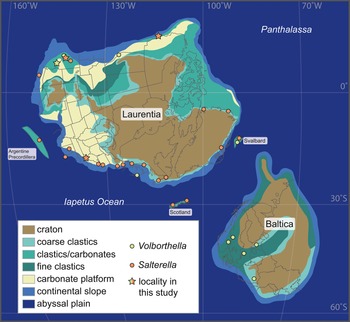
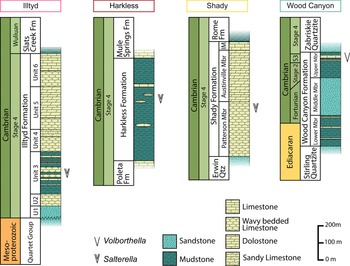
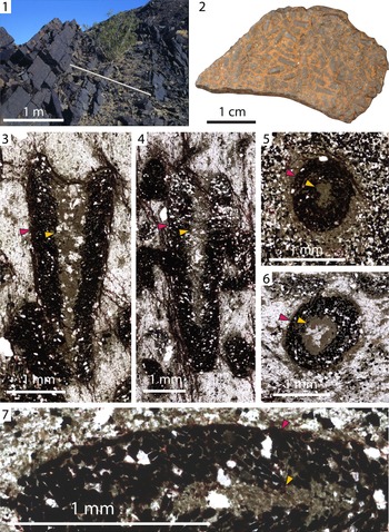
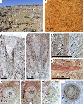
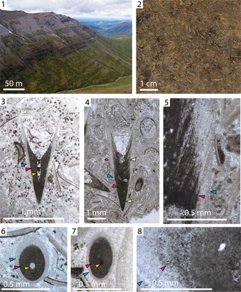
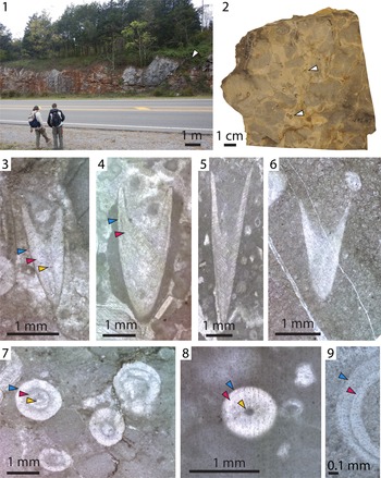


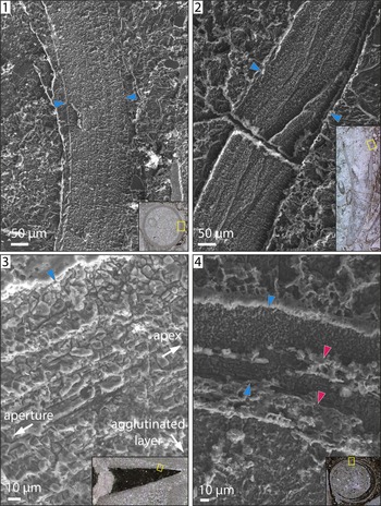
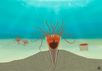

 Open access

