


Introduction
Diffuse gliomas are primary brain tumours that were historically defined by histopathological subtype and graded on a scale of 2–4. With recent molecular advancements, the 2021 World Health Organization Classification of Central Nervous System Tumours describes a classification system that centres on the integration of molecular markers and, in some cases, DNA methylome profiling for the diagnosis and categorization of gliomas. Reference Louis, Perry and Wesseling1 To date, molecular testing has revealed the clinical and prognostic significance of isocitrate dehydrogenase (IDH) mutation status. IDH mutant gliomas have a distinct biology that is associated with a superior prognosis compared to adult-type gliomas that are IDH wildtype. Reference Cairncross, Wang and Shaw2 Clinically, patients with IDH-mutated tumours have improved overall survival and progression-free survival compared to IDH wild-type tumours. Reference Leu, von Felten and Frank3–Reference Eckel-Passow, Lachance and Molinaro6 IDH mutation status therefore appears to be closely linked to tumour behaviour and may be more important for prognostication than grade. Reference Cairncross, Wang and Shaw2
To date, the literature suggests that IDH mutation is an early event in tumorigenesis and appears to be largely stable over time, though there have been reports of changes from wildtype to mutated IDH at tumour progression. Reference Juratli, Kirsch and Robel7–Reference Agnihotri, Aldape and Zadeh9 Conversely, one study identified a subset of recurrent IDH mutant tumours with copy number alterations at the IDH1 locus, leading to an IDH wild-type phenotype, a higher-grade tumour and a reprogrammed epigenome at tumour recurrence, resulting in a poor prognosis. Reference Mazor, Chesnelong and Pankov10 Similarly, a case report on the multi-regional sampling of a patient with a grade 4 IDH mutant astrocytoma at primary and recurrent disease identified extensive mutational and copy number heterogeneity, including a loss of the clonal IDH1 mutation in the recurrent tumour and poor outcome. Reference Favero, McGranahan and Salm11 Furthermore, IDH-mutated models are notoriously difficult to study both in vitro and in vivo, due to additional mutations or deletions that occur leading to alteration of the IDH1 allele, and the low efficiency of xenograft formation, respectively. Reference Han, Liu and Cai12 Therefore, a change in IDH status from mutant to wildtype may be secondary to altered tumour genetics, leading to increased aggressiveness and a poor prognosis.
The purpose of this study is to report on the institutional experience concerning the stability of IDH mutations in glioma treatment and tumour progression. To do this, we reviewed all glial tumours archived at our institution from 2009–2018, corresponding with our institution’s adaptation of IDH mutation testing after the initial discovery in 2008, Reference Parsons, Jones and Zhang13 and identified four patients with conflicting IDH mutation status longitudinally. These patients all had a loss of IDH mutation status as determined by our institution’s standardized practice of IHC testing of IDH mutations using formalin-fixed-paraffin-embedded (FFPE) tissue. In order to investigate these discrepancies in IDH mutation status, we performed methylation profiling including copy number variation, Sanger sequencing and ddPCR on longitudinal fresh frozen tissue samples and repeated IHC testing for IDH mutations using FFPE from all patients. All IDH mutation discrepancies were explained and resolved through multi-platform analysis. Our results support the concept that IDH mutation is a stable event throughout glioma evolution and highlight the role of accurate intraoperative tumour sampling and DNA methylome profiling as an adjunct diagnostic technique.
Methods
Patients and Clinical Samples
The study was approved by the University of Toronto research ethics board. Our brain tumour bank consisting of over 2500 CNS tumours catalogued from 2009 to 2018 was queried for glioma patients with matched longitudinal tumour samples (Fig. 1a). A total of 178 patients with longitudinal glioma tumour samples were identified and reviewed. This included 78 patients with IDH mutations. Four patients with IDH mutation discrepancies as documented from our institution’s routine IDH1 R132H mutation testing using IHC were identified. This antibody test recognizes the IDH1 R132H mutation which is present in approximately 90% of mutated cases but does not recognize other non-canonical IDH1 or IDH2 mutations. Reference Capper, Zentgraf, Balss, Hartmann and von Deimling14 Fresh frozen tissue from each patient was collected at all available surgical timepoints (9 samples total, approximately 20 mg–25 mg tissue per sample). A neuropathologist reviewed all available tissue blocks for all 9 samples, and IDH1 R132H IHC was repeated on IDH discrepant samples. Patient demographics, clinical information and pathology reports were collected and reviewed from electronic medical records. All diagnoses were based on the WHO 2021 CNS Guidelines. Reference Louis, Perry and Wesseling1
Investigating discrepancies in IDH mutation status in longitudinal glioma samples. Flowchart documenting the sample and patient review for the study. a ) Sanger sequencing of the IDH1 and IDH2 genes focusing on the regions directly surrounding codon 132 and 172, respectively. The presence of two peaks (arrows) demonstrates a heterozygous mutation in each of the mutated samples. b ) Droplet digital PCR identification of both an IDH1 wt sample (above) and IDH1 R132H mutation (below). Black dots (bottom left) represent double negative droplets, green dots (bottom right) represent IDH wt droplets, blue dots (top left) represent IDH1 R132H mutant droplets, and orange dots (top right) represent double positive droplets. c ) DKFZ CNS methylation classifier example showing the calibrated scores with the final diagnosis and the copy number variation plot generated.

DNA Preparation
All fresh frozen tumour samples underwent DNA extraction using the DNeasy Blood and Tissue Kit (Qiagen). The Qubit Assay Kit (Thermo Fisher Scientific) was used to quantify and measure the concentration of DNA. All kits were used according to the manufacturers’ instructions.
IDH Mutation Testing
A total of 5 ng of tumour DNA were used from all 9 tumour samples for each method of IDH mutation testing. Gold standard Sanger sequencing was performed for both IDH1 and IDH2 genes containing the regions of interest involved in IDH1 and IDH2 mutations, respectively. Primers were designed in our laboratory to flank exon 4 of both IDH1 and IDH2 genes in order to identify the presence of important mutations.
IDH1 forward primer: 5’ – TGG TGT ACT CAG AGC CTT CG – 3’
IDH1 reverse primer: 5’ – AGT TGG AAA TTT CTG GGC CA – 3’
IDH2 forward primer: 5’ – AAT TTT AGG ACC CCC GTC TG – 3’
IDH2 reverse primer: 5’ – TGT GGC CTT GTA CTG CAG AG – 3’
IDH1 R132H mutation testing was also performed using quantitative polymerase chain reaction (qPCR, Thermo Fisher) and droplet digital PCR (ddPCR, Bio-Rad) with a TaqMan SNP Genotyping Assay C_167891677_20 (Thermo Fisher).
Methylation Profiling and Copy Number Analysis
A total of 550 ng of tumour DNA from each sample was bisulphfite converted using the EZ DNA Methylation Kit (Zymo). The Infinium HumanMethylation450 BeadChip (Illumina) was then used to analyse the genome-wide methylation profiles of all 9 glioma samples. Once the raw (IDAT) files were obtained, these were uploaded to the DKFZ methylation profiling classifier at https://www.molecularneuropathology.org/mnp for methylation profiling, as shown in Figure 2. Reference Capper, Jones and Sill15 The DKFZ methylation classifier produces raw scores representing specific methylation classes. A calibration model then transforms the raw scores into calibrated scores, which represent an estimated probability measure of methylation class assignment, ranging from 0 to 1. An optimal calibrated score threshold for a single class is ≥ 0.9, and this value was used as the cut-off for all samples. Reference Capper, Jones and Sill15 Chromosomal copy number variation plots are created as part of the DKFZ methylation classifier result using the Bioconductor package conumee. Samples are compared to two sets of 50 control samples displaying a balanced copy number profile from both male and female donors. Reference Capper, Jones and Sill15 Standard copy number thresholds using the conumee package include gains or losses of ± 0.4, with less strict cut-offs of ± 0.2. Reference Capper, Jones and Sill15 Version 12.5 was used for all samples.
Integrated molecular and pathological diagnosis of IDH discordant samples from three patients. Each discordant sample has the DKFZ methylation classifier result ( a ), copy number variation plot ( b ) and repeated IDH1 R132H antibody IHC testing of pathological tissue ( c ).

Figure 3 was created with BioRender.com.
Reasons for IDH discordance and study conclusions. a ) Reasons for discrepancies in IDH mutation status over time. b ) Integrated approach between clinicians, histopathology and DNA methylation.

Results
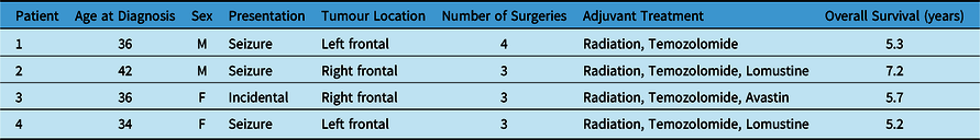
A total of 178 glioma patients with longitudinal tumour samples were identified and reviewed. All patients had routine institutional testing of IDH mutation status by IDH1 R132H antibody at time of tumour sampling. From these 178 patients, 78 patients had IDH mutations identified through IHC, and a total of four patients were found to have a mismatch in their IDH mutation status as documented from surgical pathology reports longitudinally (Fig. 1a). Table 1 summarizes the demographics and clinical course of all patients. Frozen tissue was collected from our institution’s tumour bank at all available surgical timepoints (minimum of 2 samples per patient, 9 samples total). All samples underwent methylation profiling including copy number variation and both Sanger sequencing and ddPCR testing of IDH mutation status in order to explore the discrepancies in the IHC-reported IDH status (Fig. 1b–d). All samples underwent additional pathological review, and three samples had repeat IHC performed on the most representative tissue available. Table 2 summarizes the initial histopathological and molecular findings of each tumour sample and the results of the additional analyses performed for this study.
Patient demographics and clinical course

Tumour molecular testing

*Surgery performed at outside hospital.
Our results demonstrate that all four patients had IDH mutations present at recurrent surgical resection, and no patients had a change in IDH mutation status after treatment or during glioma evolution. The first patient was found to have a rare IDH2 R172K mutation that was identified through our gene sequencing analyses (Fig. 1b) but would have been missed using only an IDH1 R132H antibody on IHC. Our methylation profiling also identified the diagnosis of this patient as an IDH mutant 1p/19q codeleted oligodendroglioma. This patient, while identified during our review as having an IDH mutation discrepancy, also had a documented 1p/19q codeletion at time of initial surgery, which is mutually inclusive of having an IDH mutation. Reference Louis, Perry and Wesseling1 Therefore, this patient was originally treated as an IDH mutant oligodendroglioma despite the negative IHC testing and does not represent a case of IDH mutation discrepancy.
Of the remaining three patients, all had documented negative IHC testing for IDH mutation at tumour recurrence, despite their tumours initially demonstrating IDH mutations on IHC testing. DNA methylome profiling and methylation classification of these discordant samples identified a combination of perilesional tissue from surgical sampling that resulted in a mixture of low recurrent tumour content mixed with either normal brain tissue, reactive tumour microenvironment, or inflammatory microenvironment (Figure 2). Overall, this represented less than a 3% sampling error rate of all samples reviewed (9/395, Fig. 1a). All patients had copy number values for IDH1 and IDH2 genes < |0.1|, demonstrating that there were no copy number variations present in any sample. IHC for R132H IDH1 mutations on these three discordant cases confirmed positive staining in all cases, with low tumour content in two and high tumour content in a subsequent tissue block that had previously not had IHC testing completed (Fig. 2).
Patient 2 (Fig. 2a) did not have a diagnostic match on the methylation classifier, with both control tissue and reactive tumour microenvironment the highest calibrated scores. This patient had a bland copy number plot suggesting non-neoplastic tissue, and IHC had positivity in only a small proportion of cells, demonstrating low tumour cell content. Patient 3 (Fig. 2b) had a methylation classifier match of 0.99 for inflammatory microenvironment and a bland copy number plot. Repeat IHC demonstrated positivity in a small proportion of cells, also demonstrating low tumour cell content. The final patient (Fig. 2c) did not have a methylation classifier match, although the highest calibrated score was in fact an adult-type diffuse glioma, with the second highest score being an IDH mutant 1p/19q codeleted diffuse glioma. The copy number plot for this sample was indicative of some tumour cells being present, with some losses observed at both 1p and 19q. IHC testing on a previously untested tissue block demonstrated positivity in a high proportion of cells.
These results support the hypothesis that IDH mutations are an early event in glioma tumorigenesis and remain unaltered throughout glioma evolution and treatment in the absence of major copy number alterations. Our research highlights the importance of accurate surgical sampling and the integrated role of DNA methylome profiling and copy number analysis as a confirmation of IDH status and its use in cases of diagnostic uncertainty.
Discussion
In this study, we investigated patients with IDH discrepancies between multiple tumour samples collected longitudinally from routine clinical testing using IHC. This included three instances of an apparent loss of IDH mutation at recurrence and one case of a rare non-canonical IDH2 mutation. In every case, we were able to identify a reason for the IDH mismatch and resolve the IDH mutation status, confirming the stability of IDH status longitudinally (Fig. 3a). All tumours exhibited an IDH mutation at recurrent surgical resection despite tumour treatment, evolution and recurrence, with no patients demonstrating copy number variations in IDH loci. Our data support the hypothesis that IDH mutation is an early event in glioma tumorigenesis and in the absence of genomic structural rearrangements, remains stable throughout the course of disease.
Previous studies have supported the concept that IDH mutation occurs early in gliomagenesis; Reference Watanabe, Nobusawa, Kleihues and Ohgaki8,Reference Agnihotri, Aldape and Zadeh9 however, multiple studies have found a subset of patients with changes in IDH mutation status at tumour recurrence. A study of 45 samples from paired low-grade astrocytomas and subsequent secondary high-grade gliomas found no changes in IDH mutation status. Reference Watanabe, Nobusawa, Kleihues and Ohgaki8 Another study of 51 lower-grade glioma patients that underwent multiple biopsies established a temporal timeline where IDH mutation precedes TP53 mutation or 1p/19q codeletion based on the proportion of patients that developed these mutations over time. A total of 41% of tumours that did not have TP53 or 1p/19q codeletions in initial samples went on to develop these in a subsequent biopsy, compared to only two patients (22%) that did not initially have IDH mutations that developed these over time. Reference Agnihotri, Aldape and Zadeh9 Another study examining 186 pairs of primary-recurrent GBMs from the EORTC 1542 study noted that three patients lost IDH mutation status at tumour recurrence. Reference Draaisma, Chatzipli and Taphoorn16 Efforts from the Glioma Longitudinal Analysis Consortium analysed longitudinal samples from 222 patients with gliomas and found that both IDH mutations and 1p/19q codeletions were not lost or gained in any patient during the surgical interval between samples. Reference Barthel, Johnson and Varn17
Few studies have explored the genetic underpinnings of apparent IDH mutation changes over time, but two studies independently found copy number alteration, potentially due to sub-clonal evolution, responsible for loss of IDH mutation at recurrence. One study reported copy number alterations in six recurrent gliomas at the IDH1 gene, which led to a loss of IDH mutation at recurrence. Reference Mazor, Chesnelong and Pankov10 This manuscript examined 50 patients with longitudinal tumour samples and found a 12% incidence of IDH mutation discrepancy over time. Reference Mazor, Chesnelong and Pankov10 They reported that these tumours recurred as higher grade with reprogrammed epigenomes and decreased 2-hydroxyglutarate. Reference Mazor, Chesnelong and Pankov10 A case report identified extensive mutational and copy number heterogeneity in a paired patient with a grade 4 IDH mutant astrocytoma, leading to the loss of IDH mutation at recurrence, hypothesized to be due to sub-clonal evolution and the evolution of a double-minute chromosome (extra-chromosomal circular DNA fragments frequently found in gliomas). Reference Favero, McGranahan and Salm11 Overall, we identified 4 patients out of 78 with longitudinally conflicting IDH mutation status, representing a 5% frequency of discrepancy, before further investigation.
It is important to note that any pathological or molecular analyses on brain tissue samples are only as good as the sample obtained. It is imperative that great care be taken as the operating surgeon to provide the most accurate biopsies and tumour samples for quick-section and for permanent pathology sections. This includes maximizing the use of image-guided sampling and providing permanent pathology and tumour bank samples in close proximity to the confirmed pathological quick-section sampling. In our study, three patients had previously documented negative IHC for IDH mutation at recurrence, suggesting a loss of IDH mutation over time. The Mib-1 (Ki-67) proliferation index and pathology report did note the presence of neoplastic cells in these specimens. DNA methylation profiling more accurately analysed these tissues and demonstrated suboptimal tissue samples including the presence of non-tumour cells identified as normal brain tissue, reactive tumour microenvironment and inflammatory tissue. Importantly, pathological review and repeat IHC testing did identify positive tumour cell staining for IDH mutation in all three samples.
Furthermore, we must also be aware of the limitations of the analyses performed on our tissue samples, including the sensitivity and specificity of the tests performed. A recent report on the R132H antibody for IHC testing compared to PCR testing identified a specificity of 100% and a sensitivity of 80%. Reference Gondim, Gener, Curless, Cohen-Gadol, Hattab and Cheng18 The previously documented negative IHC testing despite the presence of neoplastic cells may indeed be due to false negatives, and three samples out of 78 patients with paired samples (3/156) represent a less than 2% false negative rate for our institution.
In conclusion, our study utilized multiple methods and approaches for determining IDH mutation status, most notably the addition of methylation profiling with copy number variation. Our results show that DNA methylome testing can reliably be used to determine the IDH mutation status of gliomas and provide diagnostic clarity. It can also be used to interrogate and explore IDH mutation discrepancies and determine if the tissue sample provided is less than ideal. As molecular approaches are continually advancing and becoming more commonplace, it is imperative that we integrate the pathology and molecular findings, just as we have integrated our neuro-oncology approach with a multidisciplinary team (Fig. 3b).
Conclusions
All patients identified with discrepancies in their IDH mutation status were able to be resolved through methylation profiling. No patient experienced an IDH mutation alteration during the course of their treatment or at tumour recurrence. These findings support the hypothesis that IDH mutations occur early in gliomagenesis and in the absence of copy number variations at the IDH loci are stable throughout glioma evolution and treatment. This study highlights the importance of an interdisciplinary approach between the surgeon and the neuro-oncology team and an integrated approach of histopathology and novel diagnostic technologies including DNA methylome profiling for the accurate determination of IDH status and tissue diagnosis.
Statement of authorship
MRV conceptualized the manuscript. MRV performed and analysed most of the experiments. VP provided bioinformatician assistance and reviewed methylation classifier reports. AFG provided pathological expertise and performed IHC. MRV and CG wrote the manuscript. GZ oversaw the project. All authors read, edited and approved the final manuscript.
Competing interests
None.


 Open access