

Genetic and environmental factors such as sedentary lifestyle and increased energy consumption are associated with overweight, which is a major risk factor for type 2 diabetes. According to the WHO, at least 171 million individuals worldwide are affected and the prevalence will be dramatically increased over the coming decades(Reference Wilde, Roglic and Green1).
Inflammation leads to diabetic complications, being intimately linked with insulin resistance and the atherogenic process which may result in cardiovascular and cerebrovascular diseases(Reference Sell, Eckel and Dietze-Schroeder2–Reference Wei, Chen and Whaley-Connell8). Understanding the mechanisms and regulation of inflammatory pathways can be an important key to reduce diabetes-associated problems.
Cytokines such as TNF-α and IL-6, lipids, intracellular stress and excessive reactive oxygen species production are extracellular mediators enabling the initiation of inflammatory pathways(Reference Wellen and Hotamisligil3). Signals from all of these mediators come together to cause the production of additional inflammatory mediators through transcriptional regulation. These inflammatory signalling pathways stimulate a number of different kinases, including the kinase Jun-N-terminal kinase (JNK) that induces serine phosphorylation of insulin receptor substrate-I(Reference Sell, Eckel and Dietze-Schroeder2, Reference Wellen and Hotamisligil3, Reference Bastard, Maachi and Lagathu5, Reference Schenk, Saberi and Olefsky7, Reference Musi and Goodyear9). In fact, JNK plays a role in the negative regulation of insulin signalling, but the contribution of this pathway in adipose tissue, skeletal muscle or other tissue to systemic insulin resistance is currently unclear. Recent studies in mice reported that JNK inhibition in diabetes or atherosclerosis can be a viable therapy for these diseases in humans(Reference Wellen and Hotamisligil3).
Skeletal muscle is responsible for over 75 % of all insulin-mediated glucose disposal(Reference Wei, Chen and Whaley-Connell8). Therefore, mechanisms of glucose homeostasis are particularly important in this tissue. It is now believed that a negative cross-talk between adipose tissue and skeletal muscle, through adipokines, leads to disturbances in skeletal muscle insulin signalling and finally to insulin resistance, which seems to involve inflammatory components(Reference Sell, Eckel and Dietze-Schroeder2, Reference Wellen and Hotamisligil3, Reference Bastard, Maachi and Lagathu5, Reference Schenk, Saberi and Olefsky7, Reference Wei, Chen and Whaley-Connell8, Reference Goossens10–Reference Rasouli and Kern13).
Studies in a variety of species have shown that energy restriction brings some benefits to health. Rhesus monkeys submitted to energy restriction are less susceptible to CVD and diabetes mellitus(Reference Roth, Ingram and Lane14). Energy restriction in adult men and women causes many of the same metabolic adaptations that occur in energy-restricted rodents and monkeys, such as decreased metabolic, hormonal and inflammatory risk factors for diabetes, CVD and possibly cancer(Reference Fontana and Klein15). Accordingly, the characteristic up-regulation of inflammatory pathways in diabetes has also been reported to decrease with diet intervention in streptozotocin-induced diabetic rats(Reference Ugochukwu and Figgers6). Furthermore, the guidelines of the American Diabetes Association highlight the importance of medical nutrition therapy, not only for prevention, but also for metabolic control and for delaying complications of this pathology(16).
As the impairment in skeletal muscle actions may have serious effects on whole-body glucose homeostasis, the aims of the present study were to evaluate the effects of dietary restriction on systemic and skeletal muscle inflammatory signals in Goto-Kakizaki (GK) rats, and understand the role of adipose tissue in those pathways. Therefore, the last aim was to inquire about dietary restriction as a therapeutic complement to diabetes treatment.
In fact, several studies about nutritional intervention, namely energy restriction, have been done in type 2 diabetes. In the present study, rats were submitted to 50 % of dietary restriction. This approach makes a nutritional intervention easier, because the restriction did not consider the nutritional nature of the food, but the amount of food ingested. The peculiarity of GK rats also shows a new contribution in this area, because this diabetic animal model is non-obese and, consequently, the benefits shown of dietary restriction occurred outside the loss of body weight.
We demonstrated that GK rats are strongly insulin resistant, which implies large alterations in glucose metabolism in this tissue. Our data also demonstrate that our animal model has impaired adipokine physiology, although it does not have obesity or dyslipidaemia. Thus, the present study primarily shows that alterations of adipokines and insulin action are strong allies in impairing glucose metabolism in the skeletal muscle, where inflammatory pathways seem to be involved.
Materials and methods
Experimental animals
Male normal Wistar and GK (an animal model of spontaneous non-obese type 2 diabetes) rats were obtained from our local breeding colonies (Faculty of Medicine, University of Coimbra, Coimbra, Portugal).
Animals were maintained under controlled light (12 h light–12 h dark cycle), temperature (21–24°C) and humidity (50–60 %) conditions and with free access to water and a standard diet (PanLab S.I. Barcelona, Spain). Rats were separated into four experimental groups. Two groups – Wistar control (WC) and GK control (GKC) – were maintained on the standard diet for 6 months (chow intake of 26 and 24 g/rat per d, respectively). The other two groups were maintained on dietary restriction (Wistar diet-restricted (WDR) and GK diet-restricted (GKDR), at 50 % of control animals' daily chow intake) during the last 2 months of life (chow intake of 13 and 12 g/rat per d, respectively). When the study finished, all animals were aged 6 months.
All animal procedures and care were approved by the local Institutional Animal Care and Use Committee.
Sample collection and in vivo determinations
At the end of diet intervention (6 months of age), rats were fasted overnight in order to measure blood glucose levels. Fasting blood glucose and at 2 h after glucose load (1·8 mg glucose/kg) were measured on the tail vein through the glucose-oxidase method using a glucometer (Elite; Bayer, Carnaxide, Portugal) and compatible reactive test strips.
Glycated Hb (HbA1c) was also measured in the tail vein blood using an automatic analyser (DCA 2000; Bayer).
For further determinations, fasting blood samples were collected by cardiac puncture from anaesthetised animals with ketamine chloride (75 mg/kg, intramuscular; Parke-Davis, Ann Arbor, MI, USA) and chlorpromazine chloride (2·65 mg/kg, intramuscular; Laboratórios Vitória, Amadora, Portugal). For the determination of plasma and serum parameters, blood was centrifuged (2500 rpm) at 4°C and stored at − 20°C.
Muscle removal
Rats were killed by cervical displacement and skeletal muscle from the posterior face of the femur was removed and immediately frozen in liquid N2 and stored at − 80°C.
Insulin resistance indexes and NEFA
Plasma insulin was determined by an in-house competitive ELISA(Reference Nunes, Peixoto and Louro17). Insulin resistance was evaluated through the log-homeostasis model assessment (HOMA) index and the quantitative insulin-sensitivity index (QUICKI). Log-HOMA was calculated as:
where (If) is the fasting insulin level (μU/ml) and (Gf) is the fasting glucose level (mmol/l). QUICKI was calculated as 1/(log(G0)+log(I0)), where G0 is fasting glucose (mg/dl) and I0 is fasting insulin (μU/ml).
Plasma NEFA levels were measured using a commercial kit (Half-micro test; Roche Diagnostic, Mannheim, Germany).
Determination of systemic inflammatory biomarkers
Serum C-reactive protein levels were measured using the C-reactive protein ELISA kit (BD Biosciences Pharmingen, San Diego, CA, USA).
Serum leptin and adiponectin levels were measured by a Leptin Immunoassay kit (Invitrogen, Carlsbad, CA, USA) and an ELISA Phoenix Adiponectin Kit Assay (Phoenix Pharmaceuticals Inc., Belmont, CA, USA), respectively.
Muscle analysis
Frozen skeletal muscle sections were homogenised in a lysis buffer containing 25 mm-2-amino-2-(hydroxymethyl)propane-1,3-diol (Tris)-HCl, 100 mm-NaCl, 1 mm-EDTA, 1 mm-ethylene glycol-bis(2-aminoethyl)-N,N,N′,N′-tetraacetic acid, 20 mm-NaF, 10 mm-β-glycerolphophate, 2·5 mm-pyrophosphate, 2 mm-Na3VO4, 1 % Triton X-100, 10 mm-phenylmethanesulfonyl fluoride and protease inhibitor cocktail (Sigma, St Louis, MO, USA) (pH 7·4). Homogenates were centrifuged at 14 000 rpm for 20 min at 4°C. The supernatant fraction was collected and transferred to a clean tube and centrifuged at 14 000 rpm for 15 min at 4°C and the supernatant fraction was collected again. Protein concentration was determined by the Biureth method.
Samples were stored at − 80°C for subsequent analysis.
Muscle TNF-α and IL-6 levels were determined using a Rat TNF ELISA Set and a Rat IL-6 ELISA Set (BD Biosciences Pharmingen), respectively.
The expression of stress-activated protein kinase (SAPK)/JNK and phospho-SAPK/JNK was determined by the Western blot method. Samples containing 40 μg protein were loaded into 10 % acrylamide gels and transferred to Immun-Blot® polyvinylidine difluoride membranes. The membranes were blocked with 5 % bovine serum albumin–TBST solution (25 mm-Tris-HCl, 150 mm-NaCl (pH 7·6) and 0·1 % Tween) at room temperature for 1 h and then washed with TBST solution. After that, membranes were incubated overnight at 4°C with a specific primary antibody of SAPK/JNK (SAPK/JNK rabbit polyclonal antibody; Cell Signaling Technology, Danvers, MA, USA). The membranes were washed and incubated with a secondary antibody for 2 h at room temperature (anti-rabbit IgG alkaline phosphatase-linked goat antibody; GE Healthcare, Slough, Berks, UK). Membranes were washed and the immunoblots were developed with an ECF Western blotting detection system (STORM 860; GE Healthcare Life Sciences, Piscataway, NJ, USA). The same protocol was carried out for phospho-SAPK/JNK (phospho-SAPK/JNK (Thr183/Tyr185) mouse monoclonal antibody; Cell Signaling Technology).
Reagents
All the other compounds were of the purest quality available and were purchased from Sigma Chemical Co. (St Louis, MO, USA) or Merck (Darmstadt, Germany).
Statistical analysis
Data are presented as mean values with their standard errors. Statistical differences were determined by Student's t test. Results with P < 0·05 were considered statistically significant.
Results
Body weight, glycaemia and glycated Hb
As previously reported, adult diabetic GK rats of our colony were smaller than the corresponding control Wistar rats(Reference Seiça, Santos and Palmeira18). Accordingly, the body weight of diabetic rats was significantly lower (P < 0·001) and was maintained after dietary restriction, although a great reduction was observed in visceral adipose tissue. A significant decrease in body weight (P < 0·001) was observed in the WDR group (Table 1). Glycaemia, fasting and 2 h after glucose load, was significantly elevated (P < 0·001) in GKC rats when compared with normal rats (WC group). After dietary restriction, fasting glycaemia increased in both groups (P < 0·01) and glycaemia at 2 h decreased in the GKDR group (P < 0·05) (Table 1). HbA1c levels were higher in GKC rats when compared with age-matched Wistar rats (WC group) (P < 0·001) and decreased in both groups (P < 0·001) after dietary restriction (WDR and GKDR groups; Table 1).
(Mean values with their standard errors for nine to thirteen rats per group)
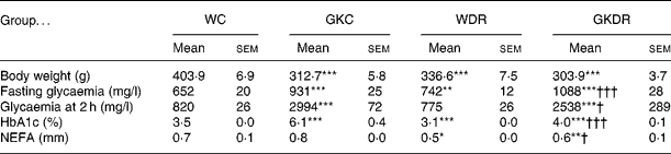
WC, Wistar control; GKC, Goto-Kakizaki control; WDR, Wistar rats with dietary restriction; GKDR, Goto-Kakizaki rats with dietary restriction.
Mean value was significantly different from that of the WC group: * P < 0·05, ** P < 0·01, *** P < 0·001.
Mean value was significantly different from that of the GKC group: † P < 0·05, ††† P < 0·001.
Dietary restriction improves insulin resistance indexes and NEFA levels
The insulin resistance index log-HOMA (Fig. 1(a)) was higher and QUICKI, an insulin-sensitivity index (Fig. 1(b)), was lower in GKC rats, when compared with normal Wistar (WC) rats (P < 0·001). Dietary restriction was able to improve insulin resistance (P < 0·01) and sensitivity (P < 0·001) in the diabetic rats (GKDR group).
Effects of dietary restriction on (a) the homeostasis model assessment index (HOMA; an index of insulin resistance) and (b) the quantitative insulin-sensitivity check index (QUICKI) in Wistar and Goto-Kakizaki (GK) rats. WC, Wistar control; GKC, GK control; WDR, Wistar rats with dietary restriction; GKDR, GK rats with dietary restriction. Values are means for nine to thirteen rats per group, with standard errors represented by vertical bars. *** Mean value was significantly different from that of the WC group (P < 0·001). Mean value was significantly different from that of the GKC group: †† P < 0·01, ††† P < 0·001.

Dietary restriction decreased plasma NEFA in both experimental groups (P < 0·05). GKDR group levels were lower (P < 0·01) than those in the WC group (Table 1).
Systemic inflammatory biomarkers
Levels of serum C-reactive protein, a systemic inflammatory marker, were significantly reduced in GKC rats (P < 0·001), when compared with normal Wistar group (WC) values and were not changed with dietary restriction in both strains (Fig. 2).
Effects of dietary restriction on serum levels of C-reactive protein (CRP) in Wistar and Goto-Kakizaki (GK) rats. WC, Wistar control; GKC, GK control; WDR, Wistar rats with dietary restriction; GKDR, GK rats with dietary restriction. Values are means for nine to thirteen rats per group, with standard errors represented by vertical bars. *** Mean value was significantly different from that of the WC group (P < 0·001).

Serum levels of leptin were significantly increased (P < 0·01) in GKC rats, when compared with normal Wistar (WC) rats. Dietary restriction decreased leptin levels in both strains (P < 0·001) (Fig. 3(a)).
Effects of dietary restriction on (a) plasma levels of leptin and (b) serum levels of adiponectin in Wistar and Goto-Kakizaki (GK) rats. WC, Wistar control; GKC, GK control; WDR, Wistar rats with dietary restriction; GKDR, GK rats with dietary restriction. Values are means for nine to thirteen rats per group, with standard errors represented by vertical bars. Mean value was significantly different from that of the WC group: * P < 0·05, ** P < 0·01, *** P < 0·001. Mean value was significantly different from that of the GKC group: †† P < 0·01, ††† P < 0·001.

Serum levels of adiponectin were lower in GKC rats when compared with age-matched normal WC rats (P < 0·05). The dietary restriction improved these levels in WDR and GKDR groups (P < 0·05 and P < 0·01, respectively). Furthermore, GKDR adiponectin levels were similar to the values of normal (WC and WDR) rats (Fig. 3(b)).
Muscular inflammatory biomarkers
Muscular levels of TNF-α were significantly reduced (P < 0·001) in GKDR rats when compared with GKC and WC values (Fig. 4(a)).
Effects of dietary restriction on muscle levels of (a) TNF-α and (b) IL-6 in Wistar and Goto-Kakizaki (GK) rats. WC, Wistar control; GKC, GK control; WDR, Wistar rats with dietary restriction; GKDR, GK rats with dietary restriction. Values are means for nine to thirteen rats per group, with standard errors represented by vertical bars. Mean value was significantly different from that of the WC group: * P < 0·05, *** P < 0·001. Mean value was significantly different from that of the GKC group: † P < 0·05, ††† P < 0·001.

The levels of IL-6 were higher in GKC rats (P < 0·05) when compared with those of the WC group and decreased in the GKDR group (P < 0·05). In contrast, dietary restriction induced an increase (P < 0·05) of IL-6 values in normal Wistar (WDR) rats (Fig. 4(b)).
Dietary restriction decreases phospho-stress-activated protein kinase/Jun-N-terminal kinase in diabetic rats
According to some studies which postulated that inflammation in diabetes mellitus can be related to the activity of the active form of SAPK/JNK, the expression of normal and phosphorylated forms of this protein was measured. As illustrated by representative images and quantitative analysis (Fig. 5), the expression of SAPK/JNK was similar in the four groups studied (Fig. 5(a) and (b)). Nevertheless, the levels of the active phospho-SAPK/JNK form decreased in GKDR rats (P < 0·05), when compared with the GKC and WC groups (Fig. 5(c) and (d)).
Effects of dietary restriction on (a, b) stress-activated protein kinase (SAPK)/Jun-N-terminal kinase (JNK) and (c, d) phospho-SAPK/JNK expression in muscular tissue in Wistar and Goto-Kakizaki (GK) rats. WC, Wistar control; GKC, GK control; WDR, Wistar rats with dietary restriction; GKDR, GK rats with dietary restriction. (a, c) Representative Western blots. (b, d) Percentage of expression of proteins in relation to the WC group by quantification of the bands using the ImageQuant program (Molecular Dynamics, Sunnyvale, CA, USA). Values are means for nine to thirteen rats per group, with standard errors represented by vertical bars. ** Mean value was significantly different from that of the WC group (P < 0·01). † Mean value was significantly different from that of the GKC group (P < 0·05).

Discussion
Data from the present study demonstrated that GK rats, a model of type 2 diabetes, compared with normal control rats, exhibited fasting hyperglycaemia and glucose intolerance, higher levels of HbA1c and insulin resistance. Serum adiponectin was lower and plasma leptin was higher, demonstrating adiponectin deficiency and the previously reported leptin resistance in this animal model(Reference Maekawa, Fujiwara and Kohno19). These results are also indicative of adipocyte dysfunction already observed in diabetes mellitus and in insulin-resistant states(Reference Ferranti and Mozaffarian20). Serum levels of C-reactive protein were lower in GK rats when compared with normal rats. These results show an intrinsic characteristic of this strain that was previously observed from our group (J Crisóstomo, L Rodrigues, P Matafome, C Amaral, E Munes, T Louro, P Monteiro and R Seiça, unpublished results) and could result from the adaptation in their selection process. This inflammatory marker, produced in liver and linked with insulin-resistance(Reference Ndumele, Pradhan and Ridker21), did not change with dietary restriction in both strains. In relation to muscular inflammatory biomarkers, IL-6 levels were higher in GK rats. The levels of TNF-α and the expression of normal and phosphorylated form of SAPK/JNK were similar in both strains.
Dietary restriction in diabetic rats was able to reduce the skeletal muscle inflammatory process and to improve glucose tolerance, insulin resistance, NEFA and HbA1c levels and adipokine profile but aggravated fasting glycaemia. This effect on glycaemia was also observed in the WDR group, suggesting a higher rate of hepatic glucose production in the fasting state, as a result of severe dietary restriction. Furthermore, dietary restriction in normal rats reduced body weight, NEFA and leptin levels, and raised adiponectinaemia. In other parameters, the levels did not change, and the levels of IL-6 even increased. Based on this, future studies are required in order to evaluate the impact of such pronounced dietary restriction on inflammation in normal models. The adipose tissue modulates its own metabolism but is also able to interfere in other tissues, in order to regulate its metabolism. Its secretory products are involved in glucose and lipid homeostasis and inflammation. In fact, it has become recently clear that adipocytes represent active secretory cells. The adipocytes were able to release NEFA by lipolysis and produce a variety of cytokines(Reference Sell, Eckel and Dietze-Schroeder2, Reference Rabe, Lehrke and Parhofer11, Reference Guerre-Millo12). Adipocyte dysfunction leads to excessive fat accumulation in non-adipose tissue and disturbed adipokine secretion(Reference Ferranti and Mozaffarian20), which may have local and systemic effects(Reference Goossens10). Adipose tissue in insulin resistance is characterised by increased levels of leptin and pro-inflammatory cytokines such as TNF-α and IL-6 and lower adiponectin expression(Reference Sell, Eckel and Dietze-Schroeder2, Reference Sesti4, Reference Goossens10).
Leptin, whose levels are proportional to overall adipose mass(Reference Sesti4), has important effects on peripheral tissue including muscle(Reference Toyoshima, Gavriola and Yakar22), but its effects are still controversial(Reference Muoio and Newgard23). In some studies leptin has increased insulin sensitivity but others have shown, however, that this adipokine impairs insulin action(Reference Sell, Eckel and Dietze-Schroeder2). Furthermore, in obese and diabetic states, it is likely that higher levels of leptin are indicative of leptin resistance(Reference Rabe, Lehrke and Parhofer11, Reference Muoio and Newgard23). In the present study we verified that GK rats have higher levels of leptin, showing the previously reported leptin resistance in this animal model(Reference Maekawa, Fujiwara and Kohno19) that decreased after dietary restriction.
A recent report suggests that adiponectin has antidiabetic properties such as stimulating glucose uptake in skeletal muscle and suppressing hepatic glucose production. Anti-inflammatory properties have also been reported(Reference Sweeney24). Adiponectin levels correlate inversely with the impairment of insulin signalling; it decreases hyperglycaemia and NEFA and affects immune and inflammatory processes throughout the body(Reference Muoio and Newgard23). Our data show that the levels of adiponectin, a positive adipokine, increased with dietary restriction in normal and diabetic rats. AdipoR1 expression predominates in skeletal muscle(Reference Roth, Ingram and Lane14). It has also been described that adiponectin antagonises many effects of TNF-α and its production is decreased by this inflammatory factor(Reference Whitehead, Richards and Hickman25). Therefore, the increase of adiponectin levels induced by dietary restriction may justify the improvement of TNF-α and IL-6 levels in skeletal muscle of diabetic rats and may be associated with the amelioration of insulin resistance and metabolic profile.
Plasma TNF-α and IL-6 are commonly elevated in type 2 diabetes(Reference Sell, Eckel and Dietze-Schroeder2, Reference Wei, Chen and Whaley-Connell8) and have a negative effect on insulin signalling, although the effect of IL-6 in skeletal muscle remains unclear(Reference Sell, Eckel and Dietze-Schroeder2, Reference Wei, Chen and Whaley-Connell8, Reference Roth, Ingram and Lane14, Reference Muoio and Newgard23).
TNF-α impairs insulin sensitivity in rodent skeletal muscle(Reference Goossens10). While the mechanism underlying desensitisation has not been fully elucidated, TNF-α decreases tyrosine phosphorylation of insulin receptor substrate (IRS)-1 and increases IRS-1 serine phosphorylation, and an improvement of insulin receptor phosphorylation in skeletal muscle is achieved with an anti-TNF-α antibody(Reference Wei, Chen and Whaley-Connell8). Moreover, TNF-α also activates an apoptotic signalling pathway that can degrade the muscle tissue and energy restriction attenuates the loss of muscle mass and function intervening on TNF-α signalling(Reference Wei, Chen and Whaley-Connell8, Reference Dirks and Leeuwenburgh26).
IL-6 can have distinct effects on different tissues. There are discrepancies between acute and long-term effects and its action depends on complex interactions with other pro- and anti-inflammatory cytokines(Reference Wei, Chen and Whaley-Connell8). In a recent study, physical exercise is associated with increased insulin action in skeletal muscle and with increased IL-6 circulating levels. The authors suggest a possible anti-inflammatory action of IL-6 in skeletal muscle(Reference Roth, Ingram and Lane14), but the real effect remains controversial. In most studies about diabetes, IL-6 seems to impair insulin action(Reference Sell, Eckel and Dietze-Schroeder2, Reference Wei, Chen and Whaley-Connell8, Reference Roth, Ingram and Lane14, Reference Muoio and Newgard23). Accordingly, with these studies, the present results show that muscle IL-6 levels were higher in diabetic rats. Dietary restriction was able to reduce both pro-inflammatory cytokines (IL-6 and TNF-α), and these observations may be related to the improvement in insulin resistance indexes after the diet.
Various studies reported that increased circulating plasma levels of TAG and NEFA contribute to insulin resistance in peripheral tissues such as skeletal muscle(Reference Sell, Eckel and Dietze-Schroeder2, Reference Schenk, Saberi and Olefsky7, Reference Petersen and Shulmam27). Furthermore, it was observed that dietary restriction increases mitochondrial biogenesis and NEFA oxidation(Reference Lange, Moreno and Silvestri28). The reduction of NEFA in GK rats induced by dietary restriction may be also related with improvement of insulin resistance.
High levels of fatty acids and pro-inflammatory cytokines activate JNK in liver, adipocyte cells and skeletal muscle, resulting in insulin-sensitivity reduction(Reference Ferranti and Mozaffarian20).
JNK is a mitogen-activated protein kinase and some previous studies made clear the role of JNK in inflammation and metabolic diseases, suggesting JNK inhibition as a viable therapeutic intervention in diabetes mellitus and atherosclerosis(Reference Wellen and Hotamisligil3).
JNK is activated by pro-inflammatory cytokines such as TNF-α and plays a negative role in insulin signalling(Reference Sell, Eckel and Dietze-Schroeder2, Reference Wellen and Hotamisligil3, Reference Bastard, Maachi and Lagathu5). The activated form, phosphorylated JNK, mediates IRS-1 serine phosphorylation and this phosphorylation can induce the dissociation of IRS proteins from the insulin receptor, impairing insulin signalling(Reference Sell, Eckel and Dietze-Schroeder2, Reference Wellen and Hotamisligil3, Reference Bastard, Maachi and Lagathu5). Our data show that dietary restriction in diabetic animals decreased the expression of the active form of JNK, and this can be related to the reduction of TNF-α and NEFA levels and the improvement of the hormone profile.
Energy restriction might affect glucose tolerance, modulating insulin action in skeletal muscle, and inducing elevated insulin-stimulated glucose transport in this tissue(Reference Park, Komatsu and Hayashi29–Reference Dean, Brozinick and Cushman31). The improvement of glucose tolerance and HbA1c in GK rats is in accordance with these studies.
In conclusion, dietary restriction in type 2 diabetes has a central role in adipocyte stress, influencing adipokines and other pro-inflammatory mediator factors and energy balance, giving rise to improved skeletal muscle inflammation and insulin sensitivity.
Acknowledgements
We thank Mário Simões from the Institute of Physiology, Faculty of Medicine of Coimbra for his technical support. We also thank Serviço de Patologia Clínica from the University Hospital of Coimbra for technical support.
The present study was supported by the University of Coimbra and University Hospitals of Coimbra. We thank SERVIER Portugal for financial support.
J. C. was the main contributor to the practical work pertaining to the present paper. L. R., P. Matafome, C. A., E. N. and T. L. contributed with technical support, including animal management and treatment. P. Monteiro and R. S. contributed with scientific advice.
There are no potential conflicts of interest.


