

Anthropometric measurements are routinely performed in paediatric settings, being used around the world as health and development indices during infancy and childhood( Reference De Onis and Blössner 1 ). In particular, height, weight and head circumference have been associated with neurodevelopmental outcomes( Reference Guerrant, DeBoer and Moore 2 ). Studies focused on head circumference have special relevance to explain neurodevelopment, where it is used as an indicator of brain size. During prenatal and early development and maturation, brain growth is the major force for the increase in intracranial volume and head size( Reference Gale, O’Callaghan and Bredow 3 , Reference Gale, O’Callaghan and Godfrey 4 ). This process continues throughout childhood and ends in early adulthood, when brain size begins to decline( Reference Borzage, Blüml and Seri 5 – Reference Shaw, Kabani and Lerch 7 ). Thus, intracranial volume is highly associated with total brain volume during early life( Reference Gale, O’Callaghan and Bredow 3 ), and therefore head circumference is used as an accurate index of brain size in normally developing children( Reference Bartholomeusz, Courchesne and Karns 8 , Reference Ivanovic, Leiva and Pérez 9 ). For children aged 6 years and below, the correlation between head circumference and total brain volume is very strong( Reference Bartholomeusz, Courchesne and Karns 8 ), but the correlation decreases among subjects aged 12 years and older( Reference Lange, Froimowitz and Bigler 10 ). Developmental changes associated with head growth and skull thickness probably contribute to this general decrease in correlation( Reference Bartholomeusz, Courchesne and Karns 8 ). Moreover, typical brain maturation encompasses distinct stages with fast and slow progressions depending on age( Reference Toga, Thompson and Sowell 11 ). Therefore, a child’s age and growth curve are crucial when estimating brain size from head circumference. In addition, head circumference has also been associated with future cognitive performance in high-risk individuals (e.g. low birth weight or premature infants)( Reference Aarnoudse-Moens, Weisglas-Kuperus and van Goudoever 12 ), as well as in full-term and normally developing infants( Reference Ivanovic, Leiva and Pérez 9 , Reference Nelson and Deutschberger 13 ). Thus, head circumference measurements at various postnatal ages are differentially associated with either motor or cognitive outcomes( Reference Cooke 14 ). Moreover, a child’s behaviour repertoire – in a given time – would be related to functional activity over a specific brain region( Reference Chugani 15 ). The ability of head circumference measurements during childhood to predict regional cortical and subcortical brain volumes is unknown, however. In fact, no study to date has demonstrated that head circumference – measured at consecutive ages – is linked to specific distributions of grey matter in healthy children.
Across lifespan, but also during fetal development, biological (e.g. sex) and environmental (e.g. family status) factors affect head and brain growth( Reference Gao, Alcauter and Smith 16 , Reference Hanson, Hair and Shen 17 ) through both genetic mechanisms or distinct nutrition and/or stimulation( Reference Veena, Krishnaveni and Wills 18 ). However, most of the studies relating head circumference and brain size have not taken into account these factors. In particular, prenatal micronutrients such as long-chain (LC) PUFA and folate are important for intra-uterine central nervous system development( Reference Morse 19 ), and their effects on head and brain growth might go beyond the fetal life( Reference Faa, Marcialis and Ravarino 20 ). Several studies have shown that prenatal supplementation with LC-PUFA( Reference Makrides, Collins and Gibson 21 ) and folate( Reference Steenweg-de Graaff, Roza and Steegers 22 ) impact prenatal, but also postnatal, head growth. Moreover, these micronutrients might enhance cognitive development, even beyond early infancy( Reference Campoy, Escolano-Margarit and Ramos 23 , Reference Catena, Muñoz-Machicao and Torres-Espínola 24 ). In addition to these confounding factors, previous studies focused on the age-dependent estimates of brain size from head circumference have been cross-sectional in nature. Head growth, as with brain maturation, occurs in a variable sequence of relatively slow and fast periods( Reference de Onis, Onyango and Borghi 25 ). Therefore, to properly examine the predictive value of head circumference to know later brain size, multiple measurements from the same individual should be acquired and several confounding factors should be taken into account.
In this study, we analysed how different early nutritional supplementations, LC-PUFA – contained in fish oil (FO) – and/or folate (5-methyltetrahydrofolate (5-MTHF)), will reflect a long-term effect on brain cortical and subcortical volumes and inner cortical surface area. In addition, we report the first structural MRI study that analyses relationships between consecutive head circumference measurements and total brain volumes at later childhood, as well as their relationship with the distribution of cortical white and grey matter within the brain and subcortical structures’ volumes in a large sample of healthy children.
Methods
Participants
The present study included eighty-five Spanish children born at term who participated in the Nutraceuticals for a healthier life EU Project NUHEAL project (registered at www.clinicaltrials.gov as NCT01180933), which studied the effects of prenatal nutrition on offspring development( Reference Krauss-Etschmann, Shadid and Campoy 26 ). Pregnant women participating in this study were supplemented with either( Reference De Onis and Blössner 1 ) a FO preparation (500 mg DHA+150 mg EPA)( Reference Guerrant, DeBoer and Moore 2 ), 5-MTHF( Reference Gale, O’Callaghan and Bredow 3 ), a combination of both supplements or( Reference Gale, O’Callaghan and Godfrey 4 ) a placebo from gestational week 22 until delivery. Detailed information on the NUHEAL project, parents’ socio-demographic data and pregnancy outcomes is reported in the online Supplementary Material. Five parents refused to participate in this new assessment. The participating children had no disorder or disability interfering with normal neurological development, nor developmental disorders or sensory disabilities. Six children were excluded from the analysis because of measuring artefacts during examination. Thus, data from seventy-four children were analysed. This study was conducted according to the guidelines laid down in the Declaration of Helsinki, and all procedures involving human subjects were approved by the Human Research Ethics Committee of the University of Granada. Written informed consent was obtained from all the participants at study entry and follow-up.
Head circumference assessment
Head circumference was obtained within 24 h of birth, at 4 years and at 10 years of age following WHO criteria( Reference de Onis, Onyango and Borghi 25 ), by passing a measuring tape around the head, placed above the ears and midway between the eyebrows and the hairline, to the occipital prominence at the back of the head and touching two head landmarks: the most anterior protuberance (approximately 0·5 cm above the eyebrows) and the most posterior one. This variable was measured three times by two trained researchers (C. M-Z. and P. B.). The largest of the three measures was considered the actual head circumference. Discrepancies between the two experts were always lower than 4 mm.
Cognitive abilities assessment
We used the Spanish adaptation of the Kaufman Assessment Battery for Children, 2nd edition (KABC-II)( Reference Kaufman and Kaufman 27 ), to assess cognitive abilities when children were 6·60 (sd 1·06) years old. The KABC-II has two scales to assess processing skills (Sequential and Simultaneous Processing Scales) as well as the combination of those scores, the Mental Processing Index (MPI) scale, which is considered equivalent to the intelligence quotient. In addition, it has the Achievement scale (not used in the present study). Raw scores are transformed into standard scores with a mean of 100 and a sd of 15.
MRI: image acquisition
Resonance images were obtained at 10 years of age, when the brain typically attained 95 % of its adult volume( Reference Webb, Ruppel Shell and Cuomo 28 ). Children were scanned using a Philips Achieva 3T MRI (Philips Medical Systems, Best, Netherlands) scanner operating with an eight-channel, phased-array head coil for reception. T1-weighted three-dimensional (3D) volumes were acquired using a T1-weighted 3D-turbo-gradient field echo sequence (3D-TFE), in sagittal orientation with 0·94×0·94×1·0-mm resolution (160 slides, Field of View (FOV)=240×240, matrix 256×256×160), with repetition time of 8 ms, echo time of 4 ms, inversion delay of 1022·6264 ms, flip angle of 8° and band width of 191 Hz/pixel. The sequence was optimal for reducing motion sensitivity, susceptibility artefacts and field inhomogeneities. Total volumes for grey and white matter (GMV and WMV) and total intracraneal volume (TIV, obtained from GM, WM and cerebrospinal fluid (CSF) voxels in native space) were computed for each child adding up to the corresponding voxel values.
Image processing
Voxel-based morphometry
The structural images were first checked for artefacts and manually re-aligned to the Anterior-Posterior Commissure (AC-PC) line. Automatic processing was carried out with the default parameters of the DARTEL toolbox( Reference Ashburner 29 ) implemented in SPM8 (http://www.fil.ion.ucl.ac.uk/spm). In brief, images were corrected for intensity inhomogeneities, tissue was then clustered into GM, WM, CSF, and registered using linear and non-linear transformations. Sample-specific GM and WM templates were created by an iterative process starting from initial template estimates using the DARTEL high-dimensional warping procedure. Next, the GM and WM individual maps were non-linearly normalised to the final sample template, and Jacobian modulated to preserve tissue amount( Reference Ashburner 29 ). The final sample GM and WM templates were registered to the Montreal Neurological Institute (MNI) space, and the individual maps were co-registered to the MNI space, using the affine transformation estimate for the GM and WM templates. Statistical analyses were carried out on GM and WM maps after smoothing them with an 8-mm full-width at half-maximum.
Cortical surface extraction
The total brain surface area (TBSA) of each child was estimated using BrainSuite 13a (available at http://www.loni.ucla.edu/Software/BrainSuite13)( Reference Joshi, Shattuck and Leahy 30 , Reference Shattuck and Leahy 31 ), which provides an automatic sequence to estimate cortical surface area: skull stripping, bias-field inhomogeneity correction, tissue classification, topological abnormalities correction, extraction of inner and pial cortical surfaces, and brain registering and labelling. Default parameters were used for each stage.
Subcortical segmentation and volumetric analysis
Seven subcortical structures for each hemisphere (hippocampus, dorsal striatum (i.e. caudate nucleus and putamen), ventral striatum (i.e. accumbens nucleus), amygdala, globus pallidus, thalamus and brainstem+4th ventricle) were segmented for each child’s scan using a semi-automated, model-based subcortical tool (FMRIB’s Integrated Registration and Segmentation Tool (FIRST) version 1.2)( Reference Nugent, Luckenbaugh and Wood 32 ) in FMRIB’s Software Library, version 4.1.4 (available at http://www.fmrib.ox.ac.uk/fsl)( Reference Smith, Jenkinson and Woolrich 33 ). A two-stage affine registration to a standard space template (MNI space) with 1-mm resolution using 12 df and a subcortical mask to exclude voxels outside the subcortical regions was performed on each participant’s Magnetization Prepared Rapid Acquisition (MPRAGE) scan. Next, subcortical structures were segmented. Erickson et al.( Reference Erickson, Boot and Basak 34 ) have reported a high test–re-test reliability of this segmentation algorithm. Segmentations were visually checked for errors. The volume of each participant’s subcortical structures was measured in mm3, and the average of bilateral subcortical brain volume values were used in subsequent analyses.
Statistical analysis
Descriptive statistics were computed for socio-demographic data, pregnancy outcomes, anthropometric variables, cognitive abilities (MPI score), brain volumes and cortical surface area. Standard deviation scores were computed for the anthropometric measures taking as reference the whole sample of the NUHEAL Project (n 316). Student’s t tests were carried out to compare total GMV, WMV, TIV and TBSA measurements between sexes.
We used an ANCOVA approach to evaluate early supplementation (four groups: FO, 5-MTHF, FO+5-MTHF and Placebo) effects on head circumference measurements and cognitive abilities (MPI score), as well as on total and regional brain volumes and inner cortical surface area. Dependent variables were submitted to a 2 (FO)×2 (5-MTHF) between-subjects ANCOVA. The covariates were sex, age in months, laterality and family socio-economic status. For the regional brain surface analysis, we computed the following lobe areas for each brain hemisphere: frontal, parietal, limbic, occipital, temporal and sub-lobar.
Statistical analyses on the relationship between head circumference measurements, cognitive abilities and MRI data were carried out using a three-level approach: global, regional and subcortical. For the global analysis, we computed partial correlations to quantify the relationships between head circumference measurements with total GMV, WMV, TIV and TBSA. We also computed four multiple regression models with head circumference as the variable of interest, and total GMV, WMV, TIV and TBSA as the dependents. For both analyses, sex, age in months, height, weight, laterality and family socio-economic status effects were partialled out. We performed consecutive analysis including head circumference at birth, at 4 years and at 10 years of age. For these regression models, sequential-Bonferroni correction (P-level≤0·05) was applied( Reference Holm 35 ). All these analyses were performed using IBM SPSS version·20 (IBM Inc.). Regional-level analysis was carried out aimed at uncovering whether head circumference measurements explained variations at regional volumes and surface areas. We used multiple linear regression models involving voxel volumes as the dependents and the same set of predictors and confounding factors listed for the global analysis. We also corrected for TIV to control for overall head size effects( Reference Peelle, Cusack and Henson 36 ) for regional volumes. For this analysis, we used an individual voxel threshold P<0·005 and a minimum cluster size of 341 voxels to correct for multiple comparisons. This yielded a corrected P-value of 0·05, as determined by a Monte Carlo simulation implemented in REST Alphasim software (available at www.restfmri.net), with the following parameters: 1000 runs, voxel P-value=0·005, full width at half maximum (FWHM)=8 mm, connectivity radius=7·10, with mask. We also used multiple linear regression involving the surface areas as the dependents and the same set of predictors and confounding factors listed for the global analysis. Sequential-Bonferroni’s correction (P-level≤0·05) was applied for statistical decisions on cortical surface areas( Reference Holm 35 ). Results were labelled according to the automatic anatomical labelling of Tzourio-Mazoyer et al.( Reference Tzourio-Mazoyer, Landeau and Papathanassiou 37 ). Finally, partial correlations were also computed to quantify relationships between head circumference measurements with subcortical bilateral volumes, controlled for sex, age, height, weight, laterality and family socio-economic status.
Results
Participants
The final cohort comprised seventy-four Caucasian children (40 (54 %) males, 68 (92 %) right-handed), aged 9·69 (sd 0·22) years. Table 1 shows the socio-demographic data and intelligence quotient, as well as pregnancy outcome, of mothers. The number of children in each supplemented group was twenty-three (FO), fourteen (5-MTHF), twenty-two (Placebo) and fifteen (FO+5-MTHF). Growth curve of the head circumference, weight and height from birth to age 10 years are presented in the online Supplementary Table S1. The mean head circumference increased from 34·9 (sd 1·4) cm at birth to 51·0 (sd 1·2) cm and 54·0 (sd 1·6) cm at 4 and at 10 years of age, respectively. At the age of 6·5 years, participants averaged around the standard mean of 100 on the MPI scale (mean 110·4 (sd 8·7)). None of the participants had a MPI score equal to or below 70, which would be indicative of a significant delay or deficit in intellectual development (minimum: 86; maximum: 137). At the age of 10 years, the total mean GMV was 692·2 (sd 51·2) cm3. The mean (unadjusted) values of TBSA, GMV, WMV and TIV for all children are shown in the online Supplementary Table S2. There were significantly greater values of brain surface area and total brain volumes in male than in female participants (P<0·005).
Socio-demographic data and intelligence quotient (IQ) of the seventy-four participating mothers, as well as pregnancy outcomes (Numbers and percentages; mean values and standard deviations)
Effects of prenatal supplementation on head circumference, measurement of cognitive abilities and neuroimaging results
Early supplementation had no significant effects on any head circumference measurement at birth, at 4 years or at 10 years of age (P>0·13). In addition, there was no effect on the MPI score at 7 years (P>0·58). The ANCOVA for the total, regional and subcortical brain volumes, as well as total inner-brain surface area, did not yield any significant difference between groups (all P>0·20) as well.
We observed a significant difference between groups in the left parietal inner surface area, F 1,66=6·02, P<0·02. Specifically, this surface area was larger for the 5-MTHF-supplemented group (180·10 cm2) than the remaining groups (FO=170·73 cm2, Placebo=170·49 cm2 and FO+5-MTHF=170·46 cm2). No differences were observed for the right parietal or for the remaining lobes.
Relationships among global neuroimaging results, head circumference and measurements of cognitive abilities
We examined the relationships between head circumference measurements, brain volumes and brain surface area by computing partial Pearson’s correlation coefficients. When age, sex, height and weight at 10 years, laterality and family socio-economic status were partialled out, all head circumference measurements significantly correlated with GMV, WMV, TIV and TBSA (see online Supplementary Table S3). Head circumference at 4 years of age showed the highest correlations with total GMV (r p =0·65), total WMV (r p =0·62), TIV (r p =0·65) and TBSA (r p =0·55) (all P-values<0·05).
Multiple regression models were performed to establish relevance of head circumference measurements predicting total brain volumes and TBSA, after controlling for age, sex, height and weight at 10 years, laterality and family socio-economic status. Head circumferences at birth at 4 years and at 10 years of age significantly predicted more than 50 % of variance of the TBSA (adj-R 2=0·59) and more than 70 % of variance of GMV, WMV and TIV (adjusted R 2=0·77, 0·72, 0·78, respectively) (see online Supplementary Table S3).
We examined relationships between head circumference measurements and MPI score by computing partial Pearson’s correlation coefficients. Head circumference at birth was not related to MPI scores (P>0·42). When age, sex, height and weight at 10 years, laterality, and family socio-economic status were partialled out, head circumference at 4 and 10 years of age correlated with MPI scores (r 0·38, P<0·001; r 0·44, P<0·001, respectively). In addition, larger GMV, WMV and total inner-brain surface area were associated with higher MPI scores (r 0·39, P=0·001; r 0·42, P<0·001; r 0·37, P=0·002, respectively).
Relationships among regional neuroimaging results, head circumference and measurements of cognitive abilities
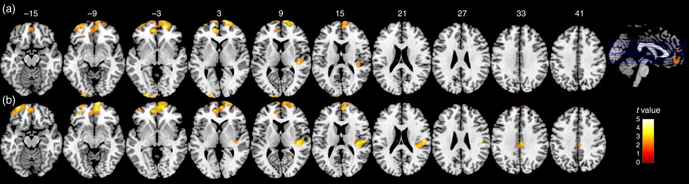
Multiple regression models were performed to establish relevance of head circumference measurements predicting voxel volumes/surface areas. We did not find results supporting the relationship between head circumference measurements at birth and local GMV at late childhood. GMV of the bilateral prefrontal cortex and right temporal and bilateral occipital areas was strongly associated to the head circumference measured at 4 years of age (Table 2, Fig. 1). Volumes of these significant GM clusters were associated with MPI scores (r 0·27, P=0·03), and, as expected, with head circumference at 4 years of age (r 0·80, P<0·001). Association between head circumference measurements at 10 years with GMV at the same time was restricted to three clusters in the frontal, temporal and limbic areas (Table 3, Fig. 1). Volumes of these significant grey matter clusters were associated with MPI scores (r 0·26, P=0·03), and, as expected, with head circumference at 10 years (r 0·77, P<0·001).
Relationships between regional grey matter volumes and head circumference. At 10 years of age, grey matter regions that showed significant positive correlations with head circumference measurements at 4 years (a) and 10 years (b) in seventy-four healthy children. No significant correlations were found at birth. The grey matter volume values in the figure were extracted from the significant clusters after sex, age, height, weight, laterality, total intracranial volume and family socio-economic status of each subject were regressed. Significant areas (corrected P-values≤0·05) are overlaid on Montreal Neurological Institute average brain axial sections (numbers indicate z coordinate). The bar on the yellowish/reddish colour scale indicates the range of t-values (lighter colours correspond to higher values).


Partial correlation results indicated that head circumference measurements at 4 years of age were significantly associated with local inner-brain surface areas, especially at bilateral frontal and temporal locations such as the cingulate gyrus, superior frontal gyrus, precentral gyrus, and superior and inferior temporal gyri (see Table 4).
Partial correlations between local inner-brain surface areas and head circumference at birth and 4 years, after age, sex, height and weight at 10 years, laterality, and family socio-economic status have been partialled out* †

L, left; R, right.
* Only significant correlations are displayed.
† All the displayed results were significant with a corrected P-value≤0·05.
Relationships among subcortical neuroimaging results, head circumference and measurements of cognitive abilities
Partial correlations coefficients indicated that head circumference measurements at birth and at 4 and 10 years of age were significantly associated with the bilateral volume of the putamen and thalamus (P<0·05). Head circumference measurements at 4 and 10 years of age were significantly associated with bilateral volume of the caudate nucleus and globus pallidus (P<0·05) (see Table 5). MPI scores were associated with bilateral volumes of the caudate nucleus, globus pallidus, putamen and thalamus (P<0·05).
Partial correlations between bilateral subcortical structure volumes and head circumference measurements (at birth, 4 years and 10 years) and the cognitive abilities index, after sex, age, height, weight, laterality and family socio-economic status have been partialled out

HC, head circumference; MPI, Mental Processing Index from the Kaufman Assessment Battery for Children, 2nd edition( Reference Kaufman and Kaufman 27 ).
* P-value<0·05.
Discussion
Head circumference during infancy and childhood reflects either biological or environmental factors, including prenatal nutritional supplementation. Thus, the measurement of head circumference is an easy, quick and inexpensive approach that may be valuable for assessing neurodevelopment. Thus far, no study had examined the relationship between head circumference and regional GMV in children exposed to high LC-PUFA and folate during their fetal lives. Some cross-sectional studies have suggested existence of a significant positive association between head circumference and total GMV( Reference Bartholomeusz, Courchesne and Karns 8 – Reference Lange, Froimowitz and Bigler 10 ). Its accuracy as an index of brain size would depend on age( Reference Bartholomeusz, Courchesne and Karns 8 , Reference Lange, Froimowitz and Bigler 10 ) and the successful control for several confounding factors, including prenatal exposures. However, such cross-sectional studies do not indicate the potential relevance of successive head circumference measurements – beginning at birth – and the role of prenatal nutrition to predict brain size in late childhood. Our results suggest that head circumference at 4 years of age is the most relevant measure to predict total and regional brain volumes at late childhood. Either LC-PUFA or folate prenatal exposition would not have a significant role predicting total and regional brain volumes at late childhood.
During fetal and early postnatal time periods, the brain is particularly vulnerable and plastic( Reference Toga, Thompson and Sowell 11 ). Thus, brain development trajectories until reaching an adult’s brain would be deeply determined not only by inherited influences but also by gestational conditions and environmental postnatal factors. Anthropometric measurements at birth, including head size, presumably represent differential prenatal exposures, whereas measurements performed during childhood presumably represent prenatal as well as postnatal exposures. In our results, head size at birth had a significant weight predicting total brain volumes and TBSA, but was not related to any regional GMV at late childhood. On the other hand, measurements of head size during childhood had a significant weight in predicting total brain volumes and TBSA, and were also predictive of specific frontal, temporal and occipital GMV at late childhood. Thus, it might be assumed that postnatal factors have the most relevant role in structural maturation of certain cortical areas since birth to late childhood. These postnatal factors imply a genetic endowment that influences a child’s growth and maturation processes and a phenotype depending on environmental circumstances, such as health status and nutrition, throughout the growing years.
The long-term signature effect of prenatal micronutrients (by means of either deficits or high exposition) on postnatal brain structure is still unclear, and more MRI studies are needed. We found that early supplementary nutrition might have an impact on brain development as measured by brain surface area. Specifically, 5-MTHF early supplementation seems to increase the size of the surface area in the parietal cortex. This lobe has been involved in sensory processing and visuomotor control( Reference Mutha, Sainburg and Haaland 38 ), attention processing( Reference Kanai, Dong and Bahrami 39 ), touch and grasping( Reference Fogassi, Ferrari and Gesierich 40 ), and other superior cognitive functions (such as numeric representation( Reference Nieder and Dehaene 41 ) or working memory( Reference Meyer, Obleser and Friederici 42 )). This increased surface area is coherent to attentional processing advantages observed in children of 5-MTHF-supplemented mothers( Reference Catena, Muñoz-Machicao and Torres-Espínola 24 ).
Postnatal neurodevelopment during the first 5 years of life is one of the most active stages of brain development( Reference Pujol, Soriano-Mas and Ortiz 43 ). This period also coincides with the surface of many developmental neuropsychiatric disorders, including autism spectrum disorders and attention deficit/hyperactivity disorder( Reference Dean, O’Muircheartaigh and Dirks 44 ). It is generally accepted that there are developmental windows that allow the brain to be moulded by experience in infancy and childhood( Reference Fox, Levitt and Nelson 45 ). This early period would be extremely relevant for neurodevelopment, and we found that, effectively, head circumference at 4 years of age is highly correlated with MRI data at 10 years of age. It was also strongly associated with total GMV, WMV and TBSA; regional GMV in the frontal, temporal and occipital areas; and with cognitive abilities index. Head circumference at 4 years of age was also positively related to subcortical structures’ volumes (caudate nucleus, globus qpallidus, putamen and thalamus) at later childhood.
Limitations
This was a randomised clinical trial study with a large longitudinal follow-up, where participants underwent several detailed anthropometric and neurodevelopment assessments, including a MRI examination. However, our findings should be interpreted in light of certain limitations; first, although the sample size of this neuroimaging study is considerable, one of the limitations was the reduced number of participants in the group supplemented with 5-MTHF. Further studies are needed to reveal the impact of early supplementation on head circumference, cognitive abilities and neuroimaging measurements along infancy and childhood. Second, we did not control for postnatal factors that have a significant impact on children’s neurodevelopment, such as fatty acids and folate intake or the richness of the child’s developmental environment( Reference Anjos, Altmäe and Emmett 46 , Reference Bann, Wallander and Do 47 ). These factors might have also affected the relationships among head circumference, cognitive abilities and neuroimaging variables evaluated in this study. Finally, we only considered in the present analysis one measurement of cognitive abilities collected during childhood, taken at a different time from neuroimaging and some of the anthropometric measurements, and only one measurement of neuroimaging variables. Several simultaneous measurements of head circumference, cognitive abilities and neuroimaging variables along infancy and childhood will help determine the precise timing of neurodevelopmental improvements and how the relationships between these variables change over time.
Conclusions
Since the nineteenth century, several diverse ways to estimate brain size from cranial vault capacity have been tested( Reference Tanner 48 ). Even though new advances in radiology and imaging have led to more accurate estimations of brain volume and its relationship to head size, no study to date has characterised the age-dependent relationships between head circumference and regional cortical and subcortical volumes. In this study, for the first time, we examined the associations of head circumference measurements at birth and childhood (4 and 10 years) with later regional cortical volumes, as well as with volumes of subcortical structures and cognitive abilities. We demonstrated that age-dependent relationships between head size and brain volumes are regionally specific, with prenatal supplementation having a minor effect on parietal surface area. We conclude that head circumference measurements, especially at the age of 4 years, are suitable to predict total and regional brain growth and might have a high potential for assessing children’s neurodevelopment.
Acknowledgements
The authors thank all the women and children who participated in this study for their collaboration and all colleagues in the study centres for their support. The authors thank Dr Leandro L. Di Stasi (Mind, Brain, and Behavior Research Center, University of Granada, Spain) for his assistance designing the graphical material.
This study was supported by the Commission of the European Community’s 7th Framework Programme (FP7/2008-2013), grant agreement no. 212652 (NUTRIMENTHE project); within the 6th Framework Programme, contract no. 007036 (EARNEST project); and supported in part by the Commission of the European Community within the 5th Framework Programme, contract no. QLK1-CT-1999-00888 (NUHEAL project). This publication is the work of the authors and does not necessarily reflect the views of the Commission of the European Community. Research by A. C. is funded by a Spanish Ministry of Economy and Competitiveness grant (State Secretariat for Research, Development and Innovation Secretary, PSI2012-39292). C. D.-P. is supported by a UGR Postdoctoral Fellowship (2013 University of Granada Research Plan). The work of B. K. is supported by the European Research Council Advanced grant no. ERC-2012-AdG – no.·322605 META-GROWTH. The funders had no role in design, analysis or writing of this article.
All authors helped in the interpretation of results and contributed to manuscript preparation. A. C. designed the study, performed the neuroimaging analysis and interpretation, the statistical analysis and wrote the manuscript; C. M-Z., F. J. T-E. and P. B. performed the examinations on children; C. D-P. performed the statistical analysis and wrote the manuscript; M. P-G. designed the study and reviewed the manuscript; T. D. and B. K. were coordinators of the NUHEAL study at Germany and Hungary, and supervised the manuscript; and C. C. supervised the NUHEAL study in Granada, designed the study and wrote the manuscript.
None of the authors has any conflicts of interest.
Supplementary Material
For supplementary material/s referred to in this article, please visit https://doi.org/10.1017/S0007114516004281


 Open access