


Introduction
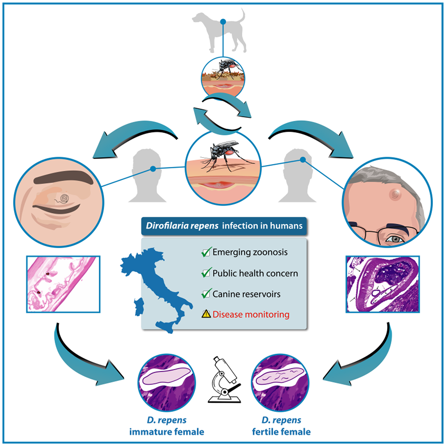
Dirofilaria immitis and Dirofilaria repens (Spirurida, Onchocercidae) are zoonotic vector-borne filarial parasites, transmitted by Culicidae mosquitoes, with dogs as the main reservoirs of infection (Capelli et al., Reference Capelli, Genchi, Baneth, Bourdeau, Brianti, Cardoso, Danesi, Fuehrer, Giannelli, Ionică, Maia, Modrý, Montarsi, Krücken, Papadopoulos, Petrić, Pfeffer, Savić, Otranto, Poppert and Silaghi2018; Genchi and Kramer, Reference Genchi and Kramer2020; Perles et al., Reference Perles, Dantas-Torres, Krücken, Morchón, Walochnik and Otranto2024), in which the nematodes may cause heartworm disease or subcutaneous nodules, respectively (Simón et al., Reference Simón, Diosdado, Siles-Lucas, Kartashev and González-Miguel2022). Data reported in recent studies shows an increase in the prevalence of both D. immitis and D. repens in Europe and in southeastern regions of Asia and Africa (McCall et al., Reference McCall, Genchi, Kramer, Guerrero and Venco2008; Capelli et al., Reference Capelli, Genchi, Baneth, Bourdeau, Brianti, Cardoso, Danesi, Fuehrer, Giannelli, Ionică, Maia, Modrý, Montarsi, Krücken, Papadopoulos, Petrić, Pfeffer, Savić, Otranto, Poppert and Silaghi2018; Genchi and Kramer, Reference Genchi and Kramer2020). In Italy, the changing distribution patterns of both D. immitis and D. repens show an increase in their prevalence in dogs from areas considered non-endemic, such as regions in southern Italy (Genchi et al., Reference Genchi, Rinaldi, Mortarino, Genchi and Cringoli2009, Reference Genchi, Rinaldi, Venco, Cringoli, Vismarra and Kramer2019; Panarese et al., Reference Panarese, Iatta, Beugnet and Otranto2022; Ciucă et al., Reference Ciucă, Caruso, Illiano, Bosco, Maurelli and Rinaldi2023; Napoli et al., Reference Napoli, De Benedetto, Ciucă, Bosco, Lia, Veneziano, Bezerra Santos, Otranto, Rinaldi and Brianti2023). Human infections are mainly due to D. repens, which remains a persistent public health problem (Szénási et al., Reference Szénási, Kovács, Pampiglione, Fioravanti, Kucsera, Tánczos and Tiszlavicz2008; Montoya-Alonso et al., Reference Montoya-Alonso, Mellado, Carretón, Cabrera-Pedrero, Morchón and Simón2010; Ciucă et al., Reference Ciucă, Simón, Rinaldi, Kramer, Genchi, Cringoli, Acatrinei, Miron and Morchon2018; Simón et al., Reference Simón, Diosdado, Siles-Lucas, Kartashev and González-Miguel2022; Perles et al., Reference Perles, Dantas-Torres, Krücken, Morchón, Walochnik and Otranto2024). Following transmission of the infecting larvae (L3) by mosquitoes, humans are usually accidental hosts for D. immitis and D. repens, although cases with fertile adults and/or microfilariaemia due to D. repens have been reported (Grandi et al., Reference Grandi, Morchón, Kramer, Kartashev and Simón2008; Poppert et al., Reference Poppert, Hodapp, Krueger, Hegasy, Niesen, Kern and Tannich2009; Sergiev et al., Reference Sergiev, Supriaga, Morozov and Zhukova2009). Zoonotic infection results in the formation of nodular structures as a consequence of the circumscribed inflammatory response mounted around dead or dying larval stages. In most patients, D. immitis localizes in the lungs, causing pulmonary dirofilariosis, while D. repens localizes in the subcutaneous tissue – causing the formation of subcutaneous nodules – or under the conjunctiva (ocular dirofilariosis). Over the years, several cases of pulmonary nodules caused by D. repens have also been reported in the Old World (Pampiglione et al., Reference Pampiglione, Rivasi and Gustinelli2009; Benzaquen et al., Reference Benzaquen, Brajon, Delord, Yin, Bittar, Toga, Berbis and Parola2015; Gabrielli et al., Reference Gabrielli, Mangano, Furzi, Oliva, Vita, Poscia, Fazii, Di Paolo, Marocco, Mastroianni, Bruschi and Mattiucci2021; Momčilović et al., Reference Momčilović, Gabrielli, Đenić, Živković, Stevanović, Krstić, Ranđelović and Tasić-Otašević2022). Pulmonary dirofilariosis can be confused with pulmonary neoplasms (benign, carcinoma, metastases), tuberculosis or fungal infections (Awe et al., Reference Awe, Mattox, Alvarez, Stork, Estrada and Greenberg1975; Ro et al., Reference Ro, Tsakalakis, White, Luna, Chang-Tung, Green, Cribbett and Ayala1989; Oliva et al., Reference Oliva, Gabrielli, Pernazza, Pagini, Daralioti, Mantovani, Mattiucci, D’Amati and Mastroianni2019) and represents a diagnostic challenge (Simón et al., Reference Simón, Diosdado, Siles-Lucas, Kartashev and González-Miguel2022). Between 1977 and 2016, more than 3500 human cases due to D. repens and 25 due to D. immitis were reported in Europe (Genchi and Kramer, Reference Genchi and Kramer2017; Capelli et al., Reference Capelli, Genchi, Baneth, Bourdeau, Brianti, Cardoso, Danesi, Fuehrer, Giannelli, Ionică, Maia, Modrý, Montarsi, Krücken, Papadopoulos, Petrić, Pfeffer, Savić, Otranto, Poppert and Silaghi2018). Since humans are not natural hosts, microfilariae are usually absent from peripheral blood. However, the parasite occasionally evades the host’s immune system to reach sexual maturity, resulting in microfilaremic infections that have so far been described in 23 human cases (Simón et al., Reference Simón, Diosdado, Siles-Lucas, Kartashev and González-Miguel2022; Tasić-Otasevic et al., Reference Tasić-Otasevic, Golubović, Trichei, Zdravkovic, Jordan and Gabrielli2023). Italy is traditionally endemic for human dirofilariosis and is one of the countries with the highest number of human cases identified so far (Muro et al., Reference Muro, Genchi, Cordero and Simón1999; Avellis et al., Reference Avellis, Kramer, Mora, Bartolino, Benedetti and Rivasi2011; Capelli et al., Reference Capelli, Genchi, Baneth, Bourdeau, Brianti, Cardoso, Danesi, Fuehrer, Giannelli, Ionică, Maia, Modrý, Montarsi, Krücken, Papadopoulos, Petrić, Pfeffer, Savić, Otranto, Poppert and Silaghi2018). In fact, the number of human cases of dirofilariosis caused by D. repens reported in the literature since Addario’s first observation in 1885 amounted to approximately 410 in 1995, 181 of which had occurred in Italy (Addario, Reference Addario1885; Pampiglione and Rivasi, Reference Pampiglione and Rivasi2000). In 2001, there was a further description of 60 cases between 1990 and 1999 (Pampiglione et al., Reference Pampiglione, Rivasi, Angeli, Boldorini, Incensati, Pastormerlo, Pavesi and Ramponi2001). More recently, eight further cases of D. repens infection were described by Gabrielli et al. (Reference Gabrielli, Mangano, Furzi, Oliva, Vita, Poscia, Fazii, Di Paolo, Marocco, Mastroianni, Bruschi and Mattiucci2021). The present study aims to describe two new human cases of D. repens infection occurring in a region of southern Italy which has recently become endemic for canine dirofilariosis (Ciucă et al., Reference Ciucă, Caruso, Illiano, Bosco, Maurelli and Rinaldi2023).
Materials and methods
Case histories and sampling of adult nematodes
First case (case 1)
In October 2023, a 33-year-old Italian-born man living in Caserta (Campania, southern Italy) was referred to the emergency hospital (Ospedale Dei Pellegrini) of Naples, with complaints of pruritus and swelling of the left upper eyelid (Figure 1A). In his description, he mentioned that he had been taking long walks along the river at Castel Volturno six months prior to his admission to the hospital. The man reported that he had been living with two dogs, aged 3 and 6 years old, for a period of 5 years. At the initial ophthalmological examination, the patient exhibited an oedema in a serpentiform shape on the upper eyelid of the left eye (Figure 1A). No evidence of swelling or similar rashes was observed in other parts of the body. The ophthalmological examination revealed that the visual acuity in both eyes was within the normal range. Given the possibility of a parasitic infection, the patient was treated with a dose of 12 mg of ivermectin. The following day, the patient was transferred to another hospital in Naples. Subsequently, he underwent the following additional diagnostic procedures: a chest X-ray, a CT scan, an ECG, blood tests, copromicroscopy examination and an IgG test for SARS-CoV-2. With the exception of the detection of Blastocystis spp. in the stool sample, all results were within the normal range. The patient was also examined by a dermatological specialist who prescribed a course of albendazole 400 mg twice a day, for 7 days. After treatment, the patient returned with a swelling of the upper eyelid in the form of a lump (Figure 1B,C). The snake-like appearance was no longer present. The patient was then referred for surgical excision of the nodule that had formed. A live and motile worm of approximately 14cm in length was found during the surgical excision of the nodule on the left upper eyelid (Figure 1D,E; supplementary file 1).
Case 1. (A) Patient with a serpiginous lump on the upper eyelid of the left eye at first presentation. (B, C) Patient with swelling of the upper eyelid of the left eye in the form of a lump after one week of treatment. (D) Patient after surgery. (E) Worm recovered after excision of the nodule from upper eyelid of the left eye.

Second case (case 2)
In September 2024, a 67-year-old Italian man from Pozzuoli, a coastal town in the Metropolitan City of Naples (southern Italy) was referred to a private dermatology clinic in Naples for an oval-shaped subcutaneous mass located in the left frontal region (Figure 2A). An ultrasound of the skin showed an oval-shaped nodule measuring 7.7 × 3.9 mm, hypoechoic with well-defined, avascular margins located in the scalp area (Figure 2B). Based on the ultrasound, a cyst or fibroma of the scalp was suspected, and surgical removal was recommended. During the surgical removal, a live worm measuring 10 cm in length was found (Figure 2C,D). The patient had no history of travel abroad in the past 3 years and owned an 18-month-old dog.
Case 2. (A) Patient with a subcutaneous mass located in the left frontal region of the scalp; (B) an ultrasound of the skin revealed an oval-shaped, hypoechoic nodule measuring 7.7 × 3.9 mm with well-defined, avascular margins in the scalp area, suggestive of the presence of a nematode; (C) patient after surgery; (D) a 10 cm-long nematode surgically removed from the nodule in the scalp area.

The 2 adult nematodes (the first along with the excised nodule from Case 1 and the second from Case 2), were sent to the Laboratory of Parasitology of the University of Naples Federico II, for histological and molecular analysis. Based on the final surgical results, there was a suspicion of infection with Dirofilaria spp. At this point, blood and serum samples were obtained from both patients to verify the presence of microfilariae in the blood and of specific antibodies against Dirofilaria spp. in the serum. Similarly blood and serum samples were collected from all dogs owned by both patients (2 from Case 1 and 1 from Case 2) and tested for the presence of microfilariae, D. immitis antigens and antibodies against D. repens somatic antigens.
The immunological tests were performed at the Department of Public Health and Infectious Diseases, University of Rome ‘Sapienza’, Rome, Italy.
Laboratory investigation of the human cases: histology, molecular analysis, modified Knott’s test and immunological tests
The worms and nodule samples (Case 1 and Case 2) were excised, then preserved in neutral buffered formalin to maintain tissue integrity. After fixation, they were embedded in paraffin, allowing for thin slicing at 3–5 μm thickness for detailed microscopic analysis. The sections were then stained with haematoxylin and eosin, a standard method for distinguishing cellular and tissue structures due to haematoxylin staining nuclei blue and eosin staining the cytoplasm and extracellular matrix pink. Digital images were captured using a Pannoramic SCAN II (3DHISTECH) digital slide scanner, enabling detailed examination and production of representative images for further analysis.
For molecular identification, genomic DNA was extracted from 25 mg of tissue from each nematode, using the DNeasy® Blood and Tissue kit (Qiagen, Germany), following the manufacturer’s instructions. Molecular analyses were performed in accordance with the multiplex PCR protocol described by Rishniw et al. (Reference Rishniw, Barr, Simpson, Frongillo, Franz and Alpizar2006) (5.8 + ITS2 region) which allows for the simultaneous detection of D. immitis and D. repens.
A modified Knott’s test was used for the detection of circulating microfilariae of D. repens (Genchi et al., Reference Genchi, Ciucă, Vismarra, Ciccone, Cringoli, Kramer and Rinaldi2021) as outlined below. One mL of EDTA blood (3× 1 mL for each tube) was mixed with 9 mL of distilled water and centrifuged for 3–5 min at 170 g. The supernatant was then removed from the tube and the contents were stained with 1–2 drops of 1% methylene blue. A drop was placed on a microscope slide covered with a cover slip and observed under an optical microscope at 100×.
The patient serum samples were analysed using an home-made ELISA test to detect the specific IgG antibody response. This involved the use of adult D. immitis and D. repens somatic antigens as described in Cabrera et al. (Reference Cabrera, Carretón, Morchón, Falcón-Cordón, Falcón-Cordón, Simón and Montoya-Alonso2018) and Mendoza-Roldán et al. (Reference Mendoza-Roldán, Gabrielli, Cascio, Manoj, Bezerra-Santos, Benelli, Brianti, Latrofa and Otranto2021). The antigens were isolated from worms obtained by necropsy of naturally infected dogs, washed, and then they were macerated and sonicated (three cycles of 70 kHz, 30 s) in sterile saline solution (Prieto et al., Reference Prieto, Venco, Simon and Genchi1997). The homogenate was centrifuged at 16 000g for 30 min. The supernatant was dialyzed against 0.01M PBS, pH 7.2. The protein concentration was measured (Bradford, Reference Bradford1976) and an ELISA microplate was coated with antigens at a final concentration of 0.8 µg/µL. The serum sample was tested in solid-phase ELISA at dilution 1:80 and 1:150 to detect anti-D. repens and anti-D. immitis IgG antibodies, respectively. Goat anti-human-IgG-antihuman IgG (H + L) conjugated to horseradish peroxidase (Sig-ma- Aldrich, MO, USA) was used as a secondary antibody at 1:40.000 dilution in both cases. The Optical density (OD) was determined at a wavelength of 492 nm (Easy-Reader, Bio Rad, CA, USA). The cut-off points (OD = 1.12 for DiSA and OD = 1.03 for DrSA) were established by calculating the mean value + 3 standard deviations (3SD) of 30 serum samples obtained from clinically healthy humans (negative controls).
Laboratory investigations on the patients’ dogs
The dogs were screened for Dirofilaria spp. microfilariae using the Knott test (only 1 mL of EDTA blood) (Genchi et al., Reference Genchi, Ciucă, Vismarra, Ciccone, Cringoli, Kramer and Rinaldi2021). Additionally, the Petcheck Heartworm Canine (IDEXX) test was used according to the manufacturer’s instructions, to detect D. immitis antigen. All dogs were also screened for antibodies against D. repens (Mendoza-Roldán et al., Reference Mendoza-Roldán, Gabrielli, Cascio, Manoj, Bezerra-Santos, Benelli, Brianti, Latrofa and Otranto2021).
Results
Histology – case 1 and case 2
In the first case, the nematode displayed a whitish, cylindrical body measuring 14 cm in length. Histological characteristics were consistent with an immature female nematode (Wong and Brummer, Reference Wong and Brummer1978; Brindicci et al., Reference Brindicci, Santoro, Signorile, Leone, Di Ciaula, Monno and Angarano2019). Additionally, a thick, intensely eosinophilic cuticle was observed, covered with multiple chitinous layers forming ridges. This ridged appearance of the cuticle was characteristic of D. repens (Figure 3A,B). The nematode’s coelomic cavity contained a monolayer of cuboidal or cylindrical cells, consistent with intestinal tissue. No microfilariae were observed in the uterus (Figure 3C,D). Macroscopically, the nodule measured 1 × 1 cm, was spheroidal in shape, firm in consistency, and smooth on the surface. Histologically, the outer skin epithelium of the nodule was diffusely hyperplastic. The dermis was significantly expanded due to chronic inflammatory infiltrate, mainly comprising numerous activated macrophages, epithelioid cells, and eosinophilic granulocytes, with a smaller number of mature lymphocytes and plasma cells (Figure 3E,F).
Hystological analysis case 1. (A,B) Longitudinal section of the parasite showing a thick, intensely eosinophilic cuticle covered with several layers of chitin raised in ridges (red arrows) (E.E. 40×); (C) longitudinal section of the parasite: caudal end. (E.E. 20×). (D) Longitudinal section of the parasite, coelomic cavity: a monolayer of cubic or cylindrical cells is observed (compatible with the intestine, red arrows). No microfilariae are observed. (E.E. 40×); (E,F) nodule. Cutis sections: the stratum corneum epithelium (blue arrow) is diffusely hyperplastic. The dermis is extensively expanded due to the presence of a chronic inflammatory infiltrate, mainly characterised by a high number of activated macrophages, epithelioid cells and eosinophilic granulocytes and a lower number of small mature lymphocytes and plasma cells (yellow arrows). There are multifocal areas of repair characterised by the presence of blood vessels (green arrows), fibroblasts associated with fibrillar matrix (fibroplasia) (E.E. 20×).

For the second case, the nematode displayed a whitish, cylindrical body and with ridged appearance of the cuticle measuring 15–16 cm in length. Histological characteristics were consistent with a fertile female nematode (Ciucă et al., Reference Ciucă, Roman, Prisco, Miron, Acatrinei, Paciello, Maurelli, Vismarra, Cringoli and Rinaldi2020; Simón et al., Reference Simón, Diosdado, Siles-Lucas, Kartashev and González-Miguel2022), showing in the transverse section the uterus with microfilariae (Figure 4A,B).
Hystological analysis case 2. (A,B) Longitudinal section of the parasite: intestine (black arrow) and uterus containing microfilariae (blue arrow) (E.E. 20×). Transverse section of the parasite: a thick, intensely eosinophilic cuticle is observed, covered by multiple layers of chitin raised into crests (red arrows), intestine (black arrow), and uterus (blue arrow) with microfilariae (green arrow) (E.E. 20×).

Knott test and multiplex PCR (case 1 and case 2)
The modified Knott’s test revealed the absence of microfilariae in the blood of both patients.
The multiplex PCR method amplified a 484-base pair fragment specific to D. repens in both patients. The sequences of the PCR products obtained are available in the GenBank database under the following accession numbers: AY693808 (Case 1) and MW242631.1 (Case 2).
The first patient (Case 1) showed a positive response to D. repens somatic antigens (OD = 1.24) and a negative response to D. immitis (OD = 0.64) somatic antigens as determined by in-house IgG.
Similarly, the second patient (Case 2) was seropositive for D. repens (OD = 1.86) while no specific IgG were detected against D. immitis antigens (OD = 0.63).
Test results for Dirofilaria spp in dogs
All dogs tested negative for Dirofilaria infection in both the modified Knott’s test and the antigen test. In addition, the ELISA IgG assay for antibodies against D. repens also yielded negative results for the three dogs tested.
Discussion
The present study unveiled two new cases of human Dirofilaria repens infection in southern Italy and involved clinical, serological, haematological and molecular analysis in the two patients and their dogs, adopting an integrated medical and veterinary approach.
Dirofilaria repens is the main etiological agent of human dirofilariosis in the Old World (Genchi and Kramer, Reference Genchi and Kramer2020). The rise in dirofilariosis cases across Europe has been linked to factors such as climate change, an increase in mosquito species capable of transmitting Dirofilaria spp., and the intensification of international dog trade and movement (Cancrini et al., Reference Cancrini, Scaramozzino, Gabrielli, Di Paolo, Toma and Romi2007; Genchi et al., Reference Genchi, Rinaldi, Mortarino, Genchi and Cringoli2009; Capelli et al., Reference Capelli, Frangipane Di Regalbono, Simonato, Cassini, Cazzin, Cancrini, Otranto and Pietrobelli2013, Reference Capelli, Genchi, Baneth, Bourdeau, Brianti, Cardoso, Danesi, Fuehrer, Giannelli, Ionică, Maia, Modrý, Montarsi, Krücken, Papadopoulos, Petrić, Pfeffer, Savić, Otranto, Poppert and Silaghi2018; Sałamatin et al., Reference Sałamatin, Pavlikovska, Sagach, Nikolayenko, Kornyushin, Kharchenko, Masny, Cielecka, Konieczna-Sałamatin, Conn and Golab2013; Simón et al., Reference Simón, González-Miguel, Diosdado, Gómez, Morchón and Kartashev2017; Perles et al., Reference Perles, Dantas-Torres, Krücken, Morchón, Walochnik and Otranto2024). In Ukraine, for instance, dirofilariosis was included in the national surveillance system for notifiable diseases in 1975, and the country has thousands of cases reported in the archives of the Ministry of Health, even if not all cases are documented in international scientific journals (Sałamatin et al., Reference Sałamatin, Pavlikovska, Sagach, Nikolayenko, Kornyushin, Kharchenko, Masny, Cielecka, Konieczna-Sałamatin, Conn and Golab2013; ESDA, 2017; Simón et al., Reference Simón, González-Miguel, Diosdado, Gómez, Morchón and Kartashev2017, Reference Simón, Diosdado, Siles-Lucas, Kartashev and González-Miguel2022).
Human dirofilariosis cases have been reported mainly from canine dirofilariosis endemic areas in the Mediterranean region (Capelli et al., Reference Capelli, Genchi, Baneth, Bourdeau, Brianti, Cardoso, Danesi, Fuehrer, Giannelli, Ionică, Maia, Modrý, Montarsi, Krücken, Papadopoulos, Petrić, Pfeffer, Savić, Otranto, Poppert and Silaghi2018; Tahir et al., Reference Tahir, Davoust and Parola2019), with Italy being the country with the highest number of reported cases to date (Pampiglione et al., Reference Pampiglione, Rivasi, Angeli, Boldorini, Incensati, Pastormerlo, Pavesi and Ramponi2001; Capelli et al., Reference Capelli, Genchi, Baneth, Bourdeau, Brianti, Cardoso, Danesi, Fuehrer, Giannelli, Ionică, Maia, Modrý, Montarsi, Krücken, Papadopoulos, Petrić, Pfeffer, Savić, Otranto, Poppert and Silaghi2018; Gabrielli et al., Reference Gabrielli, Mangano, Furzi, Oliva, Vita, Poscia, Fazii, Di Paolo, Marocco, Mastroianni, Bruschi and Mattiucci2021; Simon et al., Reference Simón, Diosdado, Siles-Lucas, Kartashev and González-Miguel2022, Reference Gabrielli, Brustenga, Morganti, Ciucă, Barlozzari, Rigamonti, Orlandi, Sforna and Veronesi2024). Considering that Italy is an endemic country for canine and human dirofilariosis (Capelli et al., Reference Capelli, Genchi, Baneth, Bourdeau, Brianti, Cardoso, Danesi, Fuehrer, Giannelli, Ionică, Maia, Modrý, Montarsi, Krücken, Papadopoulos, Petrić, Pfeffer, Savić, Otranto, Poppert and Silaghi2018; Genchi et al., Reference Genchi, Rinaldi, Venco, Cringoli, Vismarra and Kramer2019, Reference Genchi, Kramer, Venco, Ciucă and Vismarra2023; Brianti et al., Reference Brianti, Panarese, Napoli, De Benedetto, Gaglio, Bezerra-Santos, Mendoza-Roldan and Otranto2022; Napoli et al., Reference Napoli, De Benedetto, Ciucă, Bosco, Lia, Veneziano, Bezerra Santos, Otranto, Rinaldi and Brianti2023) not only is an increased number of cases of D. immitis and D. repens in both dogs and humans expected, but also a high frequency of aberrant migrations in humans, especially due to D. repens, as already shown in dogs (Pierantozzi et al., Reference Pierantozzi, Di Giulio, Traversa, Aste and Di Cesare2017; Napoli et al., Reference Napoli, De Benedetto, Di Giorgio, Sfacteria, Marino, Mazzara, Giambrone, Gaglio and Brianti2024). As an example, D. repens infections of the male genitalia have been documented in humans (Pampiglione and Rivasi, Reference Pampiglione and Rivasi2000; Simón et al., Reference Simón, Diosdado, Siles-Lucas, Kartashev and González-Miguel2022) including a case of testicular infection with D. repens recently reported in a child from northeastern Italy (Ugolini et al., Reference Ugolini, Lima, Maffi, Pierangeli, Vastano, Gargano, Varani, Gustinelli, Caffara and Fioravanti2022). Moreover, it should also be noted that two cases of D. immitis infection in humans, have already been reported in central Italy, one with localization in the right temporal bulbar subconjunctival space and the other with a 2 cm nodule in the right upper lung (Avellis et al., Reference Avellis, Kramer, Mora, Bartolino, Benedetti and Rivasi2011; Palicelli et al., Reference Palicelli, Veggiani, Rivasi, Gustinelli and Boldorini2022).
The two human cases of D. repens infection reported in this study provide further insight into the epidemiological and clinical features of human dirofilariosis in an endemic region of southern Italy, where canine dirofilariosis is prevalent (Ferrara et al., Reference Ferrara, Maglione, Ciccarelli, Mundis, Di Loria, Pisaroglo de Carvalho and Santoro2022a; Ciucă et al., Reference Ciucă, Caruso, Illiano, Bosco, Maurelli and Rinaldi2023; Napoli et al., Reference Napoli, De Benedetto, Ciucă, Bosco, Lia, Veneziano, Bezerra Santos, Otranto, Rinaldi and Brianti2023). Indeed, human dirofilariosis cases in southern Italy have been progressively increasing. The earliest reported cases date back to 1885, with 11 cases documented (Pampiglione et al., Reference Pampiglione, Rivasi, Angeli, Boldorini, Incensati, Pastormerlo, Pavesi and Ramponi2001). After a long hiatus, a new case emerged in an hospital in Naples in 2012: a 17-year-old male who presented with a pulmonary nodule that was initially suspected to be a lung tumour; histopathological examination confirmed the presence of D. repens, highlighting the diagnostic challenges posed by the atypical pulmonary location mimic malignancies (Gabrielli et al., Reference Gabrielli, Petrullo, Coppola and Cancrini2012). In 2018, Ferrari et al. reported a rare case of pulmonary dirofilariosis caused by D. repens in a 62-year-old man from Palermo, who presented with pulmonary nodules that were initially suspected to be malignant based on imaging studies. Subsequently, in 2019, a case was reported in Bari, involving an 82-year-old woman with a subcutaneous D. repens lesion on her right thigh (Brindicci et al., Reference Brindicci, Santoro, Signorile, Leone, Di Ciaula, Monno and Angarano2019). Moreover, in 2022, it was reported a case of cutaneous dirofilariosis caused by D. repens in an Italian girl from Naples (southern Italy) who spends her summer holidays in the Tuscan countryside (northern Italy) (Ferrara et al., Reference Ferrara, Barbieri, D’Auria, Castelli, Gholami Shanagolabad, Zeccolini and Esposito2022b).
Our study describes two additional autochthonous cases of subcutaneous D. repens infections in southern Italy: a 33-year-old man from Caserta (Case 1), and a 67-year-old man from Pozzuoli, a coastal town in the Metropolitan City of Naples (Case 2). These cases evidence the continuous spreading of human dirofilariosis in southern Italy and the importance of accurate diagnostic approaches. Moreover this findings, alongside our cases of subcutaneous localization, reflects the diverse manifestations of D. repens, demonstrating its potential for internal organ involvement, albeit rarely, as also documented in literature (Ro et al., Reference Ro, Tsakalakis, White, Luna, Chang-Tung, Green, Cribbett and Ayala1989; Ferrari et al., Reference Ferrari, Grisolia, Reale, Liotta, Mularoni and Bertani2018; Oliva et al., Reference Oliva, Gabrielli, Pernazza, Pagini, Daralioti, Mantovani, Mattiucci, D’Amati and Mastroianni2019). Together, these cases highlight the need for awareness of atypical presentations of dirofilariosis in endemic regions, where superficial and deep tissue nodules may both occur.
Genetic analysis has shown considerable variability among isolates from both humans and dogs in Italy, indicating potential intraspecific diversity that may influence transmission dynamics and clinical manifestations (Genchi and Kramer, Reference Genchi and Kramer2017). This genetic diversity could also explain the variety in human presentations, from superficial nodules to rare internal cases. Such variability underscores the importance of molecular diagnostics in distinguishing between different strains, as it may have implications for tracking infection sources and understanding regional differences in transmission and pathogenicity (Gabrielli et al., Reference Gabrielli, Brustenga, Morganti, Ciucă, Barlozzari, Rigamonti, Orlandi, Sforna and Veronesi2024). However, a major limitation of the present study is that the genetic variability of the strains in both cases was not characterized.
The distribution of patients by age and sex is not uniform. Most reported cases of D. repens infection occur in individuals aged 20–69, with the highest incidence observed in those aged 50–59 years and a greater prevalence in women than in men (Sałamatin et al., Reference Sałamatin, Pavlikovska, Sagach, Nikolayenko, Kornyushin, Kharchenko, Masny, Cielecka, Konieczna-Sałamatin, Conn and Golab2013; Gabrielli et al., Reference Gabrielli, Mangano, Furzi, Oliva, Vita, Poscia, Fazii, Di Paolo, Marocco, Mastroianni, Bruschi and Mattiucci2021; Simón et al., Reference Simón, Diosdado, Siles-Lucas, Kartashev and González-Miguel2022). In the current study, one patient (Case 2) falls within the age range with the highest incidence, while the other patient (Case 1) is within the broader 20–69 age range. Moreover, both patients were male, aligning with findings from other studies (Pampiglione et al., Reference Pampiglione, Del Maschio, Pagan and Rivasi1994; Pupić-Bakrač et al., Reference Pupić-Bakrač, Pupić-Bakrač, Beck, Jurković, Polkinghorne and Beck2021) and contrasting with the cases reviewed by Simón et al. (Reference Simón, Diosdado, Siles-Lucas, Kartashev and González-Miguel2022). Several studies have indicated that there is no statistically significant correlation between human subcutaneous dirofilariosis and either age or gender (Pampiglione et al., Reference Pampiglione, Rivasi, Angeli, Boldorini, Incensati, Pastormerlo, Pavesi and Ramponi2001; Fontes-Sousa et al., Reference Fontes-Sousa, Silvestre-Ferreira, Carretón, Esteves-Guimarães, Maia-Rocha, Oliveira, Lobo, Morchón, Araújo, Simón and Montoya-Alonso2019; Pupić-Bakrač et al., Reference Pupić-Bakrač, Pupić-Bakrač, Beck, Jurković, Polkinghorne and Beck2021). Nevertheless, geographic variations in age distributions have been noted. For example, However, while cases in children are rare in Italy (Bertozzi et al., Reference Bertozzi, Rinaldi, Prestipino, Giovenali and Appignani2015; Pansini et al., Reference Pansini, Magenes, Casini, Guida, De Sanctis, Canonica, Rossi, Zuccotti and Giacomet2022; Ugolini et al., Reference Ugolini, Lima, Maffi, Pierangeli, Vastano, Gargano, Varani, Gustinelli, Caffara and Fioravanti2022), in Sri Lanka it is common to find infected children under 5 years of age – possibly due to differences in clothing, customs, and vector preferences (Senanayake et al., Reference Senanayake, Infaq, Adikaram and Udagama2013; Balendran et al., Reference Balendran, Yatawara and Wickramasinghe2022) Additionally, a case in a 100-year-old individual is also unusual in Italy, perhaps reflecting the reduced effectiveness of insecticides in the Piedmont area (Pampiglione et al., Reference Pampiglione, Rivasi, Angeli, Boldorini, Incensati, Pastormerlo, Pavesi and Ramponi2001). Ultimately, the risk factors for this disease extend beyond age and gender and include the endemicity of the region (evidenced by infections in dogs and cats), areas with increased vector exposure, and lifestyle factors, such as herein presented, where both patients frequently took long walks in areas known to have canine cases. Other factors, such as individual immunological status, may also contribute, as suggested by other studies (Pupić-Bakrač et al., Reference Pupić-Bakrač, Pupić-Bakrač, Beck, Jurković, Polkinghorne and Beck2021; Simón et al., Reference Simón, Diosdado, Siles-Lucas, Kartashev and González-Miguel2022).
Almost all human cases of D. repens reported in Italy have been asymptomatic or have presented with transient localized symptoms such as swelling, erythema, rash and pruritus (Rivasi et al., Reference Rivasi, Boldorini, Criante, Leutner and Pampiglione2006; Ermakova et al., Reference Ermakova, Nagorny, Pshenichnaya, Ambalov and Boltachiev2017; Gabrielli et al., Reference Gabrielli, Mangano, Furzi, Oliva, Vita, Poscia, Fazii, Di Paolo, Marocco, Mastroianni, Bruschi and Mattiucci2021; Pupić-Bakrač et al., Reference Pupić-Bakrač, Pupić-Bakrač, Beck, Jurković, Polkinghorne and Beck2021). These symptoms are usually associated with subcutaneous or submucosal nodules in various parts of the body and can even involve the eye through the conjunctiva (Pampiglione et al., Reference Pampiglione, Canestri Trotti and Rivasi1995, Reference Pampiglione, Rivasi, Angeli, Boldorini, Incensati, Pastormerlo, Pavesi and Ramponi2001; Pampiglione and Rivasi, Reference Pampiglione and Rivasi2000; Eccher et al., Reference Eccher, Dalfior, Gobbo, Martignoni, Brunelli, Decaminada, Bonetti, Rivasi, Barbareschi and Menestrina2008; Palicelli et al., Reference Palicelli, Deambrogio, Arnulfo, Rivasi, Paganotti and Boldorini2014; Fontanelli Sulekova et al., Reference Fontanelli Sulekova, Gabrielli, De Angelis, Milardi, Magnani, Di Marco, Taliani and Cancrini2016). Only rarely the worm can migrate to deeper tissues such as muscles and lungs (Ermakova et al., Reference Ermakova, Nagorny, Pshenichnaya, Ambalov and Boltachiev2017; Gabrielli et al., Reference Gabrielli, Mangano, Furzi, Oliva, Vita, Poscia, Fazii, Di Paolo, Marocco, Mastroianni, Bruschi and Mattiucci2021; Pupić-Bakrač et al., Reference Pupić-Bakrač, Pupić-Bakrač, Beck, Jurković, Polkinghorne and Beck2021; Simón et al., Reference Simón, Diosdado, Siles-Lucas, Kartashev and González-Miguel2022; Napolitano et al., Reference Napolitano, Pallotto, Pourmolkara, Vinci and Coviello2023). The two cases in our study align with typical human presentations with subcutaneous nodules without blood microfilariae (Pampiglione and Rivasi, Reference Pampiglione and Rivasi2000; Pampiglione et al., Reference Pampiglione, Rivasi, Angeli, Boldorini, Incensati, Pastormerlo, Pavesi and Ramponi2001; Gabrielli et al., Reference Gabrielli, Mangano, Furzi, Oliva, Vita, Poscia, Fazii, Di Paolo, Marocco, Mastroianni, Bruschi and Mattiucci2021). In Case 1, the patient presented with a subcutaneous mass in the upper eyelid, while Case 2 presented with an oval-shaped nodule in the left frontal region of the scalp. Furthermore, the nematode’s presence in the subcutaneous tissue of both cases ‘resembles D. repens’, as evidenced by similar cases from Italy (Brindicci et al., Reference Brindicci, Santoro, Signorile, Leone, Di Ciaula, Monno and Angarano2019; Gabrielli et al., Reference Gabrielli, Mangano, Furzi, Oliva, Vita, Poscia, Fazii, Di Paolo, Marocco, Mastroianni, Bruschi and Mattiucci2021; Campana et al., Reference Campana, Fania, Samela, Campana, Zecchi, Ricci and Abeni2022). Both cases in the present study showed infection sites similar to those commonly reported in a Ukrainian study, which analysed clinical data from 755 cases and found that 64.6% (488) of parasitic lesions were located in the head, including 297 cases around the eyes (Sałamatin et al., Reference Sałamatin, Pavlikovska, Sagach, Nikolayenko, Kornyushin, Kharchenko, Masny, Cielecka, Konieczna-Sałamatin, Conn and Golab2013). Additionally, the eyelid is the most commonly reported anatomical site for nodules caused by D. repens, as highlighted in the recent case review by Simón et al. (Reference Simón, Diosdado, Siles-Lucas, Kartashev and González-Miguel2022). This prevalence is likely because parasitic lesions in other areas are more difficult for patients to detect.
Humans are considered accidental hosts for Dirofilaria spp., as nematodes rarely reach sexual maturity or produce microfilariae in human blood (Simón et al., Reference Simón, Siles-Lucas, Morchón, González-Miguel, Mellado, Carretón and Montoya-Alonso2012; Ermakova et al., Reference Ermakova, Nagorny, Pshenichnaya, Ambalov and Boltachiev2017). However, some studies have documented D. repens infections with microfilariae in blood or local tissues (Kłudkowska et al., Reference Kłudkowska, Pielok, Frąckowiak, Masny, Gołąb and Paul2018; Potters et al., Reference Potters, Vanfraechem and Bottieau2018; Pupić-Bakrač et al., Reference Pupić-Bakrač, Pupić-Bakrač, Beck, Jurković, Polkinghorne and Beck2021; Tasić-Otasevic et al., Reference Tasić-Otasevic, Golubović, Trichei, Zdravkovic, Jordan and Gabrielli2023).
In our study, the first patient presented with an immature adult female D. repens worm measuring 14 cm in length, lacking microfilariae in both the uterus and the blood, but exhibiting the presence of antibodies in the serum sample. Similar characteristics – including sexually immature nematodes, absence of microfilariae, and a positive IgG immune response – were reported in some of the cases described in central Italy (Gabrielli et al., Reference Gabrielli, Mangano, Furzi, Oliva, Vita, Poscia, Fazii, Di Paolo, Marocco, Mastroianni, Bruschi and Mattiucci2021).The second case was initially suspected to be a cyst or fibroma based on ultrasound findings but was later identified as a live 16–17 cm D. repens worm upon surgical excision, highlighting the challenge of distinguishing dirofilariosis from benign tumours. A similar case of subcutaneous D. repens infection in the scalp region of a patient, was also reported in Slovenia in 2017 (Kotnik et al., Reference Kotnik, Rataj and Šoba2022).
Furthermore, in both cases of the present study, the worms were encapsulated within nodules. This finding is consistent with the global cases reviewed by Simón et al. (Reference Simón, Diosdado, Siles-Lucas, Kartashev and González-Miguel2022), who reported that 96.68% (408) of D. repens worms were encapsulated within nodules, while only 3.32% (14) were free.
An intriguing aspect of our study was the discovery of a gravid female D. repens within the nodule of the second case, although no microfilariae were present in the blood and no male worm was detected. While most nematodes isolated from humans are immature, several studies have documented mature D. repens females carrying microfilariae in their uterus without releasing them into the bloodstream (Supriaga et al., Reference Supriaga, Tsybina, Denisova, Morozov, Romanenko and Starkova2004; Poppert et al., Reference Poppert, Hodapp, Krueger, Hegasy, Niesen, Kern and Tannich2009; Makaveev et al., Reference Makaveev, Galev, Marinova, Martinov, Harizanov, Rainova and Karcheva2022; Simón et al., Reference Simón, Diosdado, Siles-Lucas, Kartashev and González-Miguel2022). Furthermore, Ermakova et al. (Reference Ermakova, Nagorny, Pshenichnaya, Ambalov and Boltachiev2017) reported sexually mature D. repens in 10.4% of nodules, suggesting that humans may not be a ‘dead-end’ host for this helminth. Even though the detection of D. repens microfilariae in the bloodstream suggests the probable presence of both male and female adult worms within the human host, the conclusive identification of both sexes in human cases has yet to be confirmed. Notably, the present case is the first molecularly confirmed instance in Italy since 1992 of a gravid female D. repens without the presence of microfilariae in either the peripheral blood or local tissues. In the case documented by Pampiglione et al. (Reference Pampiglione, Schmid and Montaperto1992), a 53-year-old woman from the Campania region (Salerno province) had a parasite located in the left submammary region. The detection of microfilariae in the uterus suggested that she was also harbouring a mature male worm, although none was identified. This finding implied the potential for microfilariae release into the bloodstream, yet microfilaremia was not observed. Conversely, Fontanelli Sulekova et al. (Reference Fontanelli Sulekova, Gabrielli, De Angelis, Milardi, Magnani, Di Marco, Taliani and Cancrini2016) reported a case in Central Italy where microfilariae were detected in a fine-needle aspirate of a subcutaneous nodule, despite the absence of microfilaremia in peripheral blood samples. The infection did not become fully patent, indicating that while microfilariae can be present in local tissues, they may not always enter systemic circulation or lead to a fully developed infection.
These cases highlight the variability in human D. repens infections and suggest that the parasite’s life cycle in humans may be more complex than previously thought. The presence of gravid females and tissue microfilariae without corresponding microfilaremia challenges the notion of humans as strict dead-end hosts and underscores the need for further research to understand the transmission dynamics and host–parasite interactions of D. repens in human infections.
A recent study reviewing D. repens cases in humans with local microfilaremia or blood microfilaremia in Europe from 1992 to 2021 identified 20 patients (Pupić-Bakrač et al., Reference Pupić-Bakrač, Pupić-Bakrač, Beck, Jurković, Polkinghorne and Beck2021). Only four of these patients had a history of chronic immune disorders, while the others were either immunocompetent or had no relevant medical data available. In our study, the two patients had no history of chronic immune disorders or immunodeficiency and exhibited haematological and biochemical parameters within normal ranges. In contrast, cases of D. repens with microfilariae in the bloodstream have been associated with the development of eosinophilia (Simón et al., Reference Simón, Siles-Lucas, Morchón, González-Miguel, Mellado, Carretón and Montoya-Alonso2012; Pupić-Bakrač et al., Reference Pupić-Bakrač, Pupić-Bakrač, Beck, Jurković, Polkinghorne and Beck2021).
The diagnosis of human dirofilariosis can be challenging and difficult to achieve. Most cases present with ‘silent signs’, absence of microfilariae, or are misinterpreted as malignant tumours (Capelli et al., Reference Capelli, Genchi, Baneth, Bourdeau, Brianti, Cardoso, Danesi, Fuehrer, Giannelli, Ionică, Maia, Modrý, Montarsi, Krücken, Papadopoulos, Petrić, Pfeffer, Savić, Otranto, Poppert and Silaghi2018; Ferrari et al., Reference Ferrari, Grisolia, Reale, Liotta, Mularoni and Bertani2018; Miterpáková et al., Reference Miterpáková, Antolová, Rampalová, Undesser, Krajčovič and Víchová2022; Simón et al., Reference Simón, Diosdado, Siles-Lucas, Kartashev and González-Miguel2022). Diagnosis is primarily based on histopathological examination of excised nodules (Miterpáková et al., Reference Miterpáková, Antolová, Rampalová, Undesser, Krajčovič and Víchová2022; Simón et al., Reference Simón, Diosdado, Siles-Lucas, Kartashev and González-Miguel2022). In most cases, similar to our cases, the nematodes are identified by the typical features of striated cuticle for D. repens and smooth for D. immitis (with ridges and striae only on the ventral surface of the male caudal extremity) (Wong and Brummer, Reference Wong and Brummer1978; Simón et al., Reference Simón, Siles-Lucas, Morchón, González-Miguel, Mellado, Carretón and Montoya-Alonso2012). However, due to the presence of immature nematodes in the histology of nodules, it is sometimes difficult to distinguish them from fully developed adults at the species level. Thus, molecular analysis is a better tool to diagnose Dirofilaria spp. in humans, but also to understand the hidden diversity of Dirofilaria spp. (Rossi et al., Reference Rossi, Peix, Pavlikovskaya, Sagach, Nikolaenko, Chizh, Kartashev, Simón and Siles-Lucas2015; Palicelli et al., Reference Palicelli, Veggiani, Rivasi, Gustinelli and Boldorini2022).
Current serological tests for human dirofilariosis have limitations in sensitivity and specificity. Commercial ELISA kits, such as those from Bordier Affinity Products SA, detect antibodies against multiple filarial genera, including Dirofilaria, while home-made IgG ELISA tests target D. immitis and D. repens. These limitations are exemplified by the low OD values observed in our cases, possibly due to the immature worm in the first case. Despite this, serological tests can serve as preliminary epidemiological screening tools to assess human exposure risk, especially in regions endemic for canine dirofilariasis. Human immunological responses to Dirofilaria spp. correlate with high dirofilariosis prevalence in canine populations (Ciucă et al., Reference Ciucă, Simón, Rinaldi, Kramer, Genchi, Cringoli, Acatrinei, Miron and Morchon2018; Genchi and Kramer, Reference Genchi and Kramer2020; Mendoza-Roldán et al., Reference Mendoza-Roldán, Gabrielli, Cascio, Manoj, Bezerra-Santos, Benelli, Brianti, Latrofa and Otranto2021; Brianti et al., Reference Brianti, Panarese, Napoli, De Benedetto, Gaglio, Bezerra-Santos, Mendoza-Roldan and Otranto2022; Perles et al., Reference Perles, Dantas-Torres, Krücken, Morchón, Walochnik and Otranto2024).
A persistent challenge in the diagnosis of human dirofilariosis, despite its worldwide endemic status, is the difficulty many physicians face in recognizing clinical cases. This lack of awareness often leads to delays in diagnosis as physicians do not initially consider dirofilariosis. Such diagnostic delays are noted in numerous reports not only in Italy but worldwide, where patients struggle with the infection for a long time and often require guidance and support from specialists in parasitology to obtain a diagnosis (Pampiglione and Rivasi, Reference Pampiglione and Rivasi2000; Ilyasov et al., Reference Ilyasov, Kartashev, Bastrikov, Morchón, González-Miguel and Simón2013; Ahmed et al., Reference Ahmed, Tonelli, Labagnara, Doglioni and Pedica2022; Miterpáková et al., Reference Miterpáková, Antolová, Rampalová, Undesser, Krajčovič and Víchová2022; Simón et al., Reference Simón, Diosdado, Siles-Lucas, Kartashev and González-Miguel2022). For example, in Case 1 from our study, the patient was treated with ivermectin and albendazole for one week after the initial evaluation and subsequently referred to another hospital for further assessment – without excising the nodule as a primary intervention or recognizing dirofilariosis as a possible cause. Similarly, in Case 2, the nodule was initially suspected to be cystic in nature. Consequently, the classical presentation of D. repens infection went unrecognized at first, even though Italy is an endemic area for dirofilariosis in both dogs and humans. In southern Italy in particular, the prevalence of both D. repens and D. immitis is increasing, with prevalence in shelter dogs ranging from 10% to 75% (Ferrara et al., Reference Ferrara, Maglione, Ciccarelli, Mundis, Di Loria, Pisaroglo de Carvalho and Santoro2022a; Ciucă et al., Reference Ciucă, Caruso, Illiano, Bosco, Maurelli and Rinaldi2023; Napoli et al., Reference Napoli, De Benedetto, Ciucă, Bosco, Lia, Veneziano, Bezerra Santos, Otranto, Rinaldi and Brianti2023).
However, it should be noted that the dogs owned by both patients in this study tested negative for D. repens microfilariae. This finding indicates that the infections in humans were not directly linked to their own pets, despite close contact with potential reservoir hosts highlights the complexity of the transmission dynamics and the importance of considering the wider dog population in endemic areas as well as environmental exposure to the mosquito vectors such as Aedes albopictus and Culex pipiens (Capelli et al., Reference Capelli, Genchi, Baneth, Bourdeau, Brianti, Cardoso, Danesi, Fuehrer, Giannelli, Ionică, Maia, Modrý, Montarsi, Krücken, Papadopoulos, Petrić, Pfeffer, Savić, Otranto, Poppert and Silaghi2018; Ferrara et al., Reference Ferrara, Maglione, Ciccarelli, Mundis, Di Loria, Pisaroglo de Carvalho and Santoro2022a).
In contrast to our study, a case of D. repens in subcutaneous tissue was described in Croatia in which the patients’ own dogs were microfilaremic (Pupić-Bakrač et al., Reference Pupić-Bakrač, Pupić-Bakrač, Beck, Jurković, Polkinghorne and Beck2021). This highlights the variability of zoonotic transmission dynamics and underlines the importance of comprehensive diagnostic approaches in both humans and their pets.
In conclusion, Italy is facing increasing prevalence rates of dirofilariosis in dogs (Capelli et al., Reference Capelli, Frangipane Di Regalbono, Simonato, Cassini, Cazzin, Cancrini, Otranto and Pietrobelli2013; Genchi and Kramer, Reference Genchi and Kramer2020; Brianti et al., Reference Brianti, Panarese, Napoli, De Benedetto, Gaglio, Bezerra-Santos, Mendoza-Roldan and Otranto2022; Ciucă et al., Reference Ciucă, Caruso, Illiano, Bosco, Maurelli and Rinaldi2023; Napoli et al., Reference Napoli, De Benedetto, Ciucă, Bosco, Lia, Veneziano, Bezerra Santos, Otranto, Rinaldi and Brianti2023) and a steady increase incidence in humans, with the highest number of reported cases in Europe (Pampiglione et al., Reference Pampiglione, Rivasi, Angeli, Boldorini, Incensati, Pastormerlo, Pavesi and Ramponi2001; Rivasi et al., Reference Rivasi, Boldorini, Criante, Leutner and Pampiglione2006; Gabrielli et al., Reference Gabrielli, Mangano, Furzi, Oliva, Vita, Poscia, Fazii, Di Paolo, Marocco, Mastroianni, Bruschi and Mattiucci2021; Mendoza-Roldán et al., Reference Mendoza-Roldán, Gabrielli, Cascio, Manoj, Bezerra-Santos, Benelli, Brianti, Latrofa and Otranto2021; Pupić-Bakrač et al., Reference Pupić-Bakrač, Pupić-Bakrač, Beck, Jurković, Polkinghorne and Beck2021; Palicelli et al., Reference Palicelli, Veggiani, Rivasi, Gustinelli and Boldorini2022; Perles et al., Reference Perles, Dantas-Torres, Krücken, Morchón, Walochnik and Otranto2024). Therefore, a comprehensive and harmonized epidemiological study of human exposure to Dirofilaria spp. in Italy would be advisable to demonstrate the need for its inclusion in national disease surveillance and to better monitor the increasing number of zoonotic cases.
Supplementary material
The supplementary material for this article can be found at https://doi.org/10.1017/S0031182025000290.
Acknowledgements
We thank the medical doctors for providing us with details regarding the clinical aspects of the cases presented in this study.
Author contributions
LC and LR conceived and designed the study. LC, PF, MVP, MA, and LP collected clinical data and patient samples. SG, MOM, MPM, and LC performed molecular and immunological analyses. EN and OP conducted histological examinations and pathological assessments. LC analysed the data and drafted the manuscript. SG, OP, MPM, PF, MOM, and LP contributed to data interpretation and critically revised the manuscript. LR and SG supervised the study and provided critical revisions. All authors read and approved the final manuscript.
Financial support
This study was supported by EU funding within the NextGeneration EU-MUR PNRR Extended Partnership initiative on Emerging Infectious Diseases (Project no. PE00000007, INF-ACT).
Competing interests
The authors declare there are no conflicts of interest.
Ethical standards
This study was conducted in accordance with the ethical standards of the institutional and national research committees and with the 1964 Helsinki Declaration and its later amendments. Ethical approval for this study was obtained from the Ethics Committee of the University of Naples Federico II under approval number 0010200. All procedures performed involving human participants were in accordance with the ethical standards of the institutional and national research committees. Oral informed consent was obtained from all individual participants included in the study. Participants were provided with detailed information about the study’s objectives, procedures, potential risks and benefits and assured of their right to withdraw at any time without any consequences.


 Open access