


Introduction
Brachiopods are among the most important faunal components of Palaeozoic marine communities, and have a long geological history dating back to the early Cambrian (Terreneuvian, Stage 2) (Sepkoski et al., Reference Sepkoski, Bambach, Raup and Valentine1981; Holmer et al., Reference Holmer, Popov, Wrona and Gaździcki1996; Bassett et al., Reference Bassett, Popov and Holmer1999; Carlson, Reference Carlson2016; Z.F. Zhang et al., Reference Zhang, Li, Holmer, Brock, Balthasar, Skovsted, Fu, Zhang, Wang, Butler, Zhang, Cao, Han, Liu and Shu2016; Harper et al., Reference Harper, Popov and Holmer2017). Many fossil brachiopods have been recovered from Cambrian Konservat-Lagerstätten (Z.F. Zhang et al., Reference Zhang, Robson, Emig and Shu2008, Reference Zhang, Zhang, Holmer and Li2015; Chen et al., Reference Chen, Zhang, Betts, Zhang and Liu2019). Of these, the Cambrian Stage 4 Shipai biota yields a diverse soft-bodied fossil assemblage, including Vetulicola, Cambrorhytium, the palaeoscolecidan Maotianshania, Wronascolex, orthothecid hyoliths (Yang and Zhang, Reference Yang and Zhang2016; Liu et al., Reference Liu, Chen, Chen and Zhang2017, Reference Liu, Skovsted, Topper, Zhang and Shu2020), and an undescribed brachiopod with tubular attachments (Zhang and Hua, Reference Zhang and Hua2005). Zhang et al. (Reference Zhang, Zhang, Holmer and Li2015) described a fauna of linguloid brachiopods (Palaeobolus, Eoobolus, Lingulellotreta) from the Shipai Formation in the Wangjiaping and Aijiahe sections, showing some general similarities in preservation with the exceptionally preserved brachiopods from the Cambrian Series 2 Chengjiang Lagerstätten. Liu et al. (Reference Liu, Chen, Chen and Zhang2017) also documented, but did not formally describe, brachiopods from the Shipai biota in the Xiachazhuang section.
Cambrian brachiopods are widely used for biostratigraphy and correlation (Holmer et al., Reference Holmer, Popov, Wrona and Gaździcki1996; Skovsted and Holmer, Reference Skovsted and Holmer2005; Popov et al., Reference Popov, Holmer, Hughes, Ghobadi Pour and Myrow2015; Z.F. Zhang et al., Reference Zhang, Li, Holmer, Brock, Balthasar, Skovsted, Fu, Zhang, Wang, Butler, Zhang, Cao, Han, Liu and Shu2016). Cambrian Stage 4 brachiopod assemblages had a global distribution and have been reported from Antarctica (Holmer et al., Reference Holmer, Popov, Wrona and Gaździcki1996; Claybourn et al., Reference Claybourn, Skovsted, Holmer, Pan, Myrow, Topper and Brock2020), Australia (Jago et al., Reference Jago, Zang, Sun, Brock, Paterson and Skovsted2006, Reference Jago, Gehling, Paterson, Brock and Zang2012; Smith et al., Reference Smith, Brock and Paterson2015; Betts et al., Reference Betts, Paterson, Jago, Jacquet, Skovsted, Topper and Brock2016, Reference Betts, Paterson, Jago, Jacquet, Skovsted, Topper and Brock2017, Reference Betts, Paterson, Jacquet, Andrew, Hall, Jago, Jagodzinski, Preiss, Crowley, Brougham, Mathewson, García-Bellido, Topper, Skovsted and Brock2018, Reference Betts, Claybourn, Brock, Jago, Skovsted and Paterson2019), Greenland (Skovsted and Holmer, Reference Skovsted and Holmer2005), Siberia (Pelman, Reference Pelman1977; Ushatinskaya and Malakhovakaya, Reference Ushatinskaya and Malakhovskaya2001; Ushatinskaya, Reference Ushatinskaya2016; Ushatinskaya and Korovnikov, Reference Ushatinskaya and Korovnikov2019), the Himalaya (Popov et al., Reference Popov, Holmer, Hughes, Ghobadi Pour and Myrow2015), Kazakhstan (Holmer et al., Reference Holmer, Popov, Koneva and Rong1997), and North China (Pan et al., Reference Pan, Skovsted, Brock, Topper, Holmer, Li and Li2019). The brachiopod assemblages previously recovered from the Cambrian Series 2 siliciclastic rocks of eastern Yunnan of China are now well known (Luo et al., Reference Luo, Li, Hu, Fu, Hou, Liu, Chen, Li, Pang and Liu2008; Hu et al., Reference Hu, Zhu, Luo, Steiner and Zhao2013; Chen et al., Reference Chen, Zhang, Betts, Zhang and Liu2019). Z.F. Zhang et al. (Reference Zhang, Zhang, Li and Holmer2016) documented the brachiopod assemblages from carbonate rocks of the upper Shuijingtuo Formation in the Yangtze Platform of western Hubei Province. These faunas also permitted detailed studies of shell ultrastructure, ontogeny, and allometric development (Zhang et al., Reference Zhang, Popov, Holmer and Zhang2018a, b, 2020). However, taxonomic diversity of the brachiopod fauna from the overlying siltstones and mudstones of the Shipai Formation (Stage 4) remains unclear.
Here, we build on this earlier work by comprehensively documenting the abundant brachiopods from the silty mudstones, siltstones, and shales of the Shipai Formation in the Xiachazhuang, Wangjiaping, and Aijiahe sections, Three Gorges area, Hubei Province. The recovered brachiopod fauna comprises six families, including Acrotretidae, Lingulellotretidae, Eoobolidae, Neobolidae, Nisusiidae, and Kutorginidae. This brachiopod fauna displays close similarity to the Guanshan fauna previously described from the Wulongqing Formation (Stage 4), eastern Yunnan (Hu et al., Reference Hu, Zhu, Luo, Steiner and Zhao2013; Zhang et al., Reference Zhang, Holmer, Liang, Chen and Duan2020a, Reference Zhang, Strotz, Topper, Chen, Chen, Liang, Zhang, Skovsted and Brockb). Taxonomic resolution of the brachiopod fauna from the lower Cambrian Shipai Formation is an important contribution to understanding of the diversification of Cambrian brachiopods and their faunal successions in South China. It is also critical for regional biostratigraphy and correlation with other lower Cambrian terranes. Additionally, the abundant and often very well-preserved acrotretoids in the Shipai Formation display important shell structural details, providing the first opportunity to describe these structures from siliciclastics.
Geological setting
The Three Gorges area in Hubei Province of South China is located on the northern margin of the Yangtze Platform (Fig. 1.1), where Neoproterozoic and lower Paleozoic successions are well developed and widely distributed around the southeastern limb of the Huangling Anticline (Fig. 1.2). Many sections here have been suggested as standard stratigraphic sections in China (Chen et al., Reference Chen, Rong, Fan, Zhan, Mitchell, Harper, Melchin, Peng, Finney and Wang2006; Wang et al., Reference Wang, Stouge, Chen, Li, Wang, Finney, Zeng, Zhou, Chen and Erdtmann2009), and the depositional succession along the Three Gorges area is regarded as an auxiliary stratotype section of the traditional lower Cambrian in South China (Wang et al., Reference Wang, Ni, Zeng, Xu, Zhou, Li and Lai1987; Zhang and Hua, Reference Zhang and Hua2005; Zhu et al., Reference Zhu, Zhang and Yang2007; X.L. Zhang et al., Reference Holmer, Popov and Streng2008). The depositional succession through the Ediacaran–Cambrian Series 2 interval yields abundant shale-hosted fossils that have contributed significantly to the study of early animal evolution (Guo et al., Reference Guo, Li and Li2014; Fu et al., Reference Fu, Tong, Dai, Liu, Yang, Zhang, Cui, Li, Yun, Wu, Sun, Liu, Pei, Gaines and Zhang2019; Topper et al., Reference Topper, Guo, Clausen, Skovsted and Zhang2019). The depositional sequence in the study area includes, in ascending order, the Ediacaran Dengying Formation, the lower Cambrian Yanjiahe Formation, Shuijingtuo Formation, Shipai Formation, Tianheban Formation, and Shilongdong Formation (Fig. 1.3).
Simplified geological map, fossil localities, and the lithostratigraphic column of the lower Cambrian in the Three Gorges area. (1) Geographic map of China showing the location of Yichang. (2) Simplified geological map of the Three Gorges area, showing localities of the study sections. (3) Stratigraphic column showing the fossil horizons of brachiopods illustrated in this paper (level marked by the black arrows).
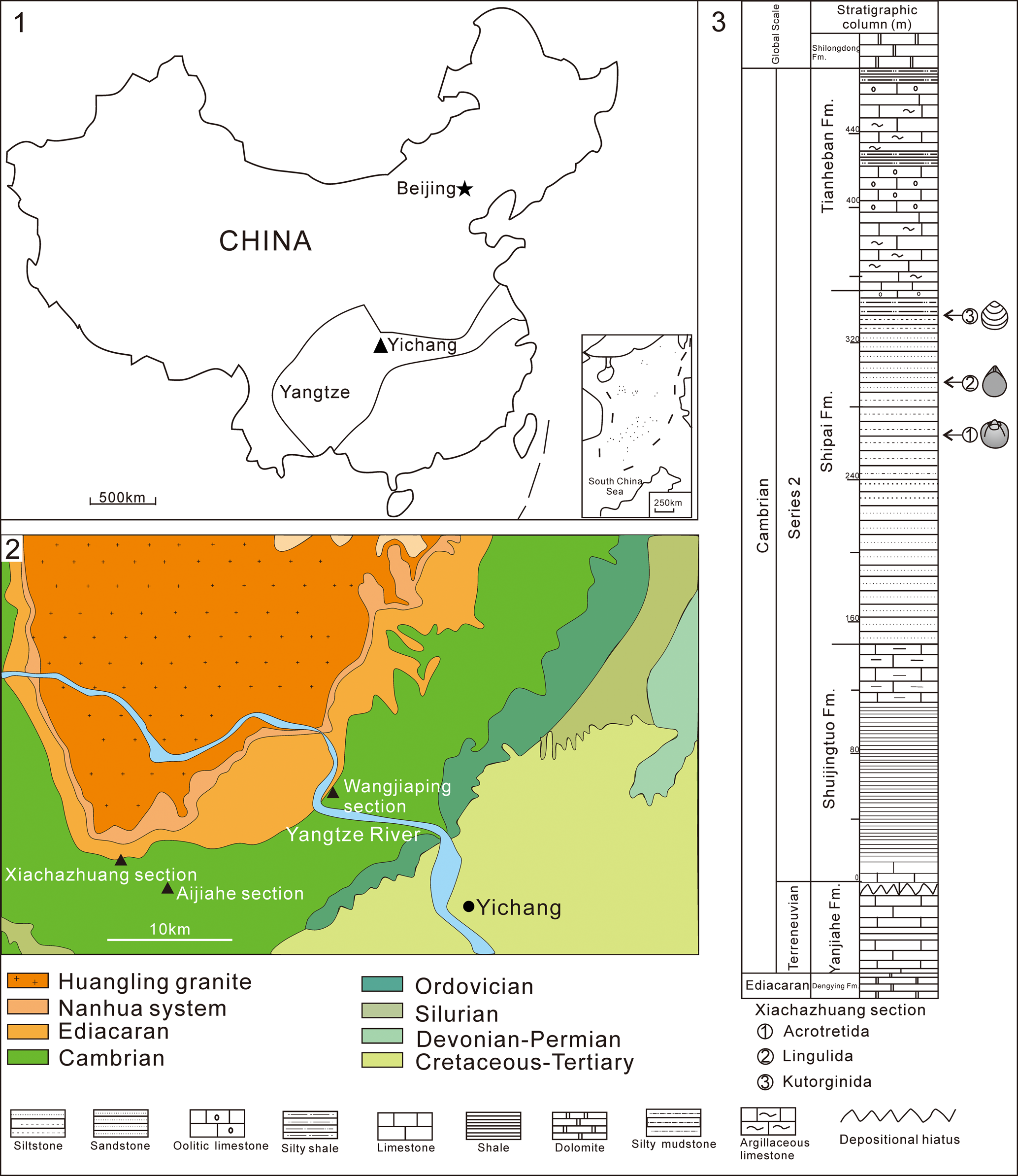
The Ediacaran Dengying Formation carbonates are disconformably overlain by Terreneuvian (Fortunian–Stage 2) lower Cambrian deposits. The lowermost Cambrian unit is the Yanjiahe Formation, containing abundant small shelly fossils (SSF) that are assigned to three SSF assemblage zones (in ascending order): the Anabarites trisulcatus-Protohertzina anabarica assemblage zone, the Purella antiqua assemblage zone, and the Aldanella yanjiaheensis assemblage zone (Guo et al., Reference Guo, Li, Han, Zhang, Zhang, Ou, Liu, Shu, Maruyama and Komiya2008, Reference Guo, Li and Li2014; Chang et al., Reference Chang, Feng, Clausen and Zhang2017, Reference Chang, Feng and Zhang2018; Steiner et al., Reference Steiner, Yang, Hohl, Zhang and Chang2020). The Shuijingtuo Formation (black shale and limestone) disconformably overlies the Yanjiahe Formation, and has yielded abundant and diverse shelly fossils, including brachiopods, in addition to the oldest eodiscoid trilobites in South China (Wang et al., Reference Wang, Ni, Zeng, Xu, Zhou, Li and Lai1987; Lin et al., Reference Lin2004; Steiner et al., Reference Steiner, Li, Qian, Zhu and Erdtmann2007; Dai and Zhang, Reference Dai and Zhang2011; Yang et al., Reference Yang, Steiner and Keupp2015; Z.F. Zhang et al., Reference Zhang, Zhang, Li and Holmer2016; Z.L. Zhang et al., Reference Zhang, Chen and Zhang2020). Conformably overlying the Shuijingtuo Formation is the Shipai Formation, which is dominated by yellow siltstone and grayish-yellow silty mudstone, intercalated by limestones. It is richly fossiliferous, including diverse trilobites, brachiopods, hyolithids, and bradoriids (Wang et al., Reference Wang, Ni, Zeng, Xu, Zhou, Li and Lai1987). The upper boundary of the Shipai Formation is marked by the contact with the argillaceous striped and oolitic limestone of the Tianheban Formation, which is itself conformably overlain by the dolomitic Shilongdong Formation (Fig. 1.3).
Materials and methods
Fossils were collected from the Shipai Formation in the Xiachazhuang, Aijiahe, and Wangjiaping sections, Three Gorges area, Hubei Province (Fig. 1). So far, >4500 individual valves have been collected from the Shipai Formation at Yichang by the work-team of the Early Life Institute (ELI), and all specimens are deposited in the Northwest University Early Life Institute, Xi'an, China. Fossils were examined under a Zeiss Smart Zoom 5 Stereo micrographic system and imaged with a Canon camera 5D Mark IV. Some specimens were analyzed with the Scanning Electron Microscope (SEM) at the State Key Laboratory of Continental Dynamics, Northwest University. When most acrotretoid specimens are cracked out, they are usually preserved as internal molds in the mudstone. In order to better display the structures, a number of latex casts were prepared with a PVB ethanol solution and latex. Some fossils and latex casts were photographed after coating with ammonium chloride (NH4Cl).
Building on the geometric morphometric work of Zhang et al (Reference Zhang, Holmer, Liang, Chen and Duan2020a), another 16 specimens from the Shipai Formation were selected for geometric morphometric analysis (Supplementary Data 1–3). Landmarks and semi-landmarks (Fig. 9) were digitized with the free software TpsDig2 v. 2.26 (Rohlf, Reference Rohlf2015). The data matrix was then analyzed using TpsRelw v. 1.65 (Rohlf, Reference Rohlf2015) to explore potential changes in morphospace and to visualize shell shape using thin plate splines. The interpretation of the Cambrian Stage 4 brachiopod faunal similarities was facilitated by multivariate cluster analysis (based on Raup-Crick similarity) (Supplementary Data 4), using the computer program PAST (version 3.06; Hammer et al., Reference Holmer, Popov, Koneva and Bassett2001).
Repository and institutional abbreviation
All fossil specimens examined in this study are housed in the following institution: The Northwest University Early Life Institute (ELI), Xi'an, China.
Systematic paleontology
Subphylum Linguliformea Williams et al., Reference Williams, Carlson, Brunton, Holmer and Popov1996
Class Lingulata Gorjansky and Popov, Reference Gorjansky and Popov1985
Order Acrotretida Kuhn, Reference Kuhn1949
Superfamily Acrotretoidea Schuchert, Reference Rowell and Caruso1893
Family Acrotretidae Schuchert, Reference Rowell and Caruso1893
Genus Linnarssonia Walcott, 1885
Type species
Original designation by Walcott (1885, p. 115); Obolella transversa Hartt in Dawson, Reference Dawson1868; middle Cambrian of New Brunswick, Canada.
Linnarssonia sapushanensis Duan et al., Reference Duan, Liang, Holmer and Zhang2021
Figures 2–5, 15–17, 20
- Reference Duan, Liang, Holmer and Zhang2021
Linnarssonia sapushanensis Duan et al., p. 41, figs. 2–4, 5.1–5.7, 6–8, 10, 11.
Ventral valves and latex cast of Linnarssonia sapushanensis from the lower Cambrian Shipai Formation at Xiachazhuang section. (1) Latex cast of ventral exterior, showing concentric growth lines on the shell surface (ELI QJP-SP-357-28); (2) ventral valve with concentric growth lines on the shell surface (ELI QJP-SP-289-7); (3, 4) internal molds (ELI QJP-SP-231, ELI QJP-SP-555-2); (5) internal view of ventral valve (ELI QJP-SP-041); (6) lateral view of (5); (7–9) internal molds showing intertrough (marked by arrows) (ELI QJP-SP-115, ELI QJP-SP-044). Scale bars = 1 mm (1–8), 500 μm (9).

Ventral valves and latex casts of Linnarssonia sapushanensis from the lower Cambrian Shipai Formation at Xiachazhuang section, and comparison to L. sapushanensis from the Wulongqing Formation (Guanshan fauna). (1) Internal mold (ELI QJP-SP-357-2); (2) latex cast of (1); (3) an enlargement of (2), showing the pedicle opening (marked by arrow); (4) internal mold (ELI QJP-SP-040); (5) close-up view of (4), note the mud-infilled internal pedicle tube; (6) one specimen of L. sapushanensis with the mud-infilled pedicle tube from the Wulongqing Formation, eastern Yunnan (ELI CLP-007-12); (7, 8) latex casts (ELI QJP-SP-357-30, ELI QJP-SP-357-30); (9) latex cast showing cardinal muscle scars (marked by arrows) and apical process with a median groove (marked by double arrows) (ELI QJP-SP-357-37); (10) latex cast showing apical process with a median groove (marked by double arrows) (ELI QJP-SP-357-9); (11) an enlargement of (8), note the latex cast of mud-infilled internal pedicle tube; (12) latex cast of L. sapushanensis from the Wulongqing Formation, eastern Yunnan (ELI CLP-183-30). Scale bars = 500 μm (1, 2, 4, 7, 8), or 200 μm (3, 5, 6, 9–12).

Dorsal valves and some relative latex casts of Linnarssonia sapushanensis from the lower Cambrian Shipai Formation at Xiachazhuang section. (1) Internal mold (ELI QJP-SP-120); (2) latex cast of (1); (3) latex cast (ELI QJP-SP-357-25); (4) lateral view of (1); (5) enlargement of (3), showing the cardinal muscle scars (marked by arrows); (6–9) latex casts (ELI QJP-SP-357-1, ELI QJP-SP-357-23, ELI QJP-SP-357-24, ELI QJP-SP-357-38); (10) close-up view of (6), showing the anterocentral muscle scars (marked by tailed arrows) and subtriangular platform-like swelling of the terminal portion of median septum (marked by double arrows). Scale bars = 1 mm (1–4, 6, 9), 500 μm (5, 8), or 200 μm (7, 10).

Schematic reconstruction of Linnarssonia sapushanensis from lower Cambrian Shipai Formation, showing location of measurements in Table 1. (1) Ventral interior; (2) dorsal interior; (3) lateral view of ventral valve; (4) lateral view of dorsal valve.

Holotype
A ventral internal mold (ELI CLP-007-12) from the Wulongqing Formation (Palaeolenus trilobite Zone, Cambrian Stage 4) in the Sapushan section at Wuding County, eastern Yunnan Province, China (Duan et al., Reference Duan, Liang, Holmer and Zhang2021, p. 41, fig. 2.8).
Description
Shell ventribiconvex, subcircular to transversely oval in outline (Fig. 2). Shell valves ornamented with concentric growth lines (Fig. 2.1, 2.2).
Ventral valve convex (Fig. 2.6), with a straight to slightly convex posterior margin, lateral and anterior margins moderately rounded (Fig. 2.3–2.5); ~90% as long as wide (Table 1), with the maximum width near to mid-valve; ventral pseudointerarea varies from catacline to procline, bisected by a slightly shallow intertrough (Fig. 2.7–2.9, marked by arrows). Ventral interior (Fig. 3.1–3.11) has an apical process, characterized by a median groove that slightly expands anteriorly (Fig. 3.9, 3.10, marked by double arrows); apical process occupying ~35% of the shell length (Table 1); the vascula lateralia are impressed as pronounced ridge-like imprints. Ventral pedicle foramen continued internally forming a pedicle tube; it is preserved as a cylindrical projection with muddy infilling (Fig. 3.4, 3.5, 3.8–3.11), which is ~80 μm in diameter. Apical pits unknown. Cardinal muscle scars oval in outline (Fig. 3.9), on posterolateral slopes of valve, occupying ~22% of the shell length and ~49% of the shell width (Table 1).
Main dimensions and ratios of ventral and dorsal valves of Linnarssonia sapushanensis from the lower Cambrian Shipai Formation in Three Gorges area. Abbreviations: V: ventral valve; D: dorsal valve; L, W: length and width of valve; La: length of ventral apical process; Lc, Wc: length and width of cardinal muscle scars; Ls: length of dorsal median septum. All measurements are in μm.

Dorsal valve subcircular in outline (Fig. 4), on average 89% as long as wide (Table 1), slightly convex in lateral profile (Fig. 4.4), with the maximum height near to the posterior one-fourth of shell length; dorsal pseudointerarea small and orthocline, characterized by a small rudimentary proparea and transversely elongate, subtriangular median groove. Dorsal valve interior with prominent median buttress; dorsal median septum well developed, starting directly anterior of median buttress and extends ~60% of the valve length, the terminal portion of the median septum forms a triangular platform-like swelling (Fig. 4.1–4.3, 4.7–4.10). Cardinal muscle scars are prominent and widely separated (Fig. 4.5), extending anterolaterally from the lateral edge of the median groove, occupying ~25% of the shell length and ~48% of the shell width (Table 1).
Materials
ELI QJP-SP-001-613, ELI AJH-SP-001-136. There are 749 slabs collected from the middle to upper part of the Shipai Formation in the Xiachazhuang and Aijiahe sections. However, the exact number of individual ventral and dorsal valves can only be approximated because many specimens overlap each other. As of now, 484 specimens have been examined and photographed.
Remarks
In the Shipai Formation, acrotretoid brachiopod shells are preserved as patchy aggregations on the bedding plane, while acrotretoids from the Wulongqing Formation form thicker shell beds (~11–13 pavements within 1 cm thick bed). Morphology of the specimens from the Shipai Formation is similar to L. sapushanensis Duan et al., Reference Duan, Liang, Holmer and Zhang2021 from the lower Cambrian Wulongqing Formation (Stage 4). Both taxa have a similar shell outline, catacline to procline ventral pseudointerarea, a pronounced dorsal median buttress, and cardinal muscle scars, as well as similar dimensions and ratios of key characters of the ventral valves (L. sapushanensis from the Wulongqing Formation: L/W = 89%, La/L = 34%, Lc/L = 21%; Duan et al., Reference Duan, Liang, Holmer and Zhang2021; data of the specimens from the Shipai Formation in Table 1, and location of measurements in Fig. 5).
Order Lingulida Waagen, Reference Waagen1885
Superfamily Linguloidea Menke, Reference Menke1828
Family Lingulellotretidae Koneva and Popov, Reference Koneva, Popov, Apollonov, Bandaletov and Ivshin1983
Genus Lingulellotreta Koneva and Popov, Reference Koneva, Popov, Apollonov, Bandaletov and Ivshin1983
Type species
Lingulellotreta ergalievi Koneva in Gorjansky and Koneva, Reference Gorjansky and Koneva1983, early Cambrian (Stage 4) Shabakty Group, Malyi Karatau, Kazakhstan.
Lingulellotreta ergalievi Koneva in Gorjansky and Koneva, Reference Gorjansky and Koneva1983
Figure 6
- Reference Koneva, Popov, Apollonov, Bandaletov and Ivshin1983
Lingulellotreta ergalievi Koneva in Gorjansky and Koneva, p. 132, figs. 1–8.
- Reference Holmer, Popov, Koneva and Rong1997
Lingulellotreta malongensis; Holmer et al., p. 581, fig. 4.1–4.14.
- Reference Li and Holmer2004
Lingulellotreta malongensis; Li and Holmer, p. 193, fig. 9.
- Reference Zhang, Zhang, Li and Holmer2016
Lingulellotreta malongensis; Z.F. Zhang et al., p. 347, fig. 10F.
The linguloid Lingulellotreta ergalievi from the lower Cambrian Shipai Formation at Xiachazhuang and Wangjiaping sections. (1) Ventral valve (Xiachazhuang section) (ELI QJP-SP-173); (2, 3) ventral valves (Wangjiaping section, from Zhang et al., Reference Zhang, Zhang, Holmer and Li2015) (ELI SPB-L002A, ELI SPB-L002B); (4) close-up view of (1) showing pseudointerarea; (5) enlargement of (2), showing the elongate oval foramen and well-developed pseudointerarea; (6) ventral valve (ELI QJP-SP-039); (7, 8) Elemental maps of (6) using micro X-ray fluorescence, showing the rich concentration of Ca and P on the conjoined shell valves. Scale bars = 1 mm (1–3, 6–8), or 500 μm (4, 5).

Holotype
A ventral valve interior, MIGSA (Museum of Geology, Institute of Geological Sciences, Almaty, Kazakhstan) from the early Cambrian (Stage 4) Shabakty Group, Malyi Karatau, Kazakhstan (Gorjansky and Koneva, Reference Gorjansky and Koneva1983, p. 132, pl. 28, fig. 1).
Description
Shell tear-shaped in outline (Fig. 6), ~142% as long as wide with maximum width anterior to mid-length; ventral valve length 4.50 mm and width 3.37 mm on average; ventral pseudointerarea orthocline and triangular (Fig. 6.4, 6.5), with well-developed flexure lines, occupying 75% of valve width and 37% of valve length; elongate oval pedicle foramen placed at posterior tip of pseudointerarea with average apical angle of 69°; foramen 0.22 mm wide on average, occupying 31% of the length and 9% of the width of the pseudointerareas; pedicle foramen usually preserved as a mud-infilled ridge or groove.
Shell surface bears weakly developed concentric growth lines. Shell shows a strong elemental abundance of Ca and P, compared with the surrounding rock in the μ-XRF study (Fig. 6.7, 6.8), suggesting that the original composition of the shell is calcium phosphate.
Materials
Eleven specimens (including fragments) collected from middle-upper part of the Shipai Formation at the Xiachazhuang and Wangjiaping sections.
Remarks
The first record of Lingulellotreta ergalievi Koneva in Gorjansky and Koneva, Reference Gorjansky and Koneva1983 was from the lower Cambrian Shabakty Group (Ushbaspis limbata Zone) of the Malyi Karatau Range, south Kazakhstan (Gorjansky and Koneva, Reference Gorjansky and Koneva1983). Holmer et al. (Reference Holmer, Popov, Koneva and Rong1997) restudied the material and made detailed morphological comparison to specimens described as “L.” malongensis (Rong, Reference Rong1974) by Jin et al. (Reference Jin, Hou and Wang1993) from the Chengjiang fauna in eastern Yunnan, and argued that “L.” malongensis from the Chengjiang fauna should be referred to Lingulellotreta, and therefore that L. malongensis be regarded as a senior synonym of L. ergalievi. Zhang et al. (Reference Zhang, Holmer, Liang, Chen and Duan2020a) compared brachiopods from the Chengjiang and Guanshan faunas, and referred the species in the Chengjiang Lagerstätte to Lingulellotreta yuanshanensis Zhang et al., Reference Zhang, Holmer, Liang, Chen and Duan2020a and the species from the Guanshan Biota to Eoobolus malongensis (Rong, Reference Rong1974). Zhang et al. (Reference Zhang, Holmer, Liang, Chen and Duan2020a) demonstrated that L. yuanshanensis from South China (Chengjiang fauna) differs from the Kazakhstan Lingulellotreta ergalievi in several characters, including the ratio of valve length and width, the apical angle, and the longer ventral pseudointerarea (Lp/L = 49% in L. yuanshanensis from Chengjiang fauna, South China; Lp/L = 34% in L. ergalievi from South Kazakhstan) (see measurements in Zhang et al., Reference Zhang, Holmer, Liang, Chen and Duan2020a).
However, specimens from the Shipai Formation described herein have striking similarities to L. ergalievi from the lower Cambrian Shabakty Group, Malyi Karatau Range of South Kazakhstan (Holmer et al., Reference Holmer, Popov, Koneva and Rong1997). These include an orthocline ventral valve pseudointerarea, as well as similar outlines and similarities in the ratios of the ventral valve (herein: L/W = 142%, Lp/L = 37%; Holmer et al., Reference Holmer, Popov, Koneva and Rong1997: L/W = 139%, Lp/L = 34% of ventral valves). Thus, the formerly so-called L. malongensis from the Shipai Formation at the Wangjiaping section (Zhang et al., Reference Zhang, Zhang, Holmer and Li2015) should be referred to L. ergalievi.
Family Eoobolidae Holmer, Popov, and Wrona, Reference Holmer, Popov, Wrona and Gaździcki1996
Genus Eoobolus Matthew, Reference Matthew1902
Type species
Obolus (Eoobolus) triparilis Matthew, Reference Matthew1902; middle Cambrian (Amgian, Bourinot Group), Cape Breton, Canada.
Eoobolus malongensis Rong, Reference Rong1974
Figures 7–10
- Reference Rong1974
Lingulepis malongensis Rong, p. 114, pl. 44, figs. 27, 32.
- non Reference Jin, Hou and Wang1993
Lingulepis malongensis (Rong); Jin et al., p. 794, figs. 5.1, 5.6, 5.7, 8.1–8.4, 9.4.
- non Reference Holmer, Popov, Koneva and Rong1997
Lingulellotreta malongensis (Rong); Holmer et al., p. 581, fig, 4.1–4.14.
- non Reference Holmer, Popov and Kaesler2000
Lingulellotreta malongensis (Rong); Holmer and Popov, p. H72, fig. 34, 1a–d.
- non Reference Zhang and Han2004
Lingulellotreta malongensis (Rong); Zhang et al., p. 4, figs. 1, 2.
- non Reference Hou, Aldridge, Bergström, Siveter, Siveter and Feng2004
Lingulellotreta malongensis (Rong); Hou et al., p. 182, fig. 17.3.
- ?Reference Li and Holmer2004
Eoobolus aff. viridis (Cobbold, Reference Cobbold1921); Li and Holmer; p. 197, figs. 6, 7.
- non Reference Li and Holmer2004
Lingulellotreta malongensis (Rong); Li and Holmer, p. 199, fig. 9.
- non Reference Zhang and Hua2005
Lingulellotreta malongensis (Rong); Zhang et al., p. 279, figs. 1f–h, 2f, g, i, j, 3b–j.
- non Reference Zhang, Han, Zhang, Liu, Guo and Shu2007a
Lingulellotreta malongensis (Rong); Zhang et al., p. 65, figs. 1–3.
- non Reference Zhang, Robson, Emig and Shu2008
Lingulellotreta malongensis (Rong); Z.F. Zhang et al., p. 243, figs. 4k–n, 6a.
- non Reference Liu, Ou, Han, Zhang, He, Yao, Fu and Shu2012
Lingulellotreta malongensis (Rong); Liu et al., p. 127, fig. 2g.
- non Reference Hu, Zhu, Luo, Steiner and Zhao2013
Lingulellotreta malongensis (Rong); Hu et al., p. 146, fig. 193.
- ?Reference Zhang, Zhang, Holmer and Li2015
Eoobolus sp.; Zhang et al., p. 175, fig. 6.
- ?Reference Zhang, Zhang, Li and Holmer2016
Eoobolus aff. viridis (Cobbold, Reference Cobbold1921); Z.F. Zhang et al., p. 347, fig. 10a–e.
- Reference Zhang, Holmer, Liang, Chen and Duan2020a
Eoobolus malongensis (Rong); Zhang et al., p. 21, figs. 2–4.
Ventral valves of the linguloid Eoobolus malongensis from the lower Cambrian Shipai Formation at Xiachazhuang section. (1) Shell concentrations (ELI QJP-SP-069); (2) ventral valve with concentric growth lines on the shell surface (ELI QJP-SP-163); (3, 4) ventral valves, (ELI QJP-SP-070, ELI QJP-SP-075); (5, 6) element maps of (4) investigated by micro X-ray fluorescence; (7, 8) close-up view of (4) and (3), respectively, showing pedicle groove (Pg) and ‘U’ shaped impression of pedicle nerve (Pn). Scale bars = 3 mm (1); or 1 mm (2–6); or 500 μm (7, 8).

Dorsal valves of the linguloid Eoobolus malongensis from the lower Cambrian Shipai Formation at the Xiachazhuang section. (1) Dorsal valve with unambiguous and faint concentric growth lines on the shell surface (ELI QJP-SP-119); (2–5) dorsal valves with variable imprints of mantle canals (ELI QJP-SP-130, ELI QJP-SP-069-2, ELI QJP-SP-216-2, ELI QJP-SP-105) (marked by double arrows); (6) close-up view of (5) showing the triangular dorsal pseudointerarea with pronounced median groove (Mg, marked by arrow) and lateral propareas as ill-defined flexure lines. Scale bars = 1 mm (1–6).

(1, 2) Definition of landmarks (marked by black circles) and semi-landmarks (marked by red circles).

Plots for RW 1–2 and RW 1–3 of the relative warp analysis, with visualized shape of thin-plate splines within RW morphospace, showing the similarities of specimens of Eoobolus from the Guanshan fauna of eastern Yunnan (Eo-GS) with those from the Shipai Formation in Yichang area (Eo-SP), and signifying their assignment to Eoobolus malongensis (see Zhang et al., Reference Zhang, Holmer, Liang, Chen and Duan2020a).

Neotype
The holotype is unfortunately lost, but was formerly housed in the Nanjing Institute of Geology and Palaeontology (NIGP 22154). Recently, a neotype was selected (ELI-CLP 012) from the Wulongqing Formation, Malong County, eastern Yunnan Province, China (Zhang et al., Reference Zhang, Holmer, Liang, Chen and Duan2020a, p. 4, fig. 2A, B).
Description
Shell ventro-biconvex, tear-shaped to elongate sub-triangular in outline (Figs. 7, 8), Shell valves ornamented with concentric growth lines (Fig. 7.2). Ventral valve acuminate (Fig. 7.2–7.6), with apical angle ~78° on average; ventral valve length 3.23 mm and width 2.45 mm on average (Table 3), with the maximum width anterior to mid-valve; ventral pseudointerarea triangular, close to orthocline, occupying 37% of valve length and 75% of valve width. Pedicle groove deep with parallel lateral margins, infilled and preserved as parallel-sided ridge; ~0.7 mm in length and ~0.2 mm in width, and extending anteriorly to ~20% of total valve length. Ventral visceral area with a ‘U’-shaped impression of pedicle nerve extending to one-third valve length (Fig. 7.7, 7.8).
Main dimensions and ratios of ventral valve of Lingulellotreta ergalievi from the lower Cambrian Shipai Formation in Three Gorges area. Abbreviations: V: ventral valve; Lp, Wp: length and width of ventral pseudointerarea; A: apical angle; All measurements are in μm.

Main dimensions and ratios of ventral and dorsal valves of Eoobolus malongensis from the lower Cambrian Shipai Formation in Three Gorges area. Abbreviations: V: ventral valve; D: dorsal valve; L, W: length and width of valve; Lp, Wp: length and width of pseudointerarea; A: apical angle. All measurements are in μm.

Dorsal valve sub-oval in outline (Fig. 8); dorsal valve length 3.00 mm and width 2.36 mm on average; dorsal pseudointerarea with broad median groove and narrow propareas (Fig. 8.6), occupying 22% of valve length and 72% of valve width; median tongue extending to 70% of valve length.
Materials
Forty-eight specimens (including fragments) from middle-upper part of the Shipai Formation in the Xiachazhuang section.
Remarks
Specimens from the Shipai Formation bear a strong resemblance to Eoobolus malongensis, described by Zhang et al (Reference Zhang, Holmer, Liang, Chen and Duan2020a) from the Cambrian Stage 4 Wulongqing Formation, Yunnan Province. Eoobolus malongensis from the Shipai Formation and Wulongqing Formation both have similar size ratios of several different morphologic characters in the ventral valve (data from the Wulongqing Formation specimens: Aa = 74°, Lp/L = 42%, Wp/W = 78%, Lpg/L = 18%; Zhang et al., Reference Zhang, Holmer, Liang, Chen and Duan2020a; data of the specimens from the Shipai Formation in the Table 3). Relative warp analysis demonstrates strong similarities between Eoobolus from the Shipai and Wulongqing formations (Fig. 10), strengthening their taxonomic assignment to Eoobolus malongensis (Zhang et al., Reference Zhang, Holmer, Liang, Chen and Duan2020a).
Eoobolus malongensis from the Shipai Formation can be distinguished from most of the species assigned to the genus in having a relatively narrow ventral pseudointerarea and a short pedicle groove with parallel lateral margins. It is also difficult to recognize any pustulose ornamentation on the postmetamorphic shell surface. However, comparison of shells from siliciclastic deposits to those acid-etched from carbonates is commonly a problem because taphonomic factors tend to alter morphological characters.
Family Neobolidae Walcott and Schuchert in Walcott, Reference Walcott1908
Neobolidae gen. indet. sp. indet.
Figure 11
Description
Shell subcircular in outline (Fig. 11.1, 11.2), ~8.9 mm in length and ~9.7 mm in width; ~91% as long as wide. Dorsal median septum well developed, extending to mid-valve or two-thirds of valve length. Shell surface may have setae (marked by white arrow in SEM image, Fig. 11.3). Shell surface bears closely spaced concentric growth lines and dark speckled marks (Fig. 11.4), which are likely to be diagenetic mineral deposits with high concentration of Fe (Fig. 11.5); shell shows a strong elemental abundance of Ca, P, and S compared with the surrounding rock in the μ-XRF study (Fig. 11.6–11.8)
Neobolidae gen. indet. sp. indet. from the lower Cambrian Shipai Formation at Xiachazhang section. (1, 2) Part and counterpart of Neobolidae gen. indet. sp. indet. with prominent dorsal median septum (marked by arrow) (ELI QJP-SP-001A, ELI QJP-SP-001B); (3) SEM image of (1) marked by the inset box, showing possible setae (marked by white arrow); (4) close-up view of (1) showing concentric growth lines of the shell surface; (5–8) micro-XRF mapping, showing the rich content of Fe on the shell dark speckled marks (5) and the concentration Ca, P, and S on the shell (6–8). Scale bars = 3 mm (1, 2, 5–8), or 1 mm (3, 4).

Materials
Three dorsal valves from the middle-upper part of the Shipai Formation in the Xiachazhuang section.
Remarks
Specimens from the Shipai Formation show a distinctive surface ornamentation with dense, regular concentric fila, and have a prominent dorsal median septum that indicates affinities with the Neobolidae. It is most similar to Neobolus wulongqingensis Zhang, Strotz, Topper, and Brock in Zhang et al., Reference Zhang, Strotz, Topper, Chen, Chen, Liang, Zhang, Skovsted and Brock2020b from the lower Cambrian Wulongqing Formation, eastern Yunnan (Zhang et al., Reference Zhang, Strotz, Topper, Chen, Chen, Liang, Zhang, Skovsted and Brock2020b), but without data on the ventral valve and more abundant materials, the discrimination of the material remains uncertain. Hence the specimens are referred to Neobolidae gen. indet. sp. indet. awaiting new data.
Subphylum Rhynchonelliformea Williams et al., Reference Williams, Carlson, Brunton, Holmer and Popov1996
Class Kutorginata Williams et al., Reference Williams, Carlson, Brunton, Holmer and Popov1996
Order Kutorginida Kuhn, Reference Kuhn1949
Superfamily Nisusioidea Walcott and Schuchert in Walcott, Reference Walcott1908
Family Nisusiidae Walcott and Schuchert in Walcott, Reference Walcott1908
Genus Nisusia Walcott, Reference Walcott1905
Type species
By original designation Orthisina festinata Billings, Reference Billings1861, from unnamed Cambrian Stage 4 (Bonnia-Olenellus Zone) of USA and Canada.
Nisusia liantuoensis Zeng in Wang et al., Reference Wang, Ni, Zeng, Xu, Zhou, Li and Lai1987
Figure 12
- Reference Zeng and Wang1987
Nisusia liantuoensis Zeng in Wang et al., p. 213, pl. 9, figs. 13–16.
- Reference Zhang, Liu and Zhao2008
Nisusia liantuoensis; Z.F. Zhang et al., p. 243, fig. 2c.
Nisusia liantuoensis from the lower Cambrian Shipai Formation at Xiachazhuang section. (1) Posterior view of ventral valve (ELI QJP-SP-015); (2, 3) ventral valves (ELI QJP-SP-045, ELI QJP-SP-006); (4) close-up view of (1) showing the apical foramen (fo, marked by arrow), developed pseudointerarea, deltidium (de, marked by arrow), and posterior median opening (marked by double arrows); (5) an enlargement of (2), showing the pedicle foramen (marked by arrow); (6) a fragment of one ventral valve, showing the radial lines on the shell surface (ELI QJP-SP-045); (7–9) dorsal valves (ELI QJP-SP-037, ELI QJP-SP-013, ELI QJP-SP-008). Scale bars = 3 mm (1, 7–9), 2 mm (2), 4 mm (3), or 1 mm (4–6).

Holotype
A ventral valve (LSP11-IV45951) from the Shipai Formation (unnamed Cambrian Series 2) of Liantuo, Yichang City, western Hubei Province, South China (Wang et al., Reference Wang, Ni, Zeng, Xu, Zhou, Li and Lai1987, pl. 9, fig. 15).
Emended diagnosis
Shell subequally biconvex, semicircular to transverse-oval in outline; hinge line slightly shorter than or equal to the maximum width. Cardinal extremities slightly obtuse to almost rectangular. Ventral valve moderately convex, ventral umbo strongly raised; ventral interarea high, catacline. Dorsal valve with anacline interarea. Radial ornament with 6–11 ribs per 5 mm, rib crests bearing hollow spines.
Description
Shell biconvex, sub-circular to transverse, sub-rectangular in outline, length about three-quarters of width. Hinge line equal to or slightly shorter than maximum shell width (~95% of the maximum width). Cardinal extremities form right angles. Shell surface bears numerous fine radial ribs that bifurcate in the adult phase to ~6–11 costae per 5 mm along the anterior margin.
Ventral valve semicircular or transversely sub-rectangular in outline, ~67% as long as wide; ventral valve moderately convex with the maximum height at the apex; apex pointed and raised, perforated by a round suproapical foramen (~0.54 mm in diameter) (Fig. 12.4, 12.5, marked by arrows). Ventral interarea high, with a triangular pseudodelthyrium occupying about one-third of interarea width (Fig. 12.4). Shell bears prominent radial lines and vague concentric lines; rib crests bearing hollow spines.
Dorsal valve subquadrate, ~78% as long as wide, with a small swelling at the umbo (Fig. 12.7–12.9); apex recurved toward the posterior. Ornament on valve surface consists of radial costae and fine, closely set, concentric growth lines. Ventral and dorsal interior not observed.
Materials
Seven ventral valves and five dorsal valves from the upper part of the Shipai Formation in the Xiachazhuang section. In addition, >10 specimens were also collected from the Shipai Formation from the Xiachazhuang section, but it is difficult to distinguish ventral or dorsal valves because all are incomplete shells.
Remarks
Nisusia liantuoensis was first recorded from the Shipai Formation of Liantuo, Yichang City, South China (Zeng, Reference Zeng and Wang1987). Spinose ornament is unclear in Zeng (Reference Zeng and Wang1987), hence Holmer et al. (Reference Holmer, Kebria-ee Zadeh, Popov, Ghobadi Pour, Álvaro, Hairapetian and Zhang2019) suggested this species designation may be questionable. Specimens from the Shipai Formation at Xiachazhuang section bear a strong resemblance to N. liantuoensis from Liantuo (shell subequally biconvex, semicircular in outline; ventral umbo strongly raised; ventral interarea high; the maximum height of the ventral valve at apex), and the new material from Xiachazhuang section also preserves the characteristic hollow spines of Nisusia.
Specimens from the Shipai Formation in the Xiachazhuang section are also similar to Nisusia grandis Roberts and Jell, Reference Roberts and Jell1990, from the Coonigan Formation (Wuliuan Stage) of western New South Wales; both have well-defined concentric lamellae and ventral valve interareas. But the new material differs from Nisusia grandis in having a rectimarginate anterior commissure and in lacking a ventral sulcus.
As discussed by Holmer et al. (Reference Holmer, Zhang, Topper, Popov and Claybourn2017, Reference Holmer, Popov, Pour, Claybourn, Zhang, Brock and Zhang2018), Nisusia has two pedicle openings, an apical foramen and a posterior median opening (between the delthyrium and notothyrium). Nisusia liantuoensis from the Shipai Formation shows a well-developed apical opening (Fig. 12.4, 12.5, marked by arrows) and bears a posterior median opening (Fig. 12.4, marked by double arrows). The new material is also similar to Nisusia sulcata Rowell and Caruso, Reference Rowell and Caruso1985, from the Marjum Formation (Drumian) of western Utah, USA (Holmer et al., Reference Holmer, Popov, Pour, Claybourn, Zhang, Brock and Zhang2018, fig. 1B, E). However, N. liantuoensis differs in having the maximum height of the ventral valve at the apex rather than at the central part of the valve.
Superfamily Kutorginoidea Schuchert, Reference Schuchert1893
Family Kutorginidae Schuchert, Reference Schuchert1893
Genus Kutorgina Billings, Reference Billings1861
Type species
Kutorgina cingulate Billings, Reference Billings1861, from lower Cambrian of Labrador, Canada.
Remarks
Kutorgina holds special significance as it is one of the oldest brachiopods with a carbonate shell and primitive articulation. Kutorgina had a cosmopolitan distribution during the early to middle Cambrian (Malakhovskaya, Reference Malakhovskaya2013), and has been recovered from China (Lu, Reference Lu1979; Zhang et al., Reference Zhang, Shu, Emig, Zhang, Han, Liu and Guo2007b; Liu et al., Reference Liu, Zhao, Liu and Mao2015), Canada (Voronova et al., Reference Voronova, Drozdova, Esakova, Zhegallo, Zhuravlev, Rozanov and Ushatinskaya1987), America (Nevada) (Walcott, Reference Walcott1905), Greenland (Skovsted and Holmer, Reference Skovsted and Holmer2005), Siberia (Gorjansky and Popov, Reference Gorjansky and Popov1985), Kazakhstan (Koneva, Reference Koneva1979), Kyrgyzstan (Popov and Tikhonov, Reference Popov and Tikhonov1990), and southeast Australia (Roberts and Jell, Reference Roberts and Jell1990). The wide geographic distribution of Kutorgina in the late early Cambrian may indicate that the larvae of Kutorgina were planktotrophic (Popov et al., Reference Popov, Holmer, Rowell and Peel1997). Species of Kutorgina have few distinctive characters, and morphological features vary throughout ontogeny. The kutorginides may be easily recognized by the wide posterior margin, coarse external concentric ornamentation of sharp rugae and ridges, and growth lines following the valve outline (Malakhovskaya, Reference Malakhovskaya2013). Specimens from the Shipai Formation in the Three Gorges area with coarse, wide-spaced concentric growth lines clearly belong in Kutorgina.
Kutorgina sinensis Rong in Lu, Reference Lu1979
Figure 13
- Reference Lu1979
Kutorgina sinensis Rong in Lu, p. 72, pl. v, figs. 9–11.
- Reference Zeng and Wang1987
Iphidella? liantuoensis; Zeng, p. 213, pl. 8, figs. 14–18.
Kutorgina sinensis from the lower Cambrian Shipai Formation at Xiachazhuang section. (1) Ventral valve (ELI QJP-SP-007); (2) lateral view of (1), note the distance between growth lines (marked by double-pointed arrow); (3–5) ventral valves (ELI QJP-SP-013, ELI QJP-SP-076, ELI QJP-SP-078); (6, 7) dorsal valves (ELI QJP-SP-014, ELI QJP-SP-012); (8) close-up view of (7) showing small umbo located posterior of the posterior margin; (9) SEM image of (1) showing pyrite crystals; (10) close-up view of (9). Scale bars = 5 mm (1–4, 6–8), 3 mm (5), 100 μm (9), or 10 μm (10).

Holotype
A ventral valve from the lower Cambrian Xinji Formation, Shuiyu section, Ruicheng, Shanxi (Lu, Reference Lu1979, pl. v, fig. 9).
Emended diagnosis
Shell sub-trapezoid, with rounded anterior and lateral margins; shell width is somewhat shorter than shell length, hinge line about three-fifths of the shell width. Ventral valve moderately convex, interarea apsacline; umbo slightly raised over the posterior margin; sulcus narrow, shallow, and starts from the postmedian part of the valve. The ornamentation consists of concentric growth lamellae (~18–20 lamellae over the entire shell).
Description
Shell biconvex, rounded to sub-pentagonal. Shell surface ornamented with coarse, widely spaced concentric growth lamellae that are best developed on the postmedian part of the valve, no visible prominent micro-ornamentation. The distance between growth lamellae is 0.64 mm on average. Ventral valve rounded to sub-pentagonal in outline (Fig. 13.1–13.5), with rounded anterior and lateral margins. The ratio of shell length to width ranges from 0.84–1.10 (average 0.92). Dorsal valve moderately convex to semicircular in outline (Fig. 13.6–13.8), ~73% as long as wide. Umbo small and slightly raised over the posterior margin (Fig. 13.8). SEM shows external shell with pyrite crystals (Fig, 13.9, 13.10). No information on the internal morphology is preserved.
Materials
Thirty-four specimens comprising 14 ventral valves, six dorsal valves, and fragments, all from the upper part of the Shipai Formation at Xiachazhuang section.
Remarks
The holotype of Kutorgina sinensis Rong in Lu, Reference Lu1979 from the lower Cambrian Xinji Formation in North China was illustrated by Lu (Reference Lu1979), but was not described in detail. Figured material from Lu (Reference Lu1979) shows that the ventral valve is moderately convex, ~13 mm wide, with an apsacline interarea. New material from the Shipai Formation is similar to K. sinensis Rong in Lu, Reference Lu1979 from the Xinji Formation (Lu, Reference Lu1979). Both have a moderately convex ventral valve, similar shell size (shell width of Shipai Formation specimens ranges from 6–14 mm), as well as concentric growth lamellae (ranging from 18–20 lamellae). However, K. sinensis from the Xinji Formation is represented by a single ventral valve with no information about the morphology of the dorsal valve, so comparisons of these features is difficult.
Kutorgina sinensis from the Shipai Formation bears some similarities with K. chengjiangensis Zhang et al., Reference Zhang, Shu, Emig, Zhang, Han, Liu and Guo2007b from the Yu'anshan Formation of South China (Zhang et al., Reference Zhang, Shu, Emig, Zhang, Han, Liu and Guo2007b), which is also recovered as “crack-outs” from siliciclastic deposits. Both have strong concentric growth lamellae, as well as other closely comparable morphology, such as shell size (K. chengjiangensis: L = 9.70 mm, W = 11.12 mm on average, data from Zhang et al., Reference Zhang, Shu, Emig, Zhang, Han, Liu and Guo2007b; K. sinensis: L = 9.40 mm, W = 11.02 mm on average), and the distance between growth lines (K. chengjiangensis: 0.6–0.8 mm; K. sinensis: ~0.64 mm on average). However, K. sinensis from the Shipai Formation has a more convex and acuminate ventral valve as compared with K. chengjiangensis.
In addition, Kutorgina chengjiangensis from the Chengjiang Lagerstätte features a stout and annulated pedicle previously described as protruding from between the delthyrium and notothyrium (Zhang et al., Reference Zhang, Shu, Emig, Zhang, Han, Liu and Guo2007b). However, recent reexamination shows that the pedicle emerges from the apical foramen (Holmer et al., Reference Holmer, Popov, Pour, Claybourn, Zhang, Brock and Zhang2018). Unfortunately, K. sinensis from the Shipai Formation are preserved as exterior molds without soft tissues, which precludes detailed study of the pedicle morphology. Additionally, study of the apical foramen is problematic due to poor preservation of the ventral apex.
Kutorgina sp.
Figure 14
Description
Shell planoconvex or slightly biconvex, up to 14 mm wide; surface ornamented by concentric growth lines. No median sulcus or fold developed in the shell valve. Ventral valve transversely oval or semicircular with rounded anterior and lateral margins. Posterior of ventral valve not well preserved. Dorsal valve almost flat and slightly convex, ~75% as long as wide; posterior margin almost straight, and slightly shorter than the maximum shell width (located in the middle of the shell). No information on the internal morphology is preserved.
Kutorgina sp. from the lower Cambrian Shipai Formation at Xiachazhuang section. (1–3) Ventral valves (ELI QJP-SP-065, ELI QJP-SP-035, ELI QJP-SP-017); (4) dorsal valve of Kutorgina sp. (ELI QJP-SP-032, marked by arrow) and an fragment of K. sinensis (marked by double arrows); (5, 6) dorsal valves (ELI QJP-SP-049, ELI QJP-SP-074); (7) close-up view of (6), showing the concentric growth lines. Scale bars = 5 mm (1, 2, 4–6), 2 mm (3), or 1 mm (7).

Materials
Seven specimens comprising three ventral valves and four dorsal valves, all from the upper part of the Shipai Formation in the Xiachazhuang section.
Remarks
Kutorgina sp. has a similar shell size as K. sinensis, but it can be distinguished from K. sinensis by the ornamentation and shell shape. Kutorgina sinensis is ornamented with concentric growth lamellae (~5–7 lamellae per 5 mm), while Kutorgina sp. has 10–15 concentric growth lines per 5 mm. Kutorgina sp. is similar to K. reticulata Poulsen, Reference Poulsen1932 (Skovsted and Holmer, Reference Skovsted and Holmer2005) in having transversely oval or semicircular outline, almost flat dorsal valve with a straight posterior margin, and shell surface with concentric growth lines. But it differs in lacking developed dorsal median fold and ventral median sulcus. Further detailed comparison is difficult due to insufficient material available for the study.
Brachiopod assemblages from the Shipai Formation
Xiachazhuang section
Brachiopod assemblages from the Xiachazhuang section are much more abundant and diverse compared to those from the Aijiahe and Wangjiaping sections. Six hundred thirteen slabs with >4000 Linnarssonia sapushanensis valves have been recovered from the Shipai Formation in the Three Gorges area. Most fossils were collected from the silty mudstone and siltstone in the middle to upper part of the Shipai Formation, ~120 m above the base. Here, Linnarssonia sapushanensis are commonly aggregated as shell concentrations on the same bedding plane (Fig. 15.1–15.4). These shell beds range from loosely to densely packed (~18 valves per 1 cm2) (Fig. 15.1) with moderate degrees of fragmentation. In the L. sapushanensis shell beds in the Xiachazhuang section, the size frequency distribution of 103 shells shows that individuals with shell widths between 1.23–2.10 mm are the most frequent (up to 86%) (Fig. 16). The orientation angle of the shells of L. sapushanensis was also statistically analyzed and plotted in a rose diagram, showing that they have random orientations (Fig. 16). Many shells retain well-preserved microstructures.
Acrotretoid brachiopod shell concentrations of Linnarssonia sapushanensis from the lower Cambrian Shipai Formation at Xiachazhuang section of Hubei Province, and comparison to shell beds of L. sapushanensis from the Wulongqing Formation of Yunnan Province. (1–4) Acrotretoid shell concentrations from the Shipai Formation; (1) shell valves aggregated as high-density concentrations on the bedding plane (ELI QJP-SP-289), with inset box indicating the position of (2) and grid in upper left used to count the number of shells in 1 cm2; (2) close-up view of (1) showing acrotretoid shell valves of different sizes distributed on the bedding plane; (3) acrotretoid shell bed (ELI QJP-SP-357); (4) close-up view of (3) marked by an inset box, showing the acrotretoid shell valves distributed at different micro-layers of bedding planes (marked by white arrows); (5) multi-layered, high-density shell beds from Wulongqing Formation packed up to 2 cm thick (ELI SJJ-164); (6) longitudinally polished section of (5), showing frequent occurrences of the acrotretoid shell valves, aggregated approximately as 11–13 pavements within 1 cm thick muddy sediment; (7) micro-XRF mapping of (6), showing the rich content of Fe within the acrotretoids. Scale bars = 1 cm (1, 3, 5–7), 3 mm (2), 4 mm (4).

Size frequency distribution and rose diagram of Linnarssonia sapushanensis from the lower Cambrian Shipai Formation at Xiachazhuang section, Three Gorges area, South China.

Linguloid brachiopods are quite common in the Shipai Formation, and two species have been recognized: Lingulellotreta ergalievi and Eoobolus malongensis. The majority of the linguloid specimens were collected from the siltstone in the middle-upper part of the Shipai Formation, ~150 m above the base of the Shipai Formation (Fig. 1.3). Eoobolus malongensis, which is the most common species in this unit, is preserved as individuals or shell concentrations (Figs. 7, 8). All specimens of E. malongensis are flattened and compressed, but retain some vague concentric growth lines. Overall morphology, including the pseudointerarea of the specimens illustrated herein, is similar to the linguloid brachiopods from the Guanshan fauna (Wulongqing Formation) (Zhang et al., Reference Zhang, Holmer, Liang, Chen and Duan2020a). In the Shipai Formation, Lingulellotreta ergalievi is relatively rare, has a longer ventral pseudointerarea than Eoobolus malongensis, and has an elongate, oval-shaped pedicle foramen. In this unit, Eoobolus and Lingulellotreta are usually <5 mm wide and long. However, the Eoobolus-yielding level in the Shipai Formation also includes larger macro-morphic brachiopods, generally ~10 mm wide (8.9 mm in length and 9.7 mm in width) (Fig. 11).The shell surface of the macro-morphic brachiopods bears closely spaced concentric growth lines, and a prominent dorsal median septum is present (Fig. 11.2). The specimens (Fig. 11) are most similar to brachiopods belonging to Neobolidae, but the limited material precludes more robust taxonomic discrimination.
In the top silty shale of the Shipai Formation, ~200 m above the base of the Shipai Formation (Fig. 1.3), the fauna is dominated by calcareous-shelled kutorginates (Nisusia liantuoensis, Kutorgina sinensis, and Kutorgina sp.). All specimens of Kutorgina have a distinctive shell ornamentation, and are sub-pentagonal or semicircular with strongly spaced concentric growth lines on the surface of the shell. Nisusia has prominent radial lines and has strikingly similar morphology to those from the Wulongqing Formation (Guanshan fauna), eastern Yunnan (Hu et al., Reference Hu, Zhu, Luo, Steiner and Zhao2013; Li et al., Reference Li, Zhang, Rong, Liu, Rong, Jin, Shen and Zhan2017). In South China, the first appearance datum (FAD) of the rhynchonelliform Nisusia is in the upper silty shale of the Shipai and Wulongqing formations.
Aijiahe and Wangjiaping sections
The Wangjiaping section is the type section of the Shipai fauna (Zhang and Hua, Reference Zhang and Hua2005) and is exposed around the northern bank of the Yangtze River near Wangjiaping Village, ~40 km west of Yichang City (Fig. 1.2). Presently, the Shipai Formation at the Wangjiaping section is poorly exposed and mostly covered, making new collections difficult. Linguloid brachiopods such as Palaeobolus, Eoobolus, and Lingulellotreta have been reported from the argillaceous siltstone and silty mudstone in the middle part of the Shipai Formation in the Wangjiaping and Aijiahe sections (Zhang et al., Reference Zhang, Zhang, Holmer and Li2015). The brachiopod assemblage in the Aijiahe section consists mainly of acrotretoid brachiopods, which is similar to that from the Xiachazhuang section. Acrotretoids collected from the silty mudstone in the middle-upper part of the Shipai Formation in the Aijiahe section are usually preserved as individuals or shell concentrations (brachiopod-trilobite) (Fig. 17). Four specimens of Eoobolus malongensis have been recovered from the brick-red silty mudstone in the top of the Shipai Formation in the Aijiahe section. All the individuals of E. malongensis were preserved as flattened internal molds with similar color to the surrounding muddy matrix.
The acrotretoid brachiopod of Linnarssonia sapushanensis and fragmental trilobite Palaeobolus liantuoensis from the lower Cambrian Shipai Formation at Aijiahe section, Three Gorges area, South China. (1–3) Acrotretoids (marked by white arrows) with fragmental trilobites distributed on the bedding plane (ELI AJH-SP-130, ELI AJH-SP-119, ELI AJH-SP-110); (4, 5) ventral valves (ELI AJH-SP-110-1, ELI AJH-SP-170); (6, 7) dorsal valves (ELI AJH-SP-109, ELI AJH-SP-097). Scale bars = 4 mm (1), 2 mm (2), 6 mm (3), or 500 μm (4–7).

Discussion
Early Cambrian brachiopod assemblages in South China
Brachiopod faunas from the lower Cambrian Shuijingtuo Formation in the Three Gorges area include four linguloids (Spinobolus popovi Zhang and Holmer in Z.F. Zhang et al., Reference Zhang, Li, Holmer, Brock, Balthasar, Skovsted, Fu, Zhang, Wang, Butler, Zhang, Cao, Han, Liu and Shu2016, Eoobolus sp., Lingulellotreta ergalievi, and Palaeobolus? liantuoensis Zeng, Reference Zeng and Wang1987), one botsfordiid (Botsfordiidae gen. indet. sp. indet.), and two acrotretoids (Eohadrotreta zhenbaensis Li and Holmer, Reference Li and Holmer2004 and Palaeotreta zhujiahensis Li and Holmer, Reference Li and Holmer2004) (Z.F. Zhang et al., Reference Zhang, Li, Holmer, Brock, Balthasar, Skovsted, Fu, Zhang, Wang, Butler, Zhang, Cao, Han, Liu and Shu2016; Z.L. Zhang et al., Reference Zhang, Chen and Zhang2020). Notably, Palaeotreta from the base of the Shuijingtuo Formation in the Xiaoyangba section of southern Shaanxi Province is the oldest acrotretoid known from the carbonate deposits in South China (Li and Holmer, Reference Li and Holmer2004; Z.F. Zhang et al., Reference Zhang, Zhang, Li and Holmer2016; Z.L. Zhang et al., Reference Zhang, Zhang, Li and Holmer2016, Reference Zhang, Popov, Holmer and Zhang2018a, Reference Zhang, Zhang, Holmer and Chenb, Reference Zhang, Chen and Zhang2020). They are typified by lacking both an internal pedicle tube and apical pits in the ventral valve interior. The overlying Shipai Formation contains linguloids (Palaeobolus liantuoensis, Lingulellotreta ergalievi, Eoobolus malongensis, and Neobolidae gen. indet. sp. indet.), an acrotretoid (Linnarssonia sapushanensis) and calcareous shelled Kutorginates (Nisusia liantuoensis, Kutorgina sinensis, Kutorgina sp.) (Fig. 18). Acrotretoids (represented by Linnarssonia sapushanensis) are numerically abundant in the siliciclastic rocks from the Shipai Formation, and also constitute the dominant taxon (including Eohadrotreta zhenbaensis, Palaeotreta zhujiahensis) in the carbonate rocks from the Shuijingtuo Formation. In addition, the occurrence of the calcareous brachiopods Kutorgina and Nisusia in the Shipai Formation may represent the earliest records of this group in the Three Gorges area.
The stratigraphical ranges of brachiopods that occur in the Three Gorges area, South China.

In the light of evidence on the Guanshan biota (Wulongqing Formation) recovered from the siliciclastic rocks of eastern Yunnan of China (Luo et al., Reference Luo, Li, Hu, Fu, Hou, Liu, Chen, Li, Pang and Liu2008; Hu et al., Reference Hu, Zhu, Luo, Steiner and Zhao2013), it is clear that the assemblage belongs to Cambrian Age 4 fossil brachiopods. Early Cambrian brachiopods from eastern Yunnan are highly diverse and abundant. Diangdongia pista Rong, Reference Rong1974 occurs in the black bioclastic siltstone of the basal Yu'anshan Formation (Parabadiella Biozone) and is one of the oldest brachiopods in South China (Z.F. Zhang et al., Reference Zhang, Han, Zhang, Liu and Shu2003, Reference Zhang, Robson, Emig and Shu2008). Brachiopods diversified during Series 2, Stage 3 (Wudingaspis-Eoredlichia Biozone), and many additional brachiopod taxa are documented from the silty shales of the Yu'anshan Formation, including linguliforms such as Eoglossa chengjiangensis Jin, Hou, and Wang, Reference Jin, Hou and Wang1993, Lingulellotreta yuanshanensis, and Xianshanella haikouensis Zhang and Han, Reference Zhang and Han2004, rhynchonelliforms Kutorgina chengjiangensis, Alisina sp., and Longtancunella chengjiangensis Hou et al., Reference Hou, Bergström, Wang, Feng and Chen1999, and stem-group brachiopods Heliomedusa orienta Sun and Hou, Reference Sun and Hou1987 and Yuganotheca elegans Zhang et al., Reference Zhang, Li, Holmer, Brock, Balthasar, Skovsted, Fu, Zhang, Wang, Butler, Zhang, Cao, Han, Liu and Shu2014 (Zhang and Holmer, Reference Zhang and Holmer2013; Hou et al., Reference Hou, David, Derek, Richard, Cong, Sarah, Ma, Mark and Mark2017; Li et al., Reference Li, Zhang, Rong, Liu, Rong, Jin, Shen and Zhan2017; Chen et al., Reference Chen, Zhang, Betts, Zhang and Liu2019; Liang et al., Reference Liang, Holmer, Skovsted, Duan and Zhang2020; Zhang et al., Reference Zhang, Holmer, Liang, Chen and Duan2020a). The acrotretoid Kuangshanotreta malungensis Zhang, Holmer, and Hu in Wang et al., Reference Wang, Zhang, Holmer, Hu, Wang and Li2012 occurs in the upper siltstone of the Yu'anshan Formation (Wang et al., Reference Wang, Zhang, Holmer, Hu, Wang and Li2012). In eastern Yunnan, brachiopod assemblages in the Hongjingshao Formation (Cambrian Series 2, Stage 4) are dominated by Palaeobolus yunnanensis Rong, Reference Rong1974. Overall, brachiopod faunas in eastern Yunnan tend to be less abundant and diverse in the Duyunian, most likely due to the large amplitude eustatic changes that resulted in a major regression (evident between the Hongjingshao and Wulongqing formations) (Li et al., Reference Li, Zhang, Rong, Liu, Rong, Jin, Shen and Zhan2017; Zhu et al., Reference Zhu, Yang, Yuan, Li, Zhang, Zhao, Ahn and Miao2019). In contrast, brachiopod assemblages associated with the Guanshan Biota in the overlying Wulongqing Formation (Cambrian Stage 4) are abundant and diverse (Luo et al., Reference Luo, Li, Hu, Fu, Hou, Liu, Chen, Li, Pang and Liu2008; Hu et al., Reference Hu, Zhu, Luo, Steiner and Zhao2013). Eight brachiopod genera are reported from the Wulongqing Formation including Linnarssonia, Eoobolus, Neobolus, Schizopholis, Acanthotretella, Palaeobolus, Kutorgina, and Nisusia (which occurs in the Palaeolenus Biozone) (Hu, Reference Hu2005; Hu et al., Reference Hu, Zhu, Luo, Steiner and Zhao2013; Zhang and Holmer, Reference Zhang and Holmer2013; Zhang and Shu, Reference Zhang and Shu2014; Zhang et al., Reference Zhang, Zhang, Holmer and Li2015, Reference Zhang, Holmer, Liang, Chen and Duan2020a, Reference Zhang, Strotz, Topper, Chen, Chen, Liang, Zhang, Skovsted and Brockb; Li et al., Reference Li, Zhang, Rong, Liu, Rong, Jin, Shen and Zhan2017; Chen et al., Reference Chen, Zhang, Betts, Zhang and Liu2019).
Regional correlations of the Shipai Formation
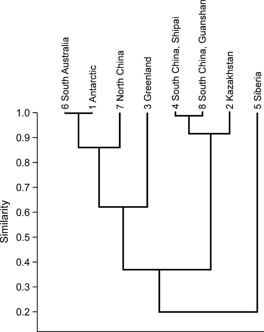
Absolute age of the Shipai Formation is poorly resolved. However, two trilobite biozones have been recognized in the Shipai Formation: the Redlichia meitanensis Zone in the lower parts of the succession and the Palaeolenus lantenosis Zone in the upper parts (Zhang et al., Reference Zhang, Lu, Zhu, Qian, Lin, Zhou, Zhang and Yuan1980; Wang et al., Reference Wang, Ni, Zeng, Xu, Zhou, Li and Lai1987; Zhang and Hua, Reference Zhang and Hua2005; X.L. Zhang et al., Reference Zhang, Liu and Zhao2008). This indicates an age of Cambrian Stage 4, similar to the Wulongqing Formation in eastern Yunnan (Wang et al., Reference Wang, Ni, Zeng, Xu, Zhou, Li and Lai1987; Zhang et al., Reference Zhang, Zhang, Holmer and Li2015). Brachiopods—particularly linguliform brachiopods (linguloids and acrotretoids)—from the Shipai Formation also corroborate a Cambrian Stage 4 age for the Shipai Formation (Z.F. Zhang et al., Reference Zhang, Li, Holmer, Brock, Balthasar, Skovsted, Fu, Zhang, Wang, Butler, Zhang, Cao, Han, Liu and Shu2016). Brachiopods from Cambrian Stage 4 are presently known from all main continents, including South Australia, Antarctic, Greenland, Kazakhstan, Siberia, and China (Pelman, Reference Pelman1977; Holmer et al., Reference Holmer, Popov, Koneva and Bassett2001; Ushatinskaya and Malakhovskaya, Reference Ushatinskaya and Malakhovskaya2001; Skovsted and Holmer, Reference Skovsted and Holmer2005; Betts et al., Reference Betts, Paterson, Jago, Jacquet, Skovsted, Topper and Brock2016, Reference Betts, Paterson, Jago, Jacquet, Skovsted, Topper and Brock2017, Reference Betts, Claybourn, Brock, Jago, Skovsted and Paterson2019; Chen et al., Reference Chen, Zhang, Betts, Zhang and Liu2019; Pan et al., Reference Pan, Skovsted, Brock, Topper, Holmer, Li and Li2019; Ushatinskaya and Korovnikov, Reference Ushatinskaya and Korovnikov2019; Claybourn et al., Reference Claybourn, Skovsted, Holmer, Pan, Myrow, Topper and Brock2020; Zhang et al., Reference Zhang, Holmer, Liang, Chen and Duan2020a, Reference Zhang, Strotz, Topper, Chen, Chen, Liang, Zhang, Skovsted and Brockb). Cluster analysis of Cambrian, Stage 4 linguliforms shows that the faunas from South China (Shipai Formation, Guanshan biota) and Kazakhstan cluster together (Fig. 19). This is defined by the occurrence of Eoobolus, Palaeobolus, Lingulellotreta, and Linnarssonia. Clustering of east Antarctica, South Australia, and North China is consistent with the biostratigraphic correlation of Claybourn et al. (Reference Claybourn, Skovsted, Holmer, Pan, Myrow, Topper and Brock2020) based on brachiopods.
Results of the pair-group cluster analysis for the Cambrian Stage 4 linguliform genera from 8 localities (Raup-Crick similarity).
The brachiopod fauna from the Shipai Formation is dominated by the acrotretoid Linnarssonia sapushanensis, which are commonly aggregated as patchy concentrations of shell valves on the same bedding plane (Fig. 15.1–15.4), notably in the Xiachazhuang section. In contrast, the acrotretoids from the Wulongqing Formation form thicker shell beds (~11–13 pavements within a 1 cm thick bed) (Fig. 15.5–15.7). Differential accumulation styles of acrotretoid valves highlight differences between sedimentary paleoenvironments and energy regimes of the Shipai and Wulongqng formations. In the Wulongqing Formation of eastern Yunnan, where acrotretoid brachiopod shells form dense stacks, the shells were probably affected by high energy currents, and were briefly suspended before their final deposition on the sea floor. In contrast, acrotretoidshell beds from the Shipai Formation in the Hubei Province are characterized by lower density shell concentrations, probably the result of deposition in a deeper environment where current energy was minimal.
Similarities between Linnarssonia shell beds in the middle Shipai Formation in the Three Gorges area and the lower to middle Wulongqing Formation in Wuding area, eastern Yunnan suggest that these two successions may be roughly correlated. This is further corroborated by the first appearance datum (FAD) of the rhynchonelliform calcareous-shelled brachiopod Nisusia in the silty mudstone of both the Shipai and Wulongqing formations.
Shell structures of acrotretoid brachiopods from fine siliciclastics
Organophosphatic brachiopod shells usually consist of an organic periostracum, a mineralized laminar primary layer, and a secondary columnar layer (Holmer, Reference Holmer1989; Williams and Holmer, Reference Williams and Holmer1992; Williams et al., Reference Williams, Brunton and Carlson1997; Holmer et al., Reference Holmer, Popov and Streng2008; Streng et al., Reference Streng, Holmer, Popov and Budd2008). The thin organic periostracum is usually exfoliated during taphonomic processes. The primary layer is generally very thin, usually not much more than 1 μm thick, and is easily lost during transportation and burial, resulting in the exposure of the secondary layer (Williams and Holmer, Reference Williams and Holmer1992; Williams et al., Reference Williams, Brunton and Carlson1997). The secondary layer is mainly composed of an alternating arrangement of lamina and columns. The columnar shell structure is characteristic of acrotretid brachiopods (Holmer, Reference Holmer1989; Williams and Holmer, Reference Williams and Holmer1992), but has been demonstrated to occur in several lingulid brachiopods, including Lingulellotretidae, Dysoristidae, and Kyrshabaktellidae (Cusack et al., Reference Cusack, Williams and Buckman1999; Skovsted and Holmer, Reference Skovsted and Holmer2006). Similar shell structures are also present in Canalilatus (Streng et al., Reference Streng, Holmer, Popov and Budd2008), the stem lineage of brachiopods Mickwitzia (Skovsted and Holmer, Reference Skovsted and Holmer2003; Holmer et al., Reference Holmer, Popov and Streng2008), Micrina (Williams and Holmer, Reference Williams and Holmer2002), and the tommotiid Tannuolina (Skovsted et al., Reference Skovsted, Clausen, Álvaro and Ponlevé2014).
Although the shell structure of acrotretoid brachiopods is well known from examples preserved in carbonate deposits (Poulsen, Reference Poulsen1971; Popov and Ushatinskaya, Reference Popov and Ushatinskaya1986; Rowell, Reference Rowell1986; Ushatinskaya et al., Reference Ushatinskaya, Zezina, Popov and Putivtseva1988; Holmer, Reference Holmer1989; Williams and Holmer, Reference Williams and Holmer1992; Holmer et al., Reference Holmer, Popov and Streng2008; Z.L. Zhang et al., Reference Zhang, Li, Holmer, Brock, Balthasar, Skovsted, Fu, Zhang, Wang, Butler, Zhang, Cao, Han, Liu and Shu2016), no details of acrotretoid shell structures preserved in siliciclastic rocks have ever been described. Most acrotretoids in muddy deposits are preserved as internal molds (e.g., Duan et al., Reference Duan, Liang, Holmer and Zhang2021), which precludes investigation of shell structural details (Mergl and Kordule, Reference Mergl and Kordule2008; Mergl, Reference Mergl2019). However, specimens of the acrotretoid Linnarssonia from the silty mudstone of the Shipai Formation have well-preserved shell ultrastructures, allowing detailed study of the acrotretoid shell ultrastructure from siliciclastic deposits for the first time.
The primary layer forming the ornamentation of the external shell surface (e.g., concentric fila) is preserved in some specimens of L. sapushanensis from the Shipai Formation (Fig. 2.1). The secondary layer is also well developed, and has a columnar structure that is mainly composed of hollow tubes (diameter = 2.5 μm on average, range 1.6–3.8 μm), with solid columns (~1 μm in diameter) that are composed of stacks of pinacoidal plates (Fig. 20.4, 20.5). The hollow tubes in L. sapushanensis are comparable morphologically with those previously documented in other acrotretoid brachiopods, such as Anglulotreta (1.5–5 μm diameter) (Table 4) (Holmer, Reference Holmer1989; Williams and Holmer, Reference Williams and Holmer1992; Streng, Reference Streng1999; Streng and Holmer, Reference Streng and Holmer2006; Streng et al., Reference Streng, Holmer, Popov and Budd2008; Z.L. Zhang et al., Reference Zhang, Zhang and Wang2016). The solid columns in the tubes (Fig. 20.5) and the solid columns that connected the laminae (Fig. 21.5) are comparable with those columnar central canals (~1 μm in diameter) (Fig. 21.9). Latex casts of these hollow tubes and thin solid columns in L. sapushanensis (Fig. 21.6, 21.7) also provide detailed molds for comparison with acid-etched material (Fig. 21.8, 21.9).
SEM images of acrotretoid Linnarssonia sapushanensis showing the secondary shell structure. (1, 2) Internal view of ventral valves (ELI QJP-SP-205-1, ELI QJP-SP-205-2), arrows indicate apical process; (3) internal view of dorsal valve (ELI QJP-SP-205-4); (4) the column structure; (5) enlarged view of (4) marked by the inset box, showing the hollow tube (marked by arrow) with a solid column (marked by arrow); (6) vertical view of columnar structure; (7) enlargement of (6), showing the circular pit on the interlaminar surface (marded by arrow on left) and external aperture of the hollow tube with a solid structure (marked by arrow on right) in vertical view; (8) columnar structure, showing the hollow tube openings on the exposed interlaminar surfaces of the secondary shell layer; (9) close-up view of (8), showing the circular pits on the interlaminar surface. Scale bars = 500 μm (1), 1 mm (2, 3), 10 μm (4, 6), 2 μm (5), 1 μm (7, 9), or 50 μm (8).

Comparison of the acrotretoid secondary shell layer from different depositional environments. (1) Internal view of ventral valves (ELI QJP-SP-205-1); (2, 3) close-up view of (1); (4) close-up view of columnar structure, note the hollow tube (marked by arrow); (5) the thin solid columns that connected the laminae; (6, 7) latex casts of (2, 3) showing the secondary columnar structure of an acrotretoid from the Shipai Formation (siliciclastic deposits); (8, 9) secondary columnar structure of an acrotretoid from the Shuijingtuo Formation (carbonate deposits) showing the columns (marked by arrow) with central canals (marked by double arrows) (ELI BE-AJH 201502-013, ELI BE-AJH 201502-014). Scale bars = 500 μm (1), 100 μm (2), 20 μm (3, 4, 6, 7, 9), or 10 μm (5, 8).

Previous studies of brachiopod column structure from dissolved limestone.

It is generally accepted that the empty intralaminar spaces in acrotretoid shells originally contained an organic matrix, and that the slots between successive laminae and columnar canals were originally occupied by sheets and strands, respectively composed of proteins or chitin (Poulsen, Reference Poulsen1971; Ushatinskaya et al., Reference Ushatinskaya, Zezina, Popov and Putivtseva1988; Holmer, Reference Holmer1989; Williams and Holmer, Reference Williams and Holmer1992). In L. sapushanensis from the Shipai Formation, the intralaminar spaces are commonly empty, thus exposing the hollow tubes in relief (Fig. 20.4), but occasionally the spaces are filled with mineralized materials (Fig. 21.4). The interlaminar surfaces of lamellae are ornamented with circular pits (Fig. 20.7, 20.9) and hollow tube openings (Fig. 20.8), usually ~500 nm and 2.5 μm, respectively. The hollow tubes may have contained unmineralized organic fibers that were lost post-mortem. Thus, the hollow tube and solid column of the acrotretoid column structure from the Shipai Formation could be the equivalent of the traditional column and central canal structure observed in shells dissolved from limestone (Fig. 22).
(1) Diagrammatic reconstruction of shell structure of the acrotretoid brachiopods, illustrating relationships between successive discrete shell layers (modified from Williams and Holmer, Reference Williams and Holmer1992; Williams et al., Reference Williams, James, Emig, Mackay, Rhodes and Kaesler2000); (2) column structure from the mudstone (gray indicates solid structure), sketch of Figure 20.4-20.5, showing the hollow tube with a solid column (2.1), and the longitudinal section of the hollow tube (2.2); (3) column structure from the dissolving limestone (gray indicates solid structure), sketch of Figure 21.8–21.9, showing the column with a central canal (cn) (3.1), and the longitudinal section of a column (3.2).

Conclusion
This is the first comprehensive description of the brachiopod faunas and their systematic diversity from the Shipai Formation (Stage 4) in the Three Gorges area of South China. This assemblage includes the representatives of the subphylum Linguliformea: linguloids (Lingulellotreta ergalievi, Eoobolus malongensis, and Neobolidae gen. indet. sp. indet.), an acrotretoid (Linnarssonia sapushanensis), and calcareous-shelled rhynchonelliforms (Kutorgina sinensis, Kutorgina sp., and Nisusia liantuoensis). Cluster analysis of linguliform, Cambrian Stage 4 brachiopods shows that the faunas of South China (Shipai Formation and the Wulongqing Formation) group closely with those from Kazakhstan.
The brachiopod fauna from the Shipai and Wulongqing formations both include the rhynchonelliform Nisusia, and preserve shell concentrations of the acrotretoid L. sapushanensis. In the Shipai Formation, L. sapushanensis are preserved in patchy aggregations on the same bedding plane, whereas in the Wulongqing Formation they form thick shell beds. This suggests that the Wulongqing Formation represents a slightly higher energy paleoenvironment than the quieter Shipai Formation.
In siliciclastics, brachiopods are commonly preserved as casts and molds and retention of shell material is generally rare. Brachiopods from the Shipai Formation however, retain shell material, the remarkable preservation of which is possibly due to deposition in a low energy paleoenvironment. Linnarssonia sapushanensis from the Shipai Formation has a hollow tube and solid column microstructure, which is likely to be the equivalent of traditional column and central canal-type microstructure often observed in acid-etched acrotretids. Knowledge of shell microstructures in Cambrian acrotretoids is primarily derived from specimens acid etched from limestones. This study provides the first detailed description of acrotretoid shell structures from Cambrian siliciclastics, providing an important comparison with acid-etched material.
Acknowledgments
We are grateful to C. Zabini, M. Mergl, and the Journal Associate Editor C.D. Sproat for their constructive comments and suggestions. Financial support from the National Natural Science Foundation of China (41890844, 41425008, 41621003, and 41720104002 to ZZF) and the 111 project (D17013) for continuous fossil collectioning of Xi'an group are sincerely acknowledged. L.E. Holmer's work was supported by a grant from the Swedish Research Council (VR 2018-03390). Many thanks to the early fossil collections organized by the working-team at the Early Life Institute (NWU), and joined by J.P. Zhai, F.Y. Chen, Z.L. Zhang, X.R. Wang, and H.Z. Wang working therein.
Accessibility of supplemental data
Data available from the Dryad Digital Repository: https://doi.org/10.5061/dryad.2ngf1vhmn.


 Open access