


Here we present the case of a 56-year-old woman with aneurysms from the right middle cerebral artery (MCA), left MCA bifurcation, right internal carotid artery (ICA) and two from the right anterior communicating artery. She underwent elective endovascular coiling of the unruptured, previously untreated right and left MCA aneurysms and superior right hypophyseal artery aneurysms (Figure 1). Thirteen days later, the patient presented to the emergency department with rapid-onset progressive left arm weakness over the past week, mild left Upper motor neuron (UMN) facial paresis, hyperreflexia, a positive Hoffman’s sign and slight wasting in the left arm and forearm.
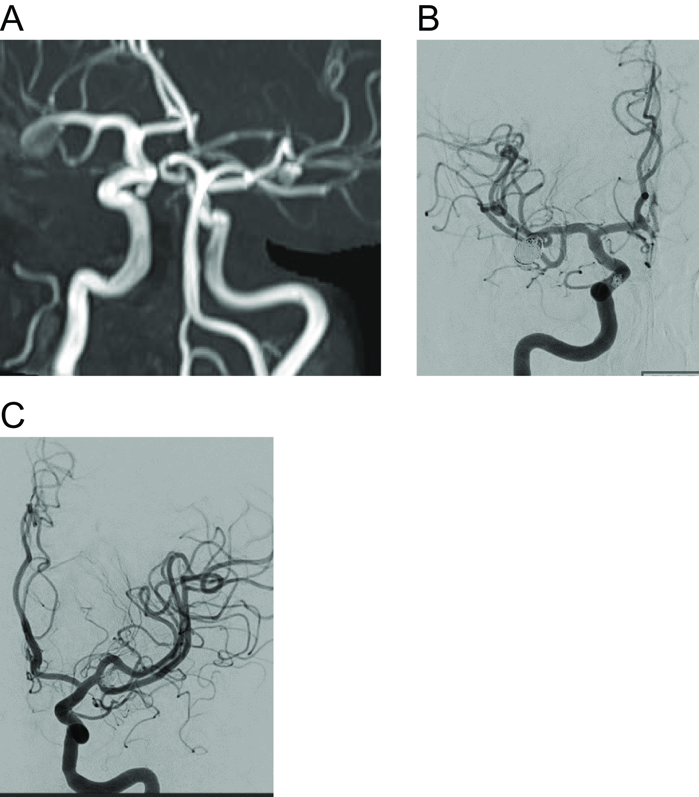
MRA images (A) demonstrate four intracranial aneurysms. The largest measures 8 × 8 × 11 mm and arising from the proximal superior M2 division of the right MCA. The second largest is at the left MCA bifurcation measuring 5 × 3 mm. The third aneurysm arises from the right internal carotid artery (ICA), measuring 4 mm in maximum size. The fourth and fifth aneurysms arise from the AComm, measuring 3 mm in maximum size. DSA images show successful coil embolization of right M2, right ICA (B) and left MCA bifurcation aneurysm (C).
CT showed multifocal areas of subcortical edema involving the cerebral hemispheres bilaterally, prominent in the frontal lobes and right temporal lobe. CTA showed no evidence of acute vascular occlusion. Subsequent contrast-enhanced MRI study showed extensive cerebral vasogenic edema with sparing of the overlying cortex, with numerous underlying punctate scattered foci of enhancement (Figure 2). The largest focus was on the right centrum semiovale, which measured 7 mm in size. Some of the lesions showed hypointense signals on gradient recalled echo (GRE) and T2-weighted images. There was some vague, ill-defined increased diffusion-weighted imaging (DWI) hyperintensity within the posterior centrum semiovale bilaterally, with possible associated apparent diffusion coefficient (ADC) signal loss; however, this finding was complicated by the presence of Nyquist ghost artifact. Parenchymal swelling contributed to right ventricle narrowing and sulcal effacement in the regions of parenchymal swelling.
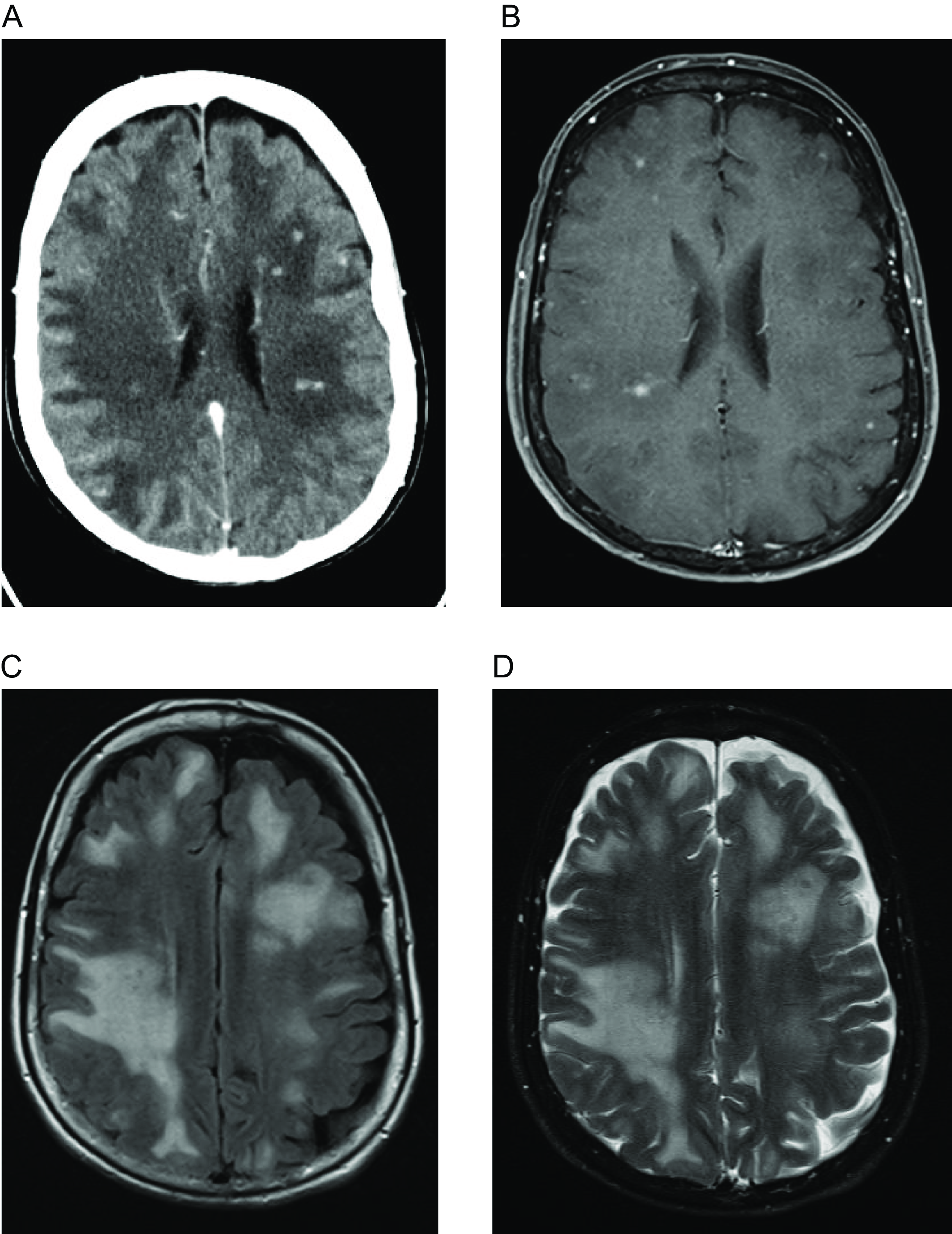
Axial post-contrast CT (A) and post-contrast T1-weighted MR images (B) demonstrate scattered multiple enhancing foci throughout the supratentorial brain parenchyma. On FLAIR (C) and T2-weighted MR images (D), there is significant vasogenic edema, resulting in the effacement of cerebral sulci. The lesions are seen as hypointense on T2-weighted images (D).
The pattern of punctate foci of enhancement could be consistent with a variety of diagnoses, including ischemic events, septic emboli, vasculitis, cerebral amyloid angiopathy-related inflammation, posterior reversible encephalopathy syndrome and non-ischemic cerebral enhancing (NICE) lesions. Reference Ridwan, Kandyba, Schug, Malsagov, Karageorgos and Hans1–Reference Ulus, Yakupoglu, Kararslan, Ilak, Siva and Koçer7 The patient was given the presumptive diagnosis of NICE lesions based on a combination of imaging and clinical history. However, the patient’s recent history of aneurysm repair, as well as the pattern of inflammatory lesions within watershed regions of the parent arteries, is characteristic of NICE lesions. NICE lesions represent a rare complication of the endovascular treatment for cerebral aneurysms. Reference Forestier, Escalard and Sedat8 The estimated incidence of NICE lesions is between 0.05% and 2.3%. Reference Bakola, Papagiannopoulou and Palaiodimou3 However, the actual incidence may be higher than this estimate, given that lesions can develop asymptomatically.
The etiology and pathogenesis of NICE lesions are unclear. One theory posits that shedding of the hydrophilic coating of the microcatheters used in endovascular procedures causes a granulomatous foreign-body reaction presenting as T1 and T2 iso-hypointense punctate lesions on neuroimaging. Evidence for this theory comes from research demonstrating that repeated friction can damage the inner wall of microcatheters, forming conglomerated fragments that can become downstream emboli. Reference Oh, Shin, Lee, Kim and Kim9
While early case studies documenting NICE lesions report the use of antibiotic therapies and anticonvulsants as treatments, anti-inflammatory steroid therapy consistently produced favorable outcomes. Reference Ridwan, Kandyba, Schug, Malsagov, Karageorgos and Hans1–Reference Bakola, Katsanos and Palaiodimou4 Our patient started on a 4-week taper of steroids following diagnosis, and a follow-up MRI demonstrated significant resolution of the vasogenic edema. No new lesions developed, and the size and enhancement of the lesions decreased. We hope that the discussion of the following case will promote clinical awareness of NICE lesions as a potential side effect of endovascular therapy.
Author contributions
MAK wrote the first draft of the manuscript and edited the manuscript. SY, NK, SM, ZIH and CF edited the manuscript and selected the images. NK was the original interpreting radiologist.
Funding statement
No financial support was provided for this research work.
Competing interests
The authors have no conflicts of interest to disclose.


 Open access