





Introduction
The members of the family Clinostomidae Lühe, 1901 are characterized by heteroxenous life cycle including fish-eating birds as preferential definitive hosts, except the subfamily Nephrocephalinae Travassos, 1928, which prefer reptiles (Kanev et al., Reference Kanev, Radev, Fried, Gibson, Jones and Bray2002). In the last 25 years, the taxonomy has been heavily revised to reorder the high number of descriptions based only on few morphological features, as happened with species of Clinostomum Leidy, 1856 (Ukoli, Reference Ukoli1966a; Yamaguti, Reference Yamaguti1971; Feizullaev and Mirzoeva, Reference Feizullaev and Mirzoeva1983). The advent of molecular methods, integrated with morphological observation, has substantially improved species delimitation, leading to the description and re-description of multispecies of Clinostomum and Euclinostomum Travassos, 1928, and in some instances prompting generic reassignment (Gustinelli et al., Reference Gustinelli, Caffara, Florio, Otachi, Wathuta and Fioravanti2010; Caffara et al., Reference Caffara, Locke, Gustinelli, Marcogliese and Fioravanti2011, Reference Caffara, Locke, Cristanini, Davidovich, Markovich and Fioravanti2016, Reference Caffara, Locke, Halajian, Luus-Powell, Benini, Tedesco, Kasembele and Fioravanti2019; Sereno-Uribe et al., Reference Sereno-Uribe, Pinacho-Pinacho, García-Varela and Pérez-ponce de León2013, Reference Sereno-Uribe, García-Varela, Pinacho-Pinacho and Pérez-ponce de León2018, Reference Sereno-Uribe, López-Jiménez, González-García, Ortega-Olivares and García-Varela2025; Locke et al., Reference Locke, Caffara, Marcogliese and Fioravanti2015; Rosser et al., Reference Rosser, Alberson, Woodyard, Cunningham, Pote and Griffin2017, Reference Rosser, Baumgartner, Alberson, Noto, Woodyard, King, Wise and Griffin2018; Truter et al., Reference Truter, Yong, Smit, Chakona, Luus-Powell and Smit2025). A clear biogeographical separation have been observed in the distributions of species of Clinostomum (Caffara et al., Reference Caffara, Locke, Gustinelli, Marcogliese and Fioravanti2011; Locke et al., Reference Locke, Caffara, Marcogliese and Fioravanti2015 and references therein; Sereno-Uribe et al., Reference Sereno-Uribe, López-Jiménez, González-García, Ortega-Olivares and García-Varela2025; Truter et al., Reference Truter, Yong, Smit, Chakona, Luus-Powell and Smit2025), but such separation has not been identified for Euclinostomum (Caffara et al., Reference Caffara, Locke, Cristanini, Davidovich, Markovich and Fioravanti2016). Finally, it is clear that species cannot be distinguished based on host species, because host associations of all developmental stages are broad or poorly known (Lo et al., Reference Lo, Wang, Huber and Kou1982; Matthews and Cribb, Reference Matthews and Cribb1998; Hoffman, Reference Hoffman1999; Bullard and Overstreet, Reference Bullard, Overstreet, Eiras, Segner, Wahli and Kapoor2008; Locke et al., Reference Locke, Caffara, Marcogliese and Fioravanti2015).
Compared with the closely related genus Clinostomum, species of Euclinostomum have been reported less frequently in the literature, and several aspects of their taxonomy and biology remain insufficiently documented (Caffara et al., Reference Caffara, Locke, Cristanini, Davidovich, Markovich and Fioravanti2016). The taxonomic history of the genus Euclinostomum has been characterized by the erection of numerous nominal species; in total, 10 species have been proposed, 8 of which are currently regarded as invalid or of doubtful status. Caffara et al. (Reference Caffara, Locke, Cristanini, Davidovich, Markovich and Fioravanti2016) revised the genus, recognizing as valid only the Old World species such as Euclinostomum heterostomum (Rudolphi, 1809) and Euclinostomum multicaecum Tubangui & Masiluñgan, 1935, a conclusion supported by the redescription of the type species (E. heterostomum) and the incorporation of molecular data. However, at that time, only a limited number of sequences linked to detailed morphological information were available (Athokpam et al., Reference Athokpam, Jyrwa and Tandon2014; Senapin et al., Reference Senapin, Phiwsaiya, Laosinchai, Kowasupat, Ruenwongsa and Panijpant2014). More recently, the number of molecular reports on E. heterostomum has increased, although comprehensive morphological descriptions remain scarce (Shukla et al., Reference Shukla, Garain, Verma and Behera2024; Guz et al., Reference Guz, Pastuszka, Puk, Torbicz, Szarek and Oszust2025; Shigoley et al., Reference Shigoley, Kmentová, Ndegwa, Topić, Thys and Vanhove2025).
A recent study by Truter et al. (Reference Truter, Yong, Smit, Chakona, Luus-Powell and Smit2025) corroborates the previous reports by Caffara et al. (Reference Caffara, Locke, Cristanini, Davidovich, Markovich and Fioravanti2016, Reference Caffara, Locke, Echi, Halajian, Benini, Luus-Powell, Tavakol and Fioravanti2017) regarding the descriptions of Clinostomum and Euclinostomum in African publications. Most of these studies report the 2 genera without providing a complete morphological description. In some recent papers (e.g., Mahdy et al., Reference Mahdy, Abdelsalam, Abdel-Maogood, Shaalan and Salem2021), molecular analyses have been carried out using only 1 molecular marker, for instance ITS2 rDNA (Kaur et al., Reference Kaur, Qureshi and Shrivastav2012; Zimik et al., Reference Zimik, Sharma and Roy2019; Mahdy et al., Reference Mahdy, Abdelsalam, Abdel-Maogood, Shaalan and Salem2021, Reference Mahdy, Abdelsalam and Salem2023), which has shown high similarity among Clinostomum species (Gustinelli et al., Reference Gustinelli, Caffara, Florio, Otachi, Wathuta and Fioravanti2010; Sereno-Uribe et al., Reference Sereno-Uribe, Pinacho-Pinacho, García-Varela and Pérez-ponce de León2013, Reference Sereno-Uribe, García-Varela, Pinacho-Pinacho and Pérez-ponce de León2018, Reference Sereno-Uribe, López-Jiménez, González-García, Ortega-Olivares and García-Varela2025; Locke et al., Reference Locke, Caffara, Marcogliese and Fioravanti2015; Pérez-ponce de León et al., Reference Pérez-ponce de León, García-Varela, Pinacho-Pinacho, Sereno-Uribe and Poulin2016; Briosio-Aguilar et al., Reference Briosio-Aguilar, Pinto, Rodríguez-Santiago, López-García, García-Varela and de León2018).
Ethiopia has many major water bodies, especially lakes, that have an estimated volume of in more than 335 billion cubic meters (Getaneh et al., Reference Getaneh, Abera, Abegaz and Tamene2022); many of these lakes are recognized for their outstanding biological diversity and societal significance (Belete et al., Reference Belete, Diekkrüger and Roehrig2015). Lake Tana is the largest lake in Ethiopia and supports a large fishing industry mainly based on tilapias and catfish. Moreover, this country is an important resting and feeding ground for many Palearctic migrant waterbirds including the great white pelican Pelecanus onocrotalus, Linnaeus, 1758, which is a suitable definitive host for clinostomids trematodes. The co-occurrence of fish and waterbirds enables the life cycle of clinostomids for which the former are second intermediate hosts while the latter are definitive hosts.
Few studies on clinostomids in birds definitive host in Africa are old reports (Dollfus, Reference Dollfus1932, Reference Dollfus1950; Van der Kuyp, Reference Van der Kuyp1953; Ukoli, Reference Ukoli1966a,Reference Ukolib; Manter and Pritchard, Reference Manter and Pritchard1969; Dennis and Sharp, Reference Dennis and Sharp1973; Britz et al., Reference Britz, Saayman and Van As1984), but most provide exhaustive morphological description useful for identification. Few report of clinostomids in fish have been reported from Ethiopia in the last 25 years (Eshetu, Reference Eshetu2000; Yimer, Reference Yimer2000; Eshetu and Mulualem, Reference Eshetu and Mulualem2003; Yimer and Enyew, Reference Yimer and Enyew2003; Zekarias and Yimer, Reference Zekarias and Yimer2007; Gulelat et al., Reference Gulelat, Eshetu, Asmare and Bekele2013; Amare et al., Reference Amare, Alemayehu and Aylate2014; Bekele and Hussien, Reference Bekele and Hussien2015; Gebawo, Reference Gebawo2015; Reshid et al., Reference Reshid, Adugna, Redda, Awol and Teklu2015; Gebremedhn and Tsegay, Reference Gebremedhn and Tsegay2017). Most of these are epidemiological studies reporting quantitative data without any specific identification. Gustinelli et al. (Reference Gustinelli, Caffara, Florio, Otachi, Wathuta and Fioravanti2010) published the first comprehensive paper using an integrative taxonomic approach (morphology coupled with molecular analyses) to report Clinostomum cutaneum Paperna, 1964 metacercariae and adults collected in Kenya, representing the first input towards a systematic revision of this complex group of parasites. Zhokhov and Morozova (Reference Zhokhov and Morozova2020) published a study on clinostomids of fish from Lake Tana, providing exhaustive morphological descriptions but no molecular data.
The current study provides the first detailed descriptions of adult clinostomids isolated from P. onocrotalus in Ethiopia. By integrating new morphological data with phylogenetic analyses, we refine species boundaries within and between Clinostomum and Euclinostomum.
Materials and methods
Sampling and morphological study
Twenty-three adult clinostomids were collected from the oesophagus (Figure 1) of 1 specimen of P. onocrotalus collected from Bahar Dar Gulf, Tana Lake (11°35′ N 37°23′ E) and processed fresh, according to permits of the Addis Ababa University (ref. No. ALIPB IRERC/141/2016/24) and the Amhara National Regional State Environment and Forest Protection Authority (ref. No. Aka/T/B-10/01-18/03/2017). The clinostomids were washed in saline and preserved in 70% ethanol for morphological and molecular analysis. Measurements are given in micrometres unless otherwise stated and follows Matthews and Cribb (Reference Matthews and Cribb1998) and Caffara et al. (Reference Caffara, Locke, Cristanini, Davidovich, Markovich and Fioravanti2016).
Oesophagus of Pelecanus onocrotalus with several adult clinostomids.

Figure 1 Long description
Insufficient visual information to describe this element accurately.
Some specimens were processed for scanning electron microscopy (SEM) analysis; they were dehydrated through a graded ethanol series, subjected to critical point drying, sputter-coated with gold palladium, and observed using a Phenom XL G2 Desktop SEM (Thermo Fisher Scientific, Eindhoven, The Netherlands) operating at 5 kV.
Molecular study
Before the clarification in Amman’s lactophenol a small portion of the posterior end of the body were removed for DNA extraction using a PureLink Genomic DNA Kit (Life Technologies, Carlsbad, CA, USA) following the manufacturer’s protocol. Amplification of ITS rDNA employed protocols and primers of Gustinelli et al. (Reference Gustinelli, Caffara, Florio, Otachi, Wathuta and Fioravanti2010); COI mtDNA employed those of Moszczynska et al. (Reference Moszczynska, Locke, Mclaughlin, Marcogliese and Crease2009). Amplified products were electrophoresed on a 1% agarose gel stained with SYBR Safe DNA Gel Stain (Thermo Fisher Scientific, Carlsbad, CA, USA) in 0.5X TBE. For sequencing of both ITS and COI, amplicons were excised and purified by NucleoSpin Gel and PCR Cleanup (Mackerey-Nagel, Düren, Germany) and sequenced with an ABI 3730 DNA analyser at StarSEQ GmbH (Mainz, Germany). The DNA trace files were assembled with VectorNTI AdvanceTM 11 software (Invitrogen) and compared with published data by BLAST tools (https://blast.ncbi.nlm.nih.gov/Blast.cgi). Multiple sequence alignments were performed using BioEdit 7.2.5 (Hall, Reference Hall1999) (32 ITS sequences and 35 COI sequences in the final dataset), p-distance and maximum-likelihood (ML) tree (T93+I substitution model for ITS, and KHY+G+I for COI, bootstrap of 1000 replicates for both genes) were performed using MEGA version 12 (Kumar et al., Reference Kumar, Stecher, Suleski, Sanderford, Sharma and Tamura2024). Some published clinostomid sequences were excluded from the analyses, as previous studies have demonstrated a clear molecular separation between Old World and New World representatives of the genus; therefore, only taxa relevant to the present study were retained.
The sequences generated in this study were uploaded in GenBank under the accession numbers PX626916-PX626929 (ITS rDNA) and PX661153-PX661172 (cox1 mtDNA).
Results
Based on morphological features, the adults recovered from the great white pelican (P. onocrotalus) belonged to 2 genera: Euclinostomum and Clinostomum.
Morphological redescription
Euclinostomum lauroi Tendeiro, Travassos and Fazendeiro, 1974 (n = 1) (Figures 2 and 3; Table 1)
Line drawing of Euclinostomum lauroi mature specimen from Pelecanus onocrotalus. Scale bar = 370 µm.

Figure 2 Long description
An elongated, leaf-shaped outline with a pointed lower end and a rounded upper end. Near the upper end, a small circular structure sits at the tip and a larger circular structure is positioned below it. Two thick, dark longitudinal lines run down the body from near the upper region toward the lower region. Along both sides, multiple thick, dark branches extend outward from the central lines, forming repeated left and right branching patterns down most of the body length. In the lower half, several irregular, looped internal shapes appear between the central lines. A vertical scale bar is placed along the left side of the drawing. The context states: Scale bar equals 370 micrometer.
Morphological details of E. lauroi mature specimen from P. onocrotalus: (A) whole specimen, LM (Scale bar = 500 µm); (B) everted cirrus, LM; (C) detail of cirrus surface, SEM; (D) detail of sperm cells from sectioned posterior testis, SEM; (E) egg, SEM; (F) detail of cirrus pouch (cp) and anterior testis (at), LM.

Figure 3 Long description
The image A showing a grayscale whole-body specimen with a tapered lower end and a rounded upper end. Dark branching lines extend from a central vertical region toward both sides in repeated segments. A darker oval area is near the upper end and a darker elongated area is near the lower end. The image B showing a low-contrast grayscale field with a mostly smooth background and a few small dark spots. A scale bar is present at the lower right. The image C showing a grayscale close-up of an elongated, curved structure with a smooth surface and a thicker rounded end. A long horizontal scale bar is present near the lower left. The image D showing a grayscale textured surface with many tightly packed, irregular rounded and folded forms across the frame. A horizontal scale bar is present near the lower left. The image E showing a grayscale close-up with an oval raised structure on a textured surface with shallow grooves. A horizontal scale bar is present near the lower left.
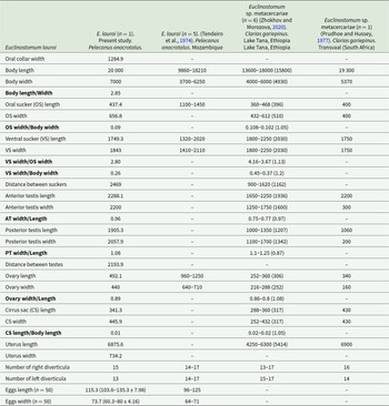
Measurements of Euclinostomum lauroi [Min–Max (Mean ± SD) µm]

Table 1 Long description
The table lists micrometre measurements and selected ratios for Euclinostomum lauroi from the present study and prior literature, plus two Euclinostomum sp. metacercariae datasets, each with different sample sizes and hosts. In the present study (one specimen from Pelecanus onocrotalus), body length is 20,000 and body width is 7,000, exceeding the earlier E. lauroi body-length range of 9,860 to 18,210 and overlapping or exceeding metacercariae lengths of 13,600 to 18,000 and a single value of 19,300. The oral sucker is smaller in the present study (length 437.4; width 656.8) than the earlier E. lauroi oral-sucker length range of 1,100 to 1,450, while ventral sucker size is broadly comparable across sources (present study length 1,749.3; width 1,843; metacercariae ranges about 1,800 to 2,250). Distances and reproductive-organ measurements are provided where available; for example, the present study reports anterior testis length 2,288.1 and posterior testis length 1,905.3, while metacercariae ranges are lower for posterior testis length (1,000 to 1,350). Diverticula counts are similar across datasets, with right-side counts around 13 to 17 and left-side counts around 13 to 17; the present study reports 15 right and 13 left. Egg measurements are only reported for the present study and Tendeiro et al., with the present study giving egg length about 115.3 and width about 73.7, generally larger than the earlier ranges (length 96 to 125; width 64 to 71). Several cells are missing in some columns, and comparisons should be interpreted cautiously because sources differ in life stage, locality, host, and sample size.
Type host: Pelecanus onocrotalus
Type locality: Maniquenique, Chibuto, Gaza, Mozambique
New locality: Bahar Dar Gulf (11°35′ N, 37°23′ E), Tana Lake, Ethiopia
Body large, slipper-shaped, slightly constricted in ventral sucker region and widest in gonadic region (Figures 2 and 3A). Anterior end invaginated to form inconspicuous oral collar. Oral sucker smaller than ventral sucker, not well evident as folded back on itself. Pharyngeal chamber not discernible. Intestine bifurcates immediately posterior to pharynx. Intestinal ceca, run laterally to ventral sucker (Figures 2 and 3A) to posterior end of body. Posterior to ventral sucker main ceca give rise to thin secondary blind diverticula, variable in number (15 at right margin and 13 at left), extending latero-posteriorly to main ceca with their external margin almost perpendicular to body side (Figures 2 and 3A). Connection between intestinal ceca and excretory system not visible. Testes tandem, intercaecal, in posterior third of body. Anterior testis in anterior part of the posterior third of body, crescent or U-shaped, with rounded and symmetrical arms embracing posterior part of uterus and cirrus pouch (Figure 3F). Posterior testis completely in posterior third of body, Y-shaped with anterior margin concave (Figure 3A). Cirrus pouch round, within arm of anterior testis (Figure 3F); genital pore not visible. Cirrus well visible and everted, finely striated in posterior half (Figure 3B, C). Ovary smooth, round, smaller than cirrus pouch, intertesticular, dextrally contiguous with posterior margin of anterior testis. Uterus in the middle third of body. Uteroduct emerging from ootype runs around left margin of anterior testis running parallel and opens into uterine sac at middle portion. Metraterm not visible. Vitelline glands distributed from ventral sucker to posterior end scattered among intestinal caeca. Tegument surface devoid of spines. Eggs smooth (Figure 3E).
The other 2 species were adults of C. phalacrocoracis (20 specimens) and C. tilapiae (2 specimens) (Figures 4 and 5).
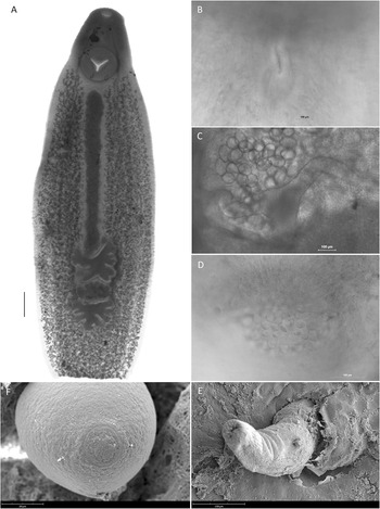
Morphological details of C. phalacrocoracis mature specimen from P. onocrotalus: (A) whole specimen, LM (scale bar = 100 µm); (B) pharynx, LM; (C) detail of vitelline reservoir and vitelline duct, LM; (D) detail of papilla-like structures at cirrus base, LM; (E) everted cirrus with small blunt tubercles at its base (arrow), SEM; (F), surface of egg showing apical operculum, SEM.

Figure 4 Long description
The image A showing a single elongated specimen on a light background, with a rounded top and a tapered bottom. A darker central region runs lengthwise through the body. A vertical scale bar is placed near the lower left. The image B showing a close-up grayscale field with a faint, narrow, vertical feature near the center. A horizontal scale bar is placed near the lower right. The image C showing a close-up with many round, bubble-like structures clustered in the upper left area. A horizontal scale bar is placed near the lower right. The image D showing a close-up with a cluster of small round structures near the center. A horizontal scale bar is placed near the lower right. The image E showing a close-up surface view with a curved, raised structure near the center on a rough, textured background. A horizontal scale bar is placed near the lower left. The image F showing a close-up of a round structure with concentric ring-like patterning on its surface. A horizontal scale bar is placed near the lower left.
Morphological details of C. tilapiae from P. onocrotalus: (A) whole mature specimen, LM (scale bar = 400 µm); (B) detail of genital pore (arrow), LM; (C) everted cirrus with small blunt tubercles at its base, SEM; (D) detail of papilla-like structures at cirrus base, SEM.

Figure 5 Long description
The image A showing a single elongated, dark gray specimen on a white background, with a rounded top end and a narrower bottom end. A darker vertical band runs down the center. A circular darker area is located near the upper third. A clustered darker region is present near the lower end. A thin vertical line is placed to the left of the specimen. The image B showing a gray textured surface with a white arrow near the center pointing toward a small darker feature. A scale bar is located near the top right. The image C showing a close-up of a ridged surface with a raised, elongated structure extending from the left toward the center-right. A scale bar is located along the bottom edge. The image D showing a close-up surface covered by many overlapping oval structures. A scale bar is located along the bottom edge.
Clinostomum phalacrocoracis (Dubois, Reference Dubois1930) adult (n = 18) (Figure 4; Table 2)
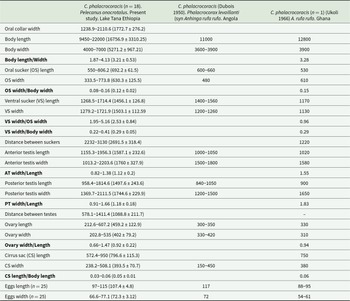
Measurements of Clinostomum phalacrocoracis [Min–Max (Mean ± SD)]

Table 2 Long description
The table lists size ranges and, for the Lake Tana sample, mean and standard deviation for multiple anatomical measurements of Clinostomum phalacrocoracis, and compares them with two earlier studies from Angola and Ghana. In the Lake Tana study (18 specimens), body length ranges from 9450 to 22000 and body width from 4000 to 7000, giving a length-to-width ratio from 1.87 to 4.13. Earlier reports show shorter body lengths (11000 in Angola and 12800 in Ghana) and narrower body widths (3600 to 3900). Sucker measurements in the Lake Tana sample include oral sucker length 550 to 806.2 and ventral sucker width 1279.2 to 1721.9; the Ghana specimen has smaller ventral sucker width (1130) and a much shorter distance between suckers (1220) than the Lake Tana range (2232 to 3130). Reproductive organ sizes in the Lake Tana sample are generally larger than the single Ghana specimen and overlap or exceed the Angola ranges, for example anterior testis length 1155.3 to 1956.3 and posterior testis width 1369.7 to 2111.5. Egg size differs by study: Lake Tana eggs are 97 to 115 long and 66.6 to 77.1 wide, Angola reports longer eggs (117 long), and Ghana reports shorter and narrower eggs (88 to 95 long; 54 to 61 wide). Some comparison cells are blank, and the Angola and Ghana columns represent limited sampling, so differences may reflect sample size, host, or locality rather than consistent species-level variation.
Body stout, slightly wider in gonadic region (Figure 4A). Oral sucker smaller than ventral one (Figure 4A), surrounded by inconspicuous oral collar. Pharynx slightly developed (Figure 4B); intestine bifurcates immediately posterior to pharynx. Intestinal caeca run laterally to ventral sucker and genital complex. Testes in tandem between middle and posterior third of body. Anterior testis, in posterior part of middle third of body, lobed, consists of 5 blunt lobes, some of which are sub-lobed. Posterior testis, in anterior part of posterior third of body, fan-shaped with anterior margin concave and with 3 major lateral lobes on each side and 1 major posterior lobe. Cirrus pouch bean-shaped, in dextral intertesticular space, anterior to ovary, with genital pore opening laterally at posterior margin of anterior testis between right and posterior lobe. Cirrus well visible and everted in 1 subject (Figure 4E), with slightly visible longitudinal ridges and small blunt tubercles at its base. Genital opening with evident small blunt tubercles along its internal edge (Figure 4D). Ovary round, smaller than cirrus pouch, located in dextral intertesticular space. Uterus runs straight from ventral sucker to anterior testis. Uteroduct runs around left margin of anterior testis and opens into uterine sac at its base. Metraterm, straight and overlapping right half of anterior testis, connects uterus. In some specimens, some of the structure composing the ootype complex were visible such as the vitelline reservoir and vitelline duct (Figure 4C). Tegument surface with numerous papillae on ventral side. Eggs smooth, operculated (Figure 4F).
Clinostomum tilapiae Ukoli 1966. Adult (1 immature and 1 mature) (Figure 5; Table 3)
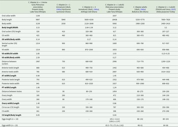
Measurements of Clinostomum tilapiae [Min–Max (Mean ± SD)]

Table 3 Long description
The table lists morphometric measurements for Clinostomum tilapiae from different hosts and locations, comparing immature and adult worms from the present study (Lake Tana, Ethiopia) with published data from Ghana and Turkey. The adult from Lake Tana is the largest entry, with body length 18,438 and body width 5,900, while the immature from the same host is smaller at 9,897 long and 3,139 wide. Published body lengths are generally lower: Ghana reports 5,250 to 5,770 in adults from Bubulcus ibis and 5,600 to 6,330 in immature worms from Anhinga rufa, while Turkey reports 7,800 to 7,820 in adults from Ardea purpurea. Sucker measurements also scale upward in the Lake Tana adult, with oral sucker length 617 and ventral sucker width 1,653, compared with smaller ranges in Ghana and Turkey. Reproductive structures are larger in the Lake Tana adult as well, including anterior testis length 1,442 and posterior testis length 1,552, versus shorter values in the other studies. Egg measurements are only provided for adults, with the Lake Tana adult showing egg length 105.1 to 112.6 and width 65.5 to 75.1, compared with smaller ranges reported from Ghana and Turkey. Several cells are blank or marked with a dash, so not all traits are comparable across studies, and sample sizes are small in multiple columns.
Body thick, elongated, thinner at anterior and terminal parts. Oral sucker subterminal, small, round, surrounded by oral collar (Figure 5A). Pharynx not well visible. Ventral sucker larger than oral sucker, in middle of anterior third of body. Intestinal caeca run lateral to ventral sucker, reaching posterior end of body. Tandem testes strongly digitated, very close to each other (Figure 5A). Anterior testis asymmetrical in medial longitudinal axis, in posterior portion of middle third of body, irregularly lobed, more developed on left side. Posterior testis located in anterior portion of posterior third of body, symmetrical, triangular, with 2 major lateral lobes, subdivided into smaller lobes, and 1 posterior lobe. Cirrus pouch oval and large, on right side, under posterior border of anterior testis. Genital pore medial to cirrus pouch, at level of posterior margin of anterior testis (Figure 5B). In immature specimens, several small blunt tubercles are present at base of everted cirrus (Figure 5C, D). Vitelline glands developed from ventral sucker to posterior end of body. Ovary round, not median, in intertesticular space posterior to cirrus pouch. Uterine sac runs straight from ventral sucker to anterior testis, partially covering, right portion of testis when filled of eggs. Uterus runs around left margin of anterior testis, forming knee-like folding before opening into uterine sac close to anterior testis. Eggs ellipsoidal, operculate.
Molecular analyses
The ITS rDNA sequence of E. lauroi was 1025 bp long (681 bp ITS1, 159 bp 5.8S, 187 bp partial ITS2), showing 96.0–96.3% similarity with Euclinostomum sp. 1–4 (KC894798-KC894801, Osphoronemid fish from Thailand, Senapin et al., Reference Senapin, Phiwsaiya, Laosinchai, Kowasupat, Ruenwongsa and Panijpant2014) and 95.0–96.7% similarity with E. heterostomum (KP721439, Cichlids from Israel, Caffara et al., Reference Caffara, Locke, Cristanini, Davidovich, Markovich and Fioravanti2016), based on BLAST search. The ITS rDNA p-distance between E. lauroi and Euclinostomum spp. sequences retrieved from GenBank was 0.03% with E. heterostomum and 0.04% with Euclinostomum sp. 1–4.
The COI mtDNA sequence was 621 bp long, showing similarity values ranging from 85% (E. heterostomum; Caffara et al., Reference Caffara, Locke, Cristanini, Davidovich, Markovich and Fioravanti2016) to 86% (Euclinostomum sp. 1–3, KC894795-97, Senapin et al., Reference Senapin, Phiwsaiya, Laosinchai, Kowasupat, Ruenwongsa and Panijpant2014). The p-distance observed were 18% relative to Euclinostomum sp. 1–3 (Senapin et al., Reference Senapin, Phiwsaiya, Laosinchai, Kowasupat, Ruenwongsa and Panijpant2014) and 19.6% relative to E. heterostomum (KP721421, Caffara et al., Reference Caffara, Locke, Cristanini, Davidovich, Markovich and Fioravanti2016).
Thirteen new ITS rDNA sequences and 17 COI mtDNA for C. phalacrocoracis, and 2 ITS rDNA and 2 COI mtDNA for C. tilapiae were obtained; BLAST analysis showed 99–100% identity with published sequences of C. phalacrocoracis (e.g. KP110569 ITS; KP110522 COI) and C. tilapiae (e.g. KY649356 ITS; KY649364 COI).
The ML trees of ITS rDNA (Figure 6) and cox1 mtDNA (Figure 7) showed the same topology with Euclinostomum spp. forming a well-supported (100% bootstrap) cluster, clearly separated from the Clinostomum group. Within Euclinostomum, E. lauroi is clearly distinct and basal to this group. The 2 Clinostomum species form well-supported clusters with sequences available in GenBank.
Phylogeny of Euclinostomum spp. and Clinostomum spp. based on ITS rDNA inferred using the maximum likelihood method and Tamura 3-parameter (+I) model of nucleotide substitutions. The percentage of replicate trees in which the associated taxa clustered together (1000 replicates) is shown next to the branches. The analytical procedure encompassed 32 sequences. Evolutionary analyses were conducted in MEGA12 utilizing up to 5 parallel computing threads.

Figure 6 Long description
The phylogenetic tree is oriented vertically, displaying relationships among Clinostomum and Euclinostomum species based on ITS rDNA sequences. At the top, several sequences labeled PX626926 to PX626929 are grouped under Clinostomum phalacrocoracis, marked as 'present study.' This cluster is supported by a bootstrap value of 98. Below, additional sequences from GenBank, such as KY649556 Clinostomum tilapiae and KY856519 Clinostomum sp. morphotype 3, are shown with varying bootstrap values, indicating different levels of support. The tree shows a clear separation between Clinostomum and Euclinostomum groups. Clinostomum species, including Clinostomum chabaudi and Clinostomum sinensis, form distinct clusters. The Euclinostomum group, including PX626929 Euclinostomum lauroi, also marked as 'present study,' is distinct and basal to the group. Other Euclinostomum species, such as KC894799 Euclinostomum sp. 1 and KC894800 Euclinostomum sp. 3, are shown with high bootstrap support, indicating well-supported clusters. The scale bar at the bottom left represents 0.02 substitutions per site.
Phylogeny of Euclinostomum spp. and Clinostomum spp. based on cox1 mtDNA inferred using the maximum likelihood method and Hasegawa–Kishino–Yano (+G+I) model of nucleotide substitutions. The percentage of replicate trees in which the associated taxa clustered together (1000 replicates) is shown next to the branches. The analytical procedure encompassed 35 coding nucleotide sequences. Evolutionary analyses were conducted in MEGA12 utilizing up to 5 parallel computing threads.

Figure 7 Long description
The phylogenetic tree is oriented vertically, displaying relationships among Clinostomum and Euclinostomum species based on cox1 mitochondrial DNA. The tree is constructed using the maximum likelihood method. At the top, a cluster of sequences labeled from KP110522 to PX661163 represents Clinostomum phalacrocoracis from the present study, highlighted in purple. Below, several branches show different species and morphotypes. Clinostomum sp. morphotype 3 and Clinostomum brieni are grouped together with a bootstrap value of 80. Clinostomum tilapiae from the present study is shown in two instances, with sequences PX661162 and PX661165, clustering with Clinostomum tilapiae from GenBank. Clinostomum complanatum and Clinostomum sinensis form a separate cluster with a bootstrap value of 89. Euclinostomum species, including E. lauroi, E. heterostomum and others, form distinct clusters at the bottom of the tree, with high bootstrap support values, indicating well-supported relationships. The scale bar at the bottom represents 0.050 substitutions per site.
Discussion
The most species-rich clinostomids genus is Clinostomum, which has been the primary focus of taxonomic revision. Early studies synonymized all Clinostomum species under a single one, Clinostomum complanatum Rudolphi, 1814 (Ukoli, Reference Ukoli1966a; Feizullaev and Mirzoeva, Reference Feizullaev and Mirzoeva1983), whereas in the molecular era, 24 valid species have now been described morphologically and characterized molecularly, along with additional putative species known only from molecular data (Sereno-Uribe et al., Reference Sereno-Uribe, López-Jiménez, González-García, Ortega-Olivares and García-Varela2025). Moreover, the number of Clinostomum sequences available in GenBank has dramatically increased from 8 in 2010 to more than 1500 in 2025. For Euclinostomum, prior the present study, the only species described using a combined morphological and molecular approach was the type species, E. heterostomum. The present research provides the first molecular data for E. lauroi and additional morphological information for C. phalacrocoracis and C. tilapiae.
Ukoli (Reference Ukoli1966b) revised the genus Euclinostomum, outlining the unique morphological features to be considered for species identification: position of the genital complex, the arrangement of diverticula, and the position of the entrance of the uteroduct in the uterus. Based on these features Ukoli (Reference Ukoli1966b) considered only 2 species valid, E. heterostomum and E. multicaecum synonymizing all the other species described until then. Later, Tendeiro et al. (Reference Tendeiro, Travassos Santos Dias and Fazendeiro Do Carmo Martins1974) described E. lauroi collected from the pharynx of Pelecanus onocrotalus in Mozambique to accommodate 5 specimens with morphological features different from the type species. These features observed in the single specimen herein described resemble those of Tendeiro et al. (Reference Tendeiro, Travassos Santos Dias and Fazendeiro Do Carmo Martins1974): overall, our specimen is a little bit bigger than the Tendeiro specimens (20 000 vs 9860–18 200). Considering Ukoli (Reference Ukoli1966b) keys, the position of the genital complex of E. lauroi is in the posterior third of the body, while in E. heterostomum it is across the middle and posterior third of body; in E. lauroi, the morphology of the anterior testis is U-shaped with long symmetrical arms embracing the cirrus pouch and slightly anterior to, the posterior portion of the uterus; in E. lauroi, the anterior testis is more pronounced and bigger than in E. heterostomum (2288.15 × 2200 vs 410–460 × 430–440); in E. lauroi, the diverticula are fishbone like, while in E. heterostomum they are almost parallel to body side; the number are variable (15 right-13 left vs 7–15 right-6–14 left), but this is not considered a useful taxonomical feature for species discrimination. Finally, in E. lauroi, the anterior end of the uterus does not reach the ventral sucker (entirely in middle part of body), while in E. heterostomum it is very close to the ventral sucker; the uteroduct emerges from the ootype (not visible in our specimen due to the body’s thickness), runs along the left margin of the anterior testis, reaches the middle portion of the uterus where it runs parallel to it, and opens subterminally into the uterine sac, whereas in E. heterostomum it opens at its anterior tip. In E. lauroi of Tendeiro et al. (Reference Tendeiro, Travassos Santos Dias and Fazendeiro Do Carmo Martins1974), the uteroduct is reported to emerge at the tip of the uterine sac; however, in our specimens, this is clearly not the case. Prudhoe and Hussey (Reference Prudhoe and Hussey1977) described a metacercaria from Clarias gariepinus in South Africa whose morphology is consistent with of E. lauroi. Finkelman (Reference Finkelman1988) briefly described Euclinostomum sp. in the esophagus of some cormorants collected in Israel, identifying it as E. heterostomum. The author included 2 line drawings of the parasites and the larger (20 mm) resembles E. lauroi. The recent study by Zhokhov and Morozova (Reference Zhokhov and Morozova2020), carried out in fish from Lake Tana (Ethiopia), reported the morphological description of an undetermined species of Euclinostomum collected at the metacercarial stage from the orbital cavity of Clarias gariepinus. The authors provided a complete description, including morphometric data, a line drawing, and a stained image of the specimen. Based on all this evidence, we can state that these metacercariae represent E. lauroi. Unfortunately, neither study provided molecular data that could be compared with those obtained in our study. We exclude E. multicaecum due to its peculiar intestinal caeca, which extend into the posterior third of the body. Molecular analyses support the distinct phylogenetic position of E. lauroi, which is recovered as the basal lineage within Euclinostomum, consistent with its distinctive morphology.
Concerning the other Clinostomum species collected from the same bird, C. phalacrocoracis was first described in its adult stage from Phalacrocorax levaillanti (syn. Anhinga rufa rufa) in Angola (Africa) by Dubois (Reference Dubois1930) later by Ukoli (Reference Ukoli1966a) from the same host in Ghana, and by Tendeiro et al. (Reference Tendeiro, Travassos Santos Dias and Fazendeiro Do Carmo Martins1974) from Pelecanus onocrotalus in Mozambique. These authors provided a complete morphological description. Nevertheless, we noticed some morphological differences between our specimens and the previous reports. Despite the thickness of the specimens, the pharynx was visible as a vestigial structure of very fine fibres, mostly radial (Figure 4B) as reported by Dubois (Reference Dubois1930) and Tendeiro et al. (Reference Tendeiro, Travassos Santos Dias and Fazendeiro Do Carmo Martins1974), while Ukoli (Reference Ukoli1966a) stated its absence. The abovementioned authors reported the genital complex in the posterior third of the body; however, in all our specimens, the complex lies between middle and posterior end. The anterior testis extends across the posterior third and the middle portion, whereas the posterior one is entirely within the posterior third. This discrepancy could be due to the egg-filled uterus pushing the genital complex posteriorly, as seen in the line drawings of Dubois (Reference Dubois1930), Ukoli (Reference Ukoli1966a), and Tendeiro et al. (Reference Tendeiro, Travassos Santos Dias and Fazendeiro Do Carmo Martins1974), while in our specimens the eggs were partially released by the parasite after collection and before preservation. This arrangement was also observed by Caffara et al. (Reference Caffara, Davidovich, Falk, Smirnov, Ofek, Cummings, Gustinelli and Fioravanti2014) in metacercariae which, being immature, do not contain eggs. The cirrus is well visible only in some of our specimens and is characterized by a strong muscular layer as described only by Dubois (Reference Dubois1930). Moreover, in 1 subject, the cirrus was everted showing at light microscopy (LM) and SEM observation the presence of some tubercle-like structure at its base together with slightly visible longitudinal ridges on its surface; these characters have never been described before. Finally, the genital opening displayed small blunt tubercles along its internal edge, which were clearly visible in some specimens at LM.
Zhokhov and Morozova (Reference Zhokhov and Morozova2020) reported, among other clinostomids, C. phalacrocoracis, providing a detailed morphometric description. In our opinion, the specimens they described do not belong to C. phalacrocoracis but rather to C. tilapiae. Indeed, both the line drawing and the accompanying figure show anatomical structures that perfectly match the description provided by Caffara et al. (Reference Caffara, Locke, Echi, Halajian, Benini, Luus-Powell, Tavakol and Fioravanti2017). Notably, these include the position of the genital complex in the middle third of the body, the ovary not being median, and the characteristic uteroduct forming a knee-like bend before opening into the uterine sac near the anterior testis. The overall measures are slightly larger than those reported by Caffara et al. (Reference Caffara, Locke, Echi, Halajian, Benini, Luus-Powell, Tavakol and Fioravanti2017) but smaller than those given for C. phalacrocoracis by Caffara et al. (Reference Caffara, Davidovich, Falk, Smirnov, Ofek, Cummings, Gustinelli and Fioravanti2014).
Clinostomum tilapiae has been reported in 5 species of waterbirds along their migratory routes, with the great majority of reports among the family Ardeidae. Immature adults of C. tilapiae were first described by Ukoli (Reference Ukoli1966a) in the oesophagus of night heron (Nycticorax nycticorax) and African darter (Anhinga rufa) from Ghana. The author described morphometric changes in maturation stages during an experimental infection of cattle egret (Bubulcus ibis) (Reference Ukoli1966a). Manter and Pritchard (Reference Manter and Pritchard1969) reported adults of C. tilapiae in Goliath heron (Ardea goliath) from Democratic Republic of Congo without providing any descriptions. Later, Britz et al. (Reference Britz, Saayman and Van As1984) described 1 adult obtained after experimental infection of a grey heron chick (A. cinerea), providing detailed descriptions of the internal architectures. Recently, adults of C. tilapiae have been described from the oral cavity of a purple heron (A. purpurea) from Turkey (Öztürk and Umur, Reference Öztürk and Umur2025), reporting morphological features that overlap with our description, except that their specimens are smaller. Finkelman (Reference Finkelman1988) described adults of C. tilapiae collected in the oral cavity and oesophagus of the great white pelican (Pelecanus onocrotalus) in Israel, but providing few morphological details.
The morphometries of C. tilapiae examined in this study are overall larger than the immature and adult parasites previously described (Ukoli, Reference Ukoli1966a). Since the age of parasites obtained from naturally infected birds is unknown, morphological differences and size variations might depend on the age of the parasite and the host species (Ukoli, Reference Ukoli1966a; Öztürk and Umur, Reference Öztürk and Umur2025). In the present study we provided further details of the cirrus pouch, not described by the abovementioned authors together with specific details of the structure of the cirrus as the presence of small tubercle around the genital opening (Figure 5. LM, SEM). Contrary to Ukoli (Reference Ukoli1966a), in our specimens, the tegument spines were visible only in the ventral side of the body between the 2 suckers, probably due to the poor preservation of the specimen.
This research provides updated morphological re-descriptions and new molecular data for E. lauroi 40 years after the original report. The recognition of E. lauroi based on congruent morphological and molecular evidence provides the first robust framework for its systematic placement and contributes to resolving long-standing taxonomic uncertainty within Euclinostomum. Notably, since its initial description by Tendeiro et al. (Reference Tendeiro, Travassos Santos Dias and Fazendeiro Do Carmo Martins1974), this species has not been recorded again, despite later studies (Prudhoe and Hussey, Reference Prudhoe and Hussey1977; Finkelman, Reference Finkelman1988; Zhokhov and Morozova, Reference Zhokhov and Morozova2020) in which Euclinostomum were recorded but identified only at the genus level, although their morphological features were consistent with E. lauroi. For the previously described species C. phalacrocoracis and C. tilapiae, the present study refines and supplements the available morphological descriptions by incorporating additional characters, including SEM observations. Moreover, C. tilapiae is herein recorded for the first time from Lake Tana (Ethiopia).
Finally, as suggested also by Truter et al. (Reference Truter, Yong, Smit, Chakona, Luus-Powell and Smit2025), we hope to encourage researchers, especially those working in this geographical area, to adopt an integrated approach (morphology combined with molecular analyses) to clarify the correct taxonomic placement of this complex family of trematodes.
Data availability statement
All the data that support the findings of this study are available from the corresponding author. Molecular sequences are available in GenBank under accession numbers PX626916-PX626929 (ITS rDNA) and PX661153-PX661172 (cox1 mtDNA).
Acknowledgements
The authors thank Mr Abrham Amare from Bahirdar Fishery and Aquatic Life Research Center for his assistance with field sample collection.
Author contributions
FM: methodology, data curation; MAM: sampling, data curation; PT: SEM analyses, writing; EM: review and editing; CT and AL: technical support, molecular analyses; AG: review and editing; MC: conceptualization, data curation, methodology, writing original draft. All authors reviewed the manuscript and approved the final version.
Financial support
The SHARE_Africa PhD Scholarship – IHEA Foundation provides the funding for Marshet Adugna’s PhD stay at the University of Padua.
Competing interests
The authors declare none.
Ethical standards
The permission to collect the great white pelican was obtained from the Amhara national Reginal State Environment and Forest Protection Authority (ref. No. Aka/T/B-10/01-18/03/2017). Moreover, this study was conducted with the approval from Addis Ababa University – Aklilu Lemma Pathobiology Institutional Research Ethics Review committee (reference number: ALIPB IRERC/141/2016/24).





 Open access

