


Non-technical Summary
The size and shape of a dinosaur's brain can be estimated from the space in the skull that once held this organ. We used medical computed tomography (CT) scans of the skull to reconstruct the brain of Fostoria dhimbangunmal, a type of bipedal, herbivorous dinosaur known as an iguanodontian from the Cretaceous (approx. 100 million years ago) of Australia. We find that the brain of Fostoria was generally similar to its iguanodontian counterparts from the northern hemisphere, including features suggesting a good sense of smell. Other features, however, indicate that Fostoria and an Australian small, non-iguanodontian species of ornithopod may have evolved brains different to their northern counterparts to help them cope with their high-latitude setting.
Introduction
Paleoneurology is the study of the nervous system in extinct taxa, such as non-avian dinosaurs (hereafter ‘dinosaurs’) (Jerison, Reference Jerison1976). Because the soft tissues of the nervous system do not readily fossilize, paleoneurological studies utilize endocasts, particularly cranial endocasts, to infer brain features. Endocasts rarely occur naturally (i.e., in-filled with matrix) and are otherwise artificially created by physically or virtually casting the braincase; the latter via computed tomography (CT) (Walsh and Knoll, Reference Walsh, Knoll, Barrett and Milner2011, and references therein). In reptiles, including non-avian dinosaurs, cranial endocasts are not exact replicas of the brain; they include meninges and venous sinuses, and the brain typically does not fill the entire braincase. Nonetheless, they provide important insights into the relative shapes and sizes of the brain and its features, and are therefore critical for understanding comparative dinosaur neuroanatomy (Balanoff et al., Reference Balanoff, Bever, Colbert, Clarke and Field2016; Watanabe et al., Reference Watanabe, Gignac, Balanoff, Green, Kley and Norell2019).
Among dinosaurs, the endocast of the non-hadrosaurid iguanodontian ornithopod Iguanodon bernissartensis was among the first dinosaur “brains” described (Andrews, Reference Andrews1897). Subsequent paleoneurological work on ornithopods has focused largely on hadrosaurids (Hopson, Reference Hopson, Gans, Northcutt and Ulinski1979; Evans, Reference Evans2005, Reference Evans2006; Evans et al., Reference Evans, Ridgely and Witmer2009; Godefroit et al., Reference Godefroit, Escuillié, Bolotsky, Lauters and Godefroit2012; Saveliev et al., Reference Saveliev, Alifanov and Bolotsky2012; Lauters et al., Reference Lauters, Vercauteren, Bolotsky and Godefroit2013; Cruzado-Caballero et al., Reference Cruzado-Caballero, Fortuny, Llacer and Canudo2015; Becerra et al., Reference Becerra, Paulina-Carabajal, Cruzado-Caballero and Taborda2018). In recent years, non-hadrosaurid iguanodontians have gained increasing attention. Iguanodontian endocast descriptions include: Dryosaurus altus Marsh, Reference Marsh1894 (Galton, Reference Galton1989), Dysalotosaurus lettowvorbecki Virchow, Reference Virchow1919 (Galton, Reference Galton1989; Sobral et al., Reference Sobral, Hipsley and Müller2012; Lautenschlager and Hübner, Reference Lautenschlager and Hübner2013), Iguanodon bernissartensis Boulenger, Reference Boulenger1881 (Andrews, Reference Andrews1897; Lauters et al., Reference Lauters, Coudyzer, Vercauteren, Godefroit and Godefroit2012), Lurdusaurus arenatus Taquet and Russell, Reference Taquet and Russell1999 (Lauters et al., Reference Lauters, Coudyzer, Vercauteren, Godefroit and Godefroit2012), Mantellisaurus atherfieldensis (Hooley, Reference Hooley1925) (Lauters et al., Reference Lauters, Coudyzer, Vercauteren, Godefroit and Godefroit2012; Brasier et al., Reference Brasier, Norman, Liu, Cotton, Hiscocks, Brasier, McIlroy and McLoughlin2017), Proa valdearinnoensis McDonald et al., Reference McDonald, Espílez, Mampel, Kirkland and Alcalá2012 (Knoll et al., Reference Knoll, Lautenschlager, Kawabe, Martínez, Espílez, Mampel and Alcalá2021), and Tenontosaurus tilletti Ostrom, Reference Ostrom1970 (Galton, Reference Galton1989; Thomas, Reference Thomas2015). Notably, most non-hadrosaurid iguanodontian endocast descriptions are from the Northern Hemisphere, with fewer studies addressing their Gondwanan counterparts (e.g., Dysalotosaurus [Galton, Reference Galton1989], Lurdusaurus [Lauters et al., Reference Lauters, Coudyzer, Vercauteren, Godefroit and Godefroit2012]).
In Australia, there are only two named non-hadrosaurid iguanodontian species, neither of which is known from their neuroanatomy: Muttaburrasaurus langdoni Bartholomai and Molnar, Reference Bartholomai and Molnar1981 (Bartholomai and Molnar, Reference Bartholomai and Molnar1981; Molnar, Reference Molnar1996) from the upper Albian Mackunda and Allaru formations in Queensland and Fostoria dhimbangunmal Bell et al., Reference Bell, Brougham, Herne, Frauenfelder and Smith2019a (Bell et al., Reference Bell, Brougham, Herne, Frauenfelder and Smith2019a) from the Cenomanian Griman Creek Formation in New South Wales. We investigated the paleoneurology of F. dhimbangunmal based on computed tomographic (CT) scans of the holotype partial skull (Lightning Ridge Fossil [LRF] 3050.A, Australian Opal Centre, Lightning Ridge, New South Wales, Australia). The aim of this study is to describe the cranial endocast of F. dhimbangunmal and compare it to other iguanodontians (including hadrosaurids), thereby providing an additional perspective on Gondwanan iguanodontian endocranial anatomy. In doing so, this study also provides novel insights into the neurosensory capabilities of F. dhimbangunmal, adding to our understanding of global brain evolution in Ornithopoda.
Materials and methods
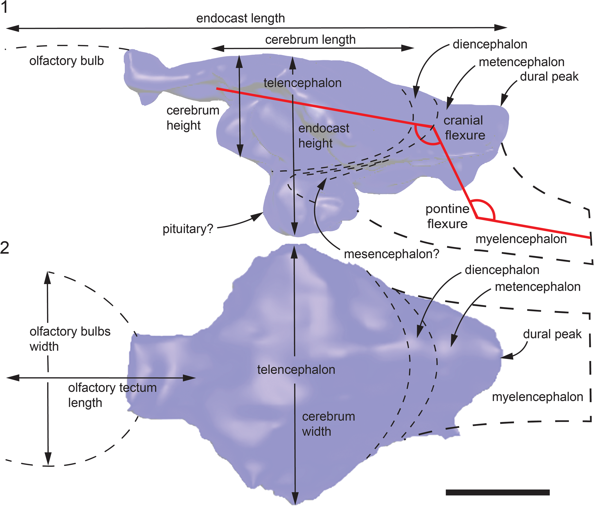
This study utilized CT scans of the braincase of Fostoria dhimbangunmal (LRF 3050.A) originally acquired by Bell et al. (Reference Bell, Brougham, Herne, Frauenfelder and Smith2019a) using a Siemens Syngo CT2012B (120 kV; slice thickness = 0.74 mm) at I-MED Radiology Armidale (formerly Armidale Radiology) (Armidale, New South Wales, Australia). The braincase and cranial endocast were volume rendered in ORS Dragonfly (version 2020.1, Comet Technologies Canada Inc., Montreal, Canada; accessed under a free academic license; https://www.theobjects.com/dragonfly). Each image in the stack was upscaled by a factor of five to increase the resolution. All linear measurements were taken in Dragonfly five times and then averaged to reduce human error. The regions of interest were manually segmented using the 3D paint tools. The thresholding function was not employed due to the inability to clearly distinguish margins between the equally attenuated opalized bone and matrix in many places. Thus, volume rendering required a degree of operator interpretation. The volume renders were exported as STL files. The meshes were repaired (to remove minor artifacts), smoothed, colorized, and organized in Meshmixer (version 3.5.474) and ZBrush (version 2022.0.2). No attempt to retrodeform the endocast was made. Figures were created using Adobe Photoshop (version 23.1.1) and Adobe Illustrator (version 26.0.3). Endocast (excluding the olfactory apparatus) and cerebrum volumes were determined by importing the meshes (repaired in Meshmixer) into the program Chitubox (version 1.9.3). Measurements taken on the cranial endocast are shown in Figure 1.
Volume-rendered cranial endocast of Fostoria dhimbangunmal in (1) lateral and (2) dorsal views. Locations of endocast measurements as indicated. Heavy dashed line indicates missing region of the hindbrain. Scale bar = 2 cm.
Repository and institutional abbreviation
The holotype skull of Fostoria dhimbangunmal (LRF 3050.A) is deposited at the Australian Opal Centre (LRF = ‘Lightning Ridge fossil’), Lightning Ridge, New South Wales, Australia.
Results
Preservation
The preserved braincase of Fostoria dhimbangunmal includes the frontals, parietal, supraoccipital, partial basisphenoid, and right paroccipital processes, as well as the incomplete left laterosphenoid and prootic (Fig. 2). The basioccipital, opisthotics, and ventral part of the right prootic are not preserved. The braincase is entirely opalized and crushed dorsoventrally, resulting in loss of details to the lateral and ventral margins of the braincase and obliteration of sutures between many of the cranial bones. This description will focus on the incomplete cranial endocast as it is preserved within the braincase (Figs. 2, 3); thus, we caution that some descriptions reflect taphonomic deformation of the endocast, including significant dorsoventral compression. Where apparent, we draw attention to these features in the description and following discussion. A detailed description of the braincase can be found in Bell et al. (Reference Bell, Brougham, Herne, Frauenfelder and Smith2019a).
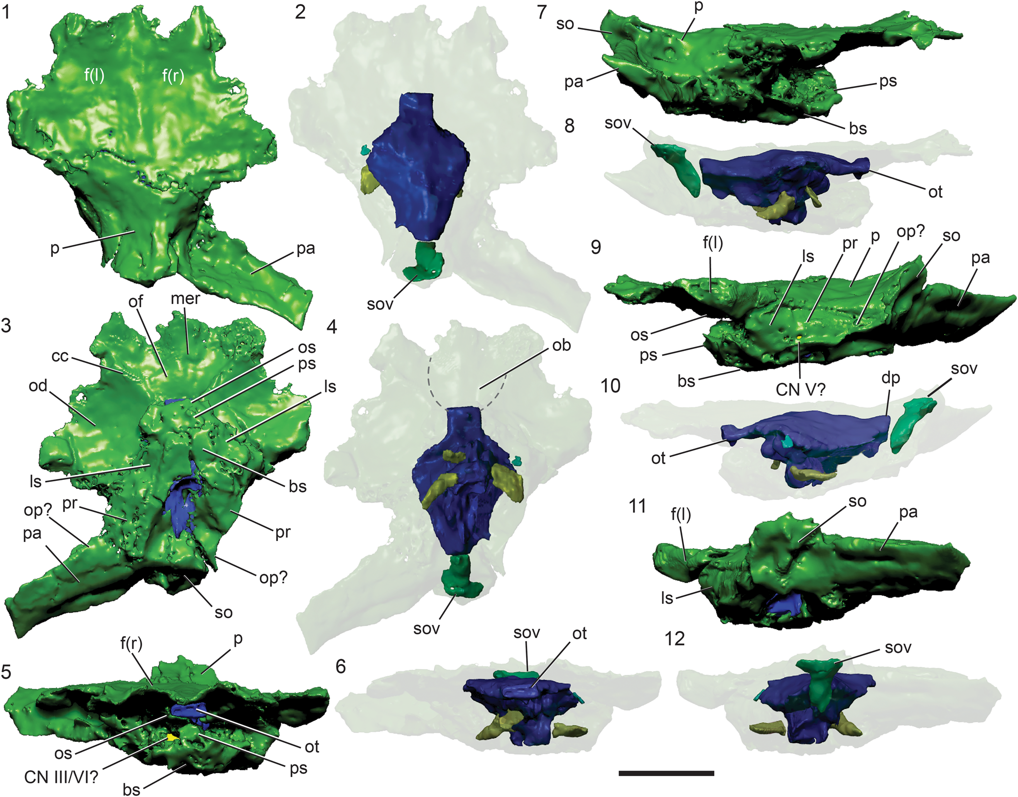
Fostoria dhimbangunmal braincase (green), cranial endocast (dark blue), and cranial nerves (yellow) in (1, 2) dorsal, (3, 4) ventral, (5, 6) anterior, (7, 8) right lateral, (9, 10) left lateral, and (11, 12) posterior views. Dashed lines in (2) and (4) represent olfactory bulb impressions on the underside of the frontals. bs = basisphenoid; cc = crista cranii; CN = cranial nerve; dp = dural peak; f (l, r) = frontal (left, right); ls = laterosphenoid region; mer = median ridge for nasal septum; ob = olfactory bulbs; od = orbital depression; of = olfactory fossa; op? = opisthotic region; os = orbiosphenoid; ot = olfactory tract; p = parietal; pa = paraoccipital process; pr = prootic region; ps = parasphenoid; so = supraoccipital; sov = supraoccipital vacuity. Scale bar = 5 cm.
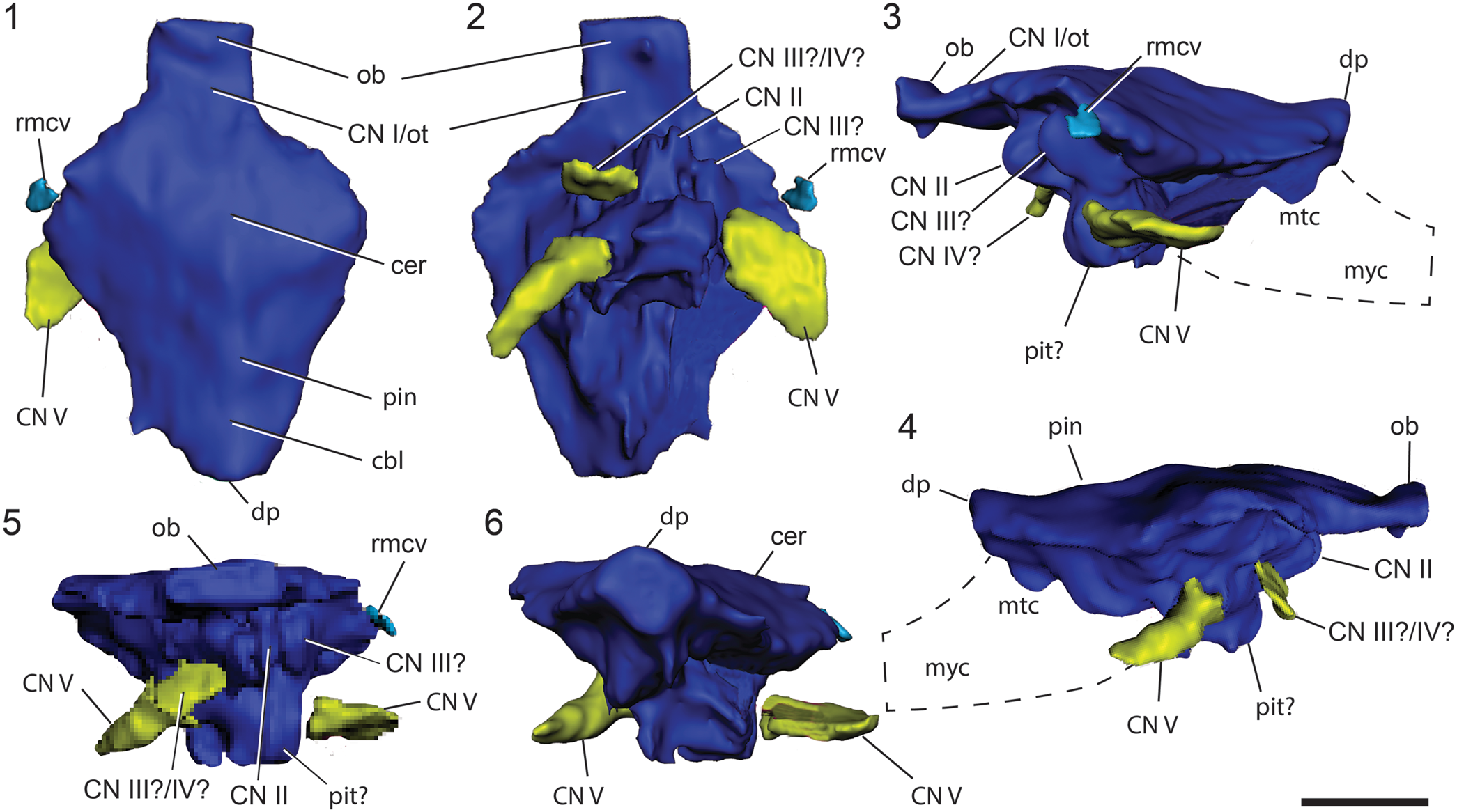
Fostoria dhimbangunmal volume-rendered neurocranial endocast (dark blue), inferred cranial nerves (yellow), and veins (light blue) in (1) dorsal, (2) ventral, (3) left lateral, (4) right lateral, (5) anterior, and (6) posterior views. The passage for cranial nerve I (the olfactory tract) is shown in dark blue. Abbreviations: cbl = cerebellum; cer = cerebrum; CN ? = possible cranial nerve; CN I/ot = cranial nerve I/olfactory tract; CN II = optic nerve; CN III? = region of the facial and vestibulocochlear nerve canals; CN III/CN IV? = region of the oculomotor and trochlear nerve canals; CN V = potential trigeminal nerve canal; dp = dural peak; mtc = metencephalon; myc = myelencephalon; ob = olfactory bulb; ot = olfactory tracts; pin = region of pineal; pit? = region of the pituitary; rmcv = rostral middle cerebral vein. Scale bar = 2 cm.
Neurocranial endocast
The dorsal aspect of the endocast is in good condition; however, the anterior, posterior, and ventral limits of the endocast, apart from the tentatively identified region of the pituitary, are incomplete. Few cranial nerves are preserved. The endocast has a maximum anteroposterior length (including the olfactory peduncles) of 75.8 mm, width of 54.0 mm, and dorsoventral height of 32.1 mm. The anteroposterior length of the endocast excluding the olfactory tracts is 63.1 mm. As preserved, the volume of the endocast is 26.3 cm3 and 25.4 cm3, with and without the olfactory apparatus, respectively. The endocast is roughly horizontally positioned within the skull, subparallel to the skull roof, which, based on the well-preserved frontals and parietals (Bell et al., Reference Bell, Brougham, Herne, Frauenfelder and Smith2019a), appears to be a real (not diagenetic) feature.
The regions of the brain represented by the endocast include the telencephalon and diencephalon of the forebrain (prosencephalon) and the posterodorsal region of the metencephalon of the hindbrain (rhombencephalon). The region of the infundibulum of the forebrain and pituitary are provisionally identified. The olfactory tracts and a posterior portion of the olfactory bulbs are preserved. The midbrain (mesencephalon), positioned between the infundibular and pituitary ventrally and hindbrain dorsally, is poorly preserved and difficult to interpret from the scans. The myelencephalon and ventral region of the metencephalon of the hindbrain are not preserved. The medulla oblongata is not preserved as a result of missing elements.
Although the endocast is dorsoventrally compressed, flexure of the brain axis is indicated. As preserved, a cranial flexure of 134° is estimated and pontine flexure of 135° is provisionally estimated, noting that the medulla oblongata is absent as a result of the missing basioccipital. In the original form, the angles of cranial and pontine flexure would have been more acute than measured. A well-developed recess is preserved posterior to the endocast within the supraoccipital and is here identified as a supraoccipital vacuity (Fig. 2). At the scan resolution available, the supraoccipital vacuity is fully enclosed in the supraoccipital and does not appear to communicate with the endocast, nor to other openings in or to the outside of the skull. It is dorsoventrally tall and anteroventrally–posterodorsally oriented, with a T-shaped profile in posterior view (Fig. 2.8, 2.10, 2.12).
The olfactory apparatus in F. dhimbangunmal is well developed (Fig. 3). The olfactory tracts form a single element that projects horizontally from the dorsal surface of the cerebrum and are surrounded by the frontals dorsally and the presphenoid ventrally and laterally (Figs. 2.4, 3.1, 3.2). The olfactory tract does not broaden in width anteriorly.
Anterior to the presphenoid, impressions of the olfactory bulbs are delineated by the divergent crista cranii on the ventral surface of the frontals, indicating that they widened and extended farther anteriorly than is suggested by the endocast alone (dashed lines in Fig. 2.4). Based on the volume-rendered endocast, the olfactory tract has a preserved anteroposterior length of 12.9 mm; however, including the anterior impressions on the frontals, the olfactory apparatus is minimally 29.3 mm in length. The maximum preserved width of the bulbs, from their anterior impressions on the underside of the frontals, is 30.7 mm.
The cerebrum is broad (Table 1) with a volume of 19.2 cm3 and makes up the largest portion of the total endocast volume, as preserved. No valleculae—endocranial vascular impressions—are preserved on the dorsal surface of the endocast, as seen in some hadrosaurids (Evans, Reference Evans2005; Lauters et al., Reference Lauters, Vercauteren, Bolotsky and Godefroit2013). However, their absence may simply be due to poor preservation/scan quality. There is no bulging or notable separation between the cerebral hemispheres. Preservation of the infundibulum and pituitary body is uncertain, but a somewhat hemispherical midline protuberance is present in the expected location of the pituitary, forming the ventral-most portion of the endocast (e.g., Galton, Reference Galton1989) (Fig. 3.3, 3.4). A dorsal protuberance is present in the region of the diencephalon at the posterior end of the forebrain, interpreted as the location of the pineal. Posteriorly from the diencephalon, the dorsal surface of the hindbrain is depressed ventrally relative to the forebrain. The width of the hindbrain narrows posteriorly. The dural peak is identified in the posterodorsal part of the hindbrain housed in the supraoccipital fossa. The midbrain, within which the optic tectum is located, is poorly preserved. The region of the myelencephalon, including the floccular fossae and lobes, is not preserved. Hence, the osseous labyrinths and cranial nerves CN VII to CN XII are not preserved.
Comparative table of iguanodontian cerebral and endocast measurements and proportions.

CRV = cerebrum volume/endocast volume; endocast volumes exclude the olfactory apparatus. *Values are tentative, calculated using the figures in Knoll et al. (Reference Knoll, Lautenschlager, Kawabe, Martínez, Espílez, Mampel and Alcalá2021).
Cranial nerves
Interpretations of the cranial nerves are challenging due to incomplete preservation of the canals in the braincase. Apart from the olfactory tracts (CN I), the largest reconstructed extension from the endocast is interpreted as the canal for the trigeminal nerve (CN V), although it is possible that these projections represent the carotid arteries. Assuming they represent the canals for CN V, the canals project posterolaterally and are visible on both sides of the endocast (Fig. 3). The three branches of the trigeminal nerve are not distinguishable, suggesting that the nerve may have split into individual branches outside the braincase, as seen in Dysalotosaurus lettowvorbecki (Lautenschlager and Hübner, Reference Lautenschlager and Hübner2013). However, such interpretations are tenuous owing to the incomplete and poor preservation of the braincase. A lateroventrally projecting canal on the right side of the endocast may have served the oculomotor (CN III) or trochlear (CN IV) nerve. Canals for the optic (CN II), abducens (CN VI), facial (CN VII), vestibulocochlear (CN VIII), glossopharyngeal (CN IX), vagus (CN X), accessory (CN XI), and hypoglossal (CN XII) nerves are not preserved, again as a result of diagenetic crushing and/or missing elements.
Discussion
The results of this study provide insights into the neuroanatomy of Fostoria dhimbangunmal and permit additional and much-needed comparative analysis between a Gondwanan iguanodontian and those known from Laurasia. Overall morphology of the endocast is similar to Laurasian non-hadrosaurid iguanodontians, including a well-developed olfactory apparatus and a large cerebrum (the broadest part of the endocast). Additionally, details of the midbrain and much of the hindbrain were missing, as is the case in most other non-hadrosaurid iguanodontians (Galton, Reference Galton1989; Lauters et al., Reference Lauters, Coudyzer, Vercauteren, Godefroit and Godefroit2012; Sobral et al., Reference Sobral, Hipsley and Müller2012; Lautenschlager and Hübner, Reference Lautenschlager and Hübner2013; Brasier et al., Reference Brasier, Norman, Liu, Cotton, Hiscocks, Brasier, McIlroy and McLoughlin2017; Knoll et al., Reference Knoll, Lautenschlager, Kawabe, Martínez, Espílez, Mampel and Alcalá2021). Notably, ventral depression of the dorsal margin of the hindbrain metencephalon (i.e., between the diencephalon and the dural peak), relative to the forebrain, is comparable to the condition in the iguanodontians Tenontosaurus tilletti (Galton, Reference Galton1989; Thomas, Reference Thomas2015), Dryosaurus altus (Galton, Reference Galton1989), and hadrosaurids (Evans et al., Reference Evans, Ridgely and Witmer2009; Lauters et al., Reference Lauters, Vercauteren, Bolotsky and Godefroit2013; Cruzado-Caballero et al., Reference Cruzado-Caballero, Fortuny, Llacer and Canudo2015). Ventral depression of the dorsal metencephalic margin is minimal in the ‘basal’ ornithopod Hypsilophodon foxii Huxley, Reference Huxley1870 (Galton, Reference Galton1989) and the non-ornithopod neornithischian Thescelosaurus neglectus Gilmore, Reference Gilmore1913 (Galton, Reference Galton1989; Button and Zanno, Reference Button and Zanno2023). In addition, the metencephalon of Fostoria dhimbangunmal is anteroposteriorly extended, as in derived ornithopods such as Tenontosaurus tilletti (Galton, Reference Galton1989; Thomas, Reference Thomas2015), Dryosaurus altus (Galton, Reference Galton1989), and hadrosaurids (Evans et al., Reference Evans, Ridgely and Witmer2009). The anteroposterior length of the metencephalon is comparatively reduced in Hypsilophodon foxii, the ‘basal’ ceratopsian Protoceratops andrewsi Granger and Gregory, Reference Granger and Gregory1923 (Brown and Schlaikjer, Reference Brown and Schlaikjer1940), and the basal neornithischian Thescelosaurus neglectus (Galton, Reference Galton1989; Button and Zanno, Reference Button and Zanno2023). Elongation of the metencephalon could be a phylogenetically useful character in the future; however, more detailed cranial endocast descriptions will be required across Ornithischia.
Degree of brain-fill
The degree to which the brain fills the endocranial cavity in dinosaurs remains unresolved. Initial paleoneurological studies assumed that dinosaur brains were similar to those of non-avian reptiles in that they filled approximately 50% of the endocranial cavity (Jerison, Reference Jerison1969; Hopson, Reference Hopson1977). However, subsequent works have demonstrated that brain-fill within the endocranium is phylogenetically dependent among non-avian dinosaurs, but can also vary with ontogeny, adult body size, and between regions of the endocast (Osmólska, Reference Osmólska2004; Evans, Reference Evans2006; Hurlbert et al., Reference Hurlburt, Ridgely, Witmer, Parrish, Molnar, Currie and Koppelhus2013; Lauters et al., Reference Lauters, Vercauteren, Godefroit, Calvey, de Sousa and Beaudet2023; Paulina-Carabajal et al., Reference Paulina-Carabajal, Bronzati, Cruzado-Caballero, Dozo, Paulina-Carabajal, Macrini and Walsh2023). Hadrosaurids are now commonly thought to have possessed a high degree of brain-fill based on the presence of valleculae on the internal surfaces of the braincase, and because the cerebral hemispheres are usually expanded, bulbous, and make up a considerable portion of the endocast volume (Evans, Reference Evans2005; Evans et al., Reference Evans, Ridgely and Witmer2009; Godefroit et al., Reference Godefroit, Escuillié, Bolotsky, Lauters and Godefroit2012; Lauters et al., Reference Lauters, Vercauteren, Godefroit, Calvey, de Sousa and Beaudet2023).
The amount to which the brain filled the endocast in F. dhimbangunmal is tentatively interpreted here, owing to the preservational state of the holotype specimen. However, like other non-hadrosaurid iguanodontians, it does not appear to possess the same degree of brain-fill within the endocranium as hadrosaurids. For instance, valleculae are not discernable on the endocast and the cerebral hemispheres are less bulbous (Evans et al., Reference Evans, Ridgely and Witmer2009; Cruzado-Caballero et al., Reference Cruzado-Caballero, Fortuny, Llacer and Canudo2015; see below), although suboptimal preservation/crushing and/or low-resolution scans could account for these differences. Further, owing to incomplete preservation of the endocast, we cannot determine whether the degree of brain-fill in F. dhimbangunmal varied across the different regions of the endocast (Evans, Reference Evans2005; Evans et al., Reference Evans, Ridgely and Witmer2009; Lauters et al., Reference Lauters, Coudyzer, Vercauteren, Godefroit and Godefroit2012; Brasier et al., Reference Brasier, Norman, Liu, Cotton, Hiscocks, Brasier, McIlroy and McLoughlin2017).
Cerebrum
As preserved, the cerebrum of F. dhimbangunmal is relatively large compared to the total endocast. The encephalization quotient, which measures the relative size of the brain (or endocast) given a particular body mass (Jerison, Reference Jerison1969) and adapted to reptiles (Hurlburt, Reference Hurlburt1996), could not be determined for F. dhimbangunmal due to the incomplete cranial endocast; its body mass is also unknown. However, cerebrum-to-endocast length and volume proportions could be obtained, providing a comparative basis (Table 1). Such values provide an estimate of relative cerebrum size, which has been linked to behavioral complexity and cognitive ability (Jerison, Reference Jerison1969; Hopson, Reference Hopson1977). Fostoria dhimbangunmal has notably high cerebral proportions, ~86% width-to-length (excluding the olfactory apparatus) and ~76% of the volume. These proportions are exaggerated due to the incomplete mid- and hindbrain regions, but they nonetheless suggest that a significant portion of the endocast was dedicated to the cerebrum. The cerebral hemispheres in F. dhimbangunmal are dorsoventrally flattened compared to the more bulbous hemispheres in other iguanodontians (e.g., Dysalotosaurus lettowvorbecki and Proa valdearinnoensis; Lautenschlager and Hübner, Reference Lautenschlager and Hübner2013; Knoll et al., Reference Knoll, Lautenschlager, Kawabe, Martínez, Espílez, Mampel and Alcalá2021), including hadrosaurids (Evans et al., Reference Evans, Ridgely and Witmer2009; Godefroit et al., Reference Godefroit, Escuillié, Bolotsky, Lauters and Godefroit2012; Cruzado-Caballero et al., Reference Cruzado-Caballero, Fortuny, Llacer and Canudo2015; Becerra et al., Reference Becerra, Paulina-Carabajal, Cruzado-Caballero and Taborda2018), but this is almost certainly the result of crushing/preservation, which is an unmistakable taphonomic feature of the Fostoria dhimbangunmal holotype.
Brain flexures
Fostoria dhimbangunmal does not possess the derived flexure pattern seen in hadrosaurids and some non-hadrosaurid iguanodontians where cranial and pontine flexures are significantly reduced or eliminated, making the endocast axis straight (Hopson, Reference Hopson, Gans, Northcutt and Ulinski1979; Giffin, Reference Giffin1989; Lauters et al., Reference Lauters, Vercauteren, Godefroit, Calvey, de Sousa and Beaudet2023). Reduced flexures (i.e., straight endocasts) are normally associated with large body size, although flexures also reduce ontogenetically in non-avian dinosaurs and crocodilians (Lautenschlager and Hübner, Reference Lautenschlager and Hübner2013; Hu et al., Reference Hu, King, Romick, Dufeau, Witmer, Stubbs, Rayfield and Benton2021). Shallow angles of flexure can also result from a rapid braincase growth rate compared to the brain (Lauters et al., Reference Lauters, Vercauteren, Godefroit, Calvey, de Sousa and Beaudet2023). In F. dhimbangunmal, both cranial and pontine flexures are subequal (~135°), as preserved; noting that diagenetic crushing has influenced the apparent flexure angles in the F. dhimbangunmal endocast and the angles would have been more acute in life. The angles of flexure are similar to those of Dysalotosaurus lettowvorbecki, which is the ancestral flexure condition observed in most dinosaur cranial endocasts (Hopson, Reference Hopson, Gans, Northcutt and Ulinski1979; Giffin, Reference Giffin1989; Lautenschlager and Hübner, Reference Lautenschlager and Hübner2013; Button and Zanno, Reference Button and Zanno2023). In the ancestral condition, where cranial and pontine flexures are subequal, the subparallel cerebral and medullary regions are separated by an oblique mid- and hindbrain region (Giffin, Reference Giffin1989). This condition also is observed in F. dhimbangunmal (Fig. 1).
Supraoccipital vacuity
A large posterior vacuity, restricted to the supraoccipital, was identified in F. dhimbangunmal. This vacuity is entirely enclosed within the supraoccipital with no clear evidence that it communicated with the endocranial space, and no other pneumatic features were observed in the skull (Fig. 2.2, 2.3, 2.10, 2.12). Although scan resolution and/or diagenetic crushing may have influenced these relationships, it nevertheless differs from the dorsal sagittal sinus in Tenontosaurus tilletti, which lies on the anterior surface of the supraoccipital (not within it) and forms the dorsal roof over the posterior part of the endocast (Thomas, Reference Thomas2015). A similar vacuity within the supraoccipital (= supraoccipital sinus) is consistently present in tyrannosaurids, although this feature is pneumatic and communicates with other cranial sinuses (Witmer and Ridgely, Reference Witmer and Ridgely2009; McKeown et al., Reference McKeown, Brusatte, Williamson, Schwab and Carr2020; Carabajal et al., Reference Carabajal, Currie, Dudgeon, Larsson and Miyashita2021). No such features have been observed in any other ornithischian, suggesting a new autapomorphy for F. dhimbangunmal. Given the relatively limited number of ornithopod skulls that have been CT scanned compared to theropods (e.g., Lauters et al., Reference Lauters, Vercauteren, Godefroit, Calvey, de Sousa and Beaudet2023), it is possible that a supraoccipital vacuity may be found in additional taxa as more specimens are investigated.
Sensory capabilities
Based on the crista cranii and the reconstructed part of the olfactory bulbs, Fostoria dhimbangunmal possessed a relatively broad olfactory apparatus (potentially as broad as the cerebral hemispheres) compared to other non-hadrosaurid iguanodontians, such as Dysalotosaurus lettowvorbecki, Iguanodon bernissartensis, Mantellisaurus atherfieldensis, and Proa valdearinnoensis (Lauters et al., Reference Lauters, Coudyzer, Vercauteren, Godefroit and Godefroit2012; Lautenschlager and Hübner, Reference Lautenschlager and Hübner2013; Knoll et al., Reference Knoll, Lautenschlager, Kawabe, Martínez, Espílez, Mampel and Alcalá2021), also differing from most hadrosaurids, except Amurosaurus riabinini Bolotsky and Kurzanov, Reference Bolotsky, Kurzanov and Moiseyenko1991 (Lauters et al., Reference Lauters, Vercauteren, Bolotsky and Godefroit2013), which had a relatively reduced olfactory apparatus compared to the cerebral hemispheres (Evans et al., Reference Evans, Ridgely and Witmer2009; Becerra et al., Reference Becerra, Paulina-Carabajal, Cruzado-Caballero and Taborda2018). The olfactory tract is relatively shorter in F. dhimbangunmal compared to D. lettowvorbecki (Lautenschlager and Hübner, Reference Lautenschlager and Hübner2013) and, as preserved, the dorsal margin of the cerebrum is close to the level of the olfactory bulbs, unlike in D. lettowvorbecki, P. valdearinnoensis, and some hadrosaurids where the cerebrum exceeds the height of the olfactory bulbs (Evans et al., Reference Evans, Ridgely and Witmer2009; Lautenschlager and Hübner, Reference Lautenschlager and Hübner2013), although we acknowledge this may have been affected by crushing in F. dhimbangunmal. Based on the relative proportions of the olfactory apparatus, F. dhimbangunmal likely possessed a strong sense of smell, assuming the olfactory apparatus is a proxy for olfaction capabilities (Zelenitsky et al., Reference Zelenitsky, Therrien and Kobayashi2008; Lauters et al., Reference Lauters, Coudyzer, Vercauteren, Godefroit and Godefroit2012; Sakagami and Kawabe, Reference Sakagami and Kawabe2020; Knoll et al., Reference Knoll, Lautenschlager, Kawabe, Martínez, Espílez, Mampel and Alcalá2021; Paulina-Carabajal et al., Reference Paulina-Carabajal, Bronzati, Cruzado-Caballero, Dozo, Paulina-Carabajal, Macrini and Walsh2023).
The midbrain, or optic tectum, was not visible in the cranial endocast of F. dhimbangunmal; however, its absence is expected given that such structures are not typically observed in ornithopod endocasts (Evans et al., Reference Evans, Ridgely and Witmer2009; Lauters et al., Reference Lauters, Coudyzer, Vercauteren, Godefroit and Godefroit2012; Lautenschlager and Hübner, Reference Lautenschlager and Hübner2013; Thomas, Reference Thomas2015; Brasier et al., Reference Brasier, Norman, Liu, Cotton, Hiscocks, Brasier, McIlroy and McLoughlin2017). Even broadly among dinosaur cranial endocasts, the midbrain is indistinguishable from other regions and obscured by the cerebellum and/or thick venous sinus(es) (Hopson, Reference Hopson, Gans, Northcutt and Ulinski1979; Thomas, Reference Thomas2015). A notable exception is in small coelurosaurian dinosaurs, in which optic lobes can be readily observed and are linked to greater visual acuity (Hopson, Reference Hopson, Gans, Northcutt and Ulinski1979; Kundrát, Reference Kundrát2007; Balanoff et al., Reference Balanoff, Bever and Norell2014). In non-iguanodontian ornithopods, similar hypotheses have been made for the cranial fragment (NMV P185990) referred to Leaellynasaura amicagraphica Rich and Vickers-Rich, Reference Rich and Vickers-Rich1989, from the Eumeralla Formation (upper Aptian–lower Albian) in Victoria (Rich and Vickers-Rich, Reference Rich and Vickers-Rich1989), noting that this referral has also been questioned based on taphonomic arguments (Herne et al., Reference Herne, Tait and Salisbury2016). Nevertheless, the optic tectum is distinguishable in this small-bodied ornithopod, suggesting comparatively strong visual capabilities, which was a possible adaptation to the prolonged dark winter months of its polar environment (Galton, Reference Galton1989; Rich and Vickers-Rich, Reference Rich and Vickers-Rich1989; Kundrát, Reference Kundrát2007; Rich et al., Reference Rich, Galton and Vickers-Rich2010; Buchholtz, Reference Buchholtz, Farlow, Brett-Surman and Holtz2012). However, F. dhimbangunmal may have been less dependent on its visual capabilities because its type locality (the Griman Creek Formation) was at a lower paleolatitude (~60°S) than the Eumeralla Formation (70–85°S) and likely experienced milder winter conditions (Rich and Vickers-Rich, Reference Rich and Vickers-Rich1989; Poropat et al., Reference Poropat, Martin, Tosolini, Wagstaff, Bean, Kear, Vickers-Rich and Rich2018; Bell et al., Reference Bell, Brougham, Herne, Frauenfelder and Smith2019a, Reference Bell, Fanti, Hart, Milan, Craven, Birch and Smithb; Kitchener et al., Reference Kitchener, Campione, Smith and Bell2019). In either case, the primitive, indistinguishable optic lobes observed in F. dhimbangunmal highlight the unique condition seen in ‘L. amicagraphica’ (NMV P185990), supporting previous visual adaptation and lifestyle hypotheses (Rich and Vickers-Rich, Reference Rich and Vickers-Rich1989), although a juvenile condition of the brain could alternatively account for exposure of the optic lobes, which may have become more embedded in the midbrain at a later ontogenetic stage (Agnolin et al., Reference Agnolin, Ezcurra, Pais and Salisbury2010). These findings further demonstrate potentially diverse evolutionary paleoneurological trajectories between penecontemporaneous Australian ornithopods and highlight the need for future endocranial reconstructions of other Gondwanan taxa.
Conclusion
This study provides the first description of the neuroanatomy of the Australian non-hadrosaurid iguanodontian, Fostoria dhimbangunmal. The braincase of F. dhimbangunmal, from which the cranial endocast was derived, is crushed dorsoventrally and only a partial cranial endocast was recovered, missing its anterior, posterior, and ventral limits. The overall morphology of the endocast is similar in some regards to other large-bodied non-hadrosaurid iguanodontians (e.g., a relatively enlarged cerebrum, anteroposteriorly extended hindbrain). However, the olfactory apparatus is well developed, possibly indicative of strong olfactory capabilities (Lauters et al., Reference Lauters, Coudyzer, Vercauteren, Godefroit and Godefroit2012; Lautenschlager and Hübner, Reference Lautenschlager and Hübner2013; Knoll et al., Reference Knoll, Lautenschlager, Kawabe, Martínez, Espílez, Mampel and Alcalá2021). The endocast of F. dhimbangunmal also differs from most hadrosaurids in its ancestral flexure pattern and less developed cerebral hemispheres, although we cannot rule out diagenetic crushing as a major influence on the observed morphology in F. dhimbangunmal. A unique vacuity within the supraoccipital may represent a new autapomorphic feature of Fostoria dhimbangunmal. The endocast of F. dhimbangunmal also differs from the only other Australian ornithopod known to have an endocast, NMV P185990 (‘Leaellynasaura amicagraphica’), highlighting the unique development of the optic lobes in the latter. We recommend that future research should investigate Gondwanan non-hadrosaurid iguanodontian paleoneurology to improve our global understanding of ornithopod behavior and brain evolution more generally.
Acknowledgments
The present research was undertaken as part of an MSc by OD and supported by the School of Environmental and Rural Science and the Destination Australia Program (DAP) Scholarship. OD would like to thank the following members of the Dinosaur Lab at the University of New England for their support and guidance: T. Frauenfelder, A. Whitebone, N. Enriquez, S. Birch, H. Henderson, E. Herbert, K. Allison, and J. Kitchener. We are indebted to J. Brammall and E. Smith (Australian Opal Centre) for permitting the scanning of Fostoria, which was performed by K. Little at I-MED Radiology Armidale (formerly Armidale Radiology) and funded by an Australian Research Council Discovery Early Career Researcher Award (project ID: DEI70101325) to PB. We thank A. Paulina-Carabajal (National Scientific and Technical Research Council, Argentina) and S. Poropat (Curtin University, Australia) who provided helpful insights and comments on a previous version of this manuscript and to J. Calede (editor), H.-D. Sues (associate editor), G. Sobral, and an anonymous reviewer for their helpful reviews that greatly improved the final version.
Competing interests
The authors declare none.
Data availability statement
Scan data available via Morphosource: https://www.morphosource.org/projects/000596887/temporary_link/VzuerxRrTFNuRqu4NbH3WKL9?locale=en


 Open access