


Introduction
An eruption on Fimmvörðuháls, the pass that lies between Mýrdalsjökul and Eyjafjallajökul glaciers in south Iceland, began in March 2010. In June, July and September of 2010, Dr. Kristján Jónasson from the Icelandic Institute of Natural History collected a number of fumarolic samples, which formed on the fresh lava at Fimmvörðuháls and sent some fragments to the X-ray Diffraction Laboratory of the Department of Geosciences and Natural Resource Management of the Copenhagen University for analyses. We recognised an unknown powder diffraction pattern in the sample collected on 28th June from a new crater called Magni. Dr. Kristján Jónasson measured the temperature at the collection spot to be 670°C.
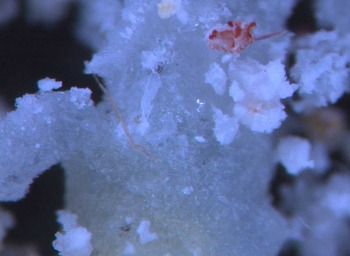
The new mineral was found in the part of the sample consisting of a seemingly glassy, greenish mass of metathénardite mixed with the new mineral and small amounts of belomarinaite (KNaSO4), aphthitalite [K3Na(SO4)2], ivsite [Na3H(SO4)2], thénardite and kröhnkite (the last two being most probably products of a transformation of metathénardite at low temperatures after collection), with several unattributed diffraction lines. Blades of belomarinaite and white globules made of tiny crystals of the new mineral overgrow the ‘glassy’ mass (Fig. 1).
White globules of kristjánite on the surface of a blade of belomarinaite, specimen 24468. The width of the photograph is 1 mm.

We named the new mineral after Kristján Jónasson. The type material is deposited in the mineral collection of the Icelandic Institute of Natural History, Urriðaholtsstræti 6–8, 210 Garðabær, Iceland, catalogue number 24468. The Commission on New Minerals, Nomenclature and Classification of the International Mineralogical Association (IMA–CNMNC) approved the mineral and its name (symbol Kjn) as IMA2022–131 (Balić-Žunić et al., Reference Balić-Žunić, Nestola and Pamato2023).
Experimental methods
Chemical analyses were performed using a Scanning Electron Microscope with Energy Dispersive Spectrometer (SEM-EDS) Quanta FEG 200 ESEM equipped with a detector from Oxford Instruments, SDD x-max 80 mm2, at the Nanocenter, Danish Technical University. The instrument has a superb resolution and is the best possible for this type of sensitive material. Conditions were: acceleration voltage = 15 kV; beam current = 0.5 nA; and beam diameter = 1.5 μm.
Powder X-ray diffraction (PXRD) measurements were carried out using a Bruker-AXS D8 Advance Powder Diffractometer, operating in the Bragg-Brentano reflection geometry. The instrument is equipped with a primary Ge111 monochromator producing a CuKα1 (1.54059 Å) radiation and a silicon-strip LynxEye detector. A small quantity of powdered material was spread on the zero-background quartz plate covered by a thin film of silica fat (to prevent the sample from gliding off the plate during measurement) embedded in a variable height sample holder. The mounted sample was gently pressed by a glass slide and adjusted on the linear focus of the instrument. The measurements were made on rotating sample in steps of 0.02°2θ with measuring time of four seconds per step, over a 2θ range of 5 to 90°.
Among the grains found in debris fallen from the sample, one hemimorphic grain showed clear X-ray diffraction and gave unit cell parameters consistent with those of the new mineral phase observed by PXRD. Single-crystal X-ray diffraction (SCXRD) was performed using a Supernova diffractometer (Rigaku Oxford Diffraction) equipped with a micro (Mo) X-ray source (wavelength 0.71073 Å) and a Pilatus Dectris 200K area detector. Details of the crystal structure, measurement and refinement are in Table 1. The CrysAlisPro 1.171.40.55a (Rigaku Oxford Diffraction, Reference Rigaku Oxford Diffraction2019) program was used for data collection, data reduction, unit cell refinement and absorption correction. Empirical absorption correction was performed using spherical harmonics, implemented in SCALE3 ABSPACK scaling algorithm. The program JANA (Petříček et al., Reference Petříček, Palatinus, Plášil and Dušek2023), including the charge-flipping method SUPERFLIP program (Oszlányi and Sütõ, Reference Oszlányi and Sütõ2004; Palatinus and Chapuis, Reference Palatinus and Chapuis2007) was used for structure solution and refinement.
Details of the crystal, SCXRD measurement and crystal structure refinement.

Results
Chemical composition
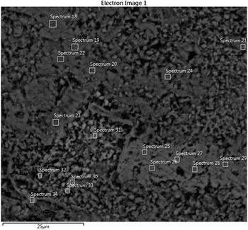
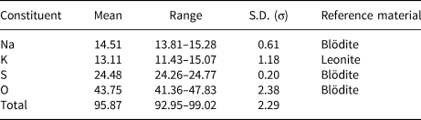
The chemical measurements were done on six spots on a polished, largely porous sample embedded in epoxy containing kristjánite and a mineral with tentative composition K2NaH(SO4)2 (Fig. 2). The results of the chemical analysis on kristjánite are given in Table 2. Besides the elements cited in the table, we also measured Mg, Al, Si, Ca, Ti, Fe and Cu. Their quantities were under the detection limits in kristjánite.
Back-scattered electron image of the sample where chemical composition of kristjánite was determined. Spots 18–20 + 22–24: kristjánite. Spots 25–29: mineral with tentative composition K2NaH(SO4)2.

Chemical data (in wt.%) for kristjánite.

S.D. = standard deviation
The empirical formula for kristjánite, calculated by the charge balance, is K0.88Na1.65HS2O7.76 (according to structural analysis, the mineral contains 1 H per formula unit). The ideal formula is KNa2H(SO4)2, which requires K 14.05 wt.%, Na 16.53 wt.%, H 0.36 wt.%, S 23.05 wt.% and O 46.01 wt.%.
Powder X-ray diffraction
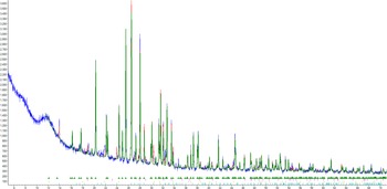
The best PXRD was obtained from the white globules from the surface of the sample. According to Rietveld quantitative calculations (using the Topas V.6 program from Bruker-AXS with fundamental parameters used to define profile shapes) it contained 86 wt.% kristjánite with 7 wt.% kröhnkite, 4 wt.% thénardite and 3 wt.% aphthitalite (Fig. 3). We suppose that kröhnkite and thénardite, which are low temperature phases, are products of the decomposition of metastable metathénardite after sample collection and preparation. The numerical PXRD data for kristjánite are in Table 3. The unit cell parameters obtained through PXRD analysis are: a = 6.9632(3) Å, b = 9.9950(4) Å, c = 11.0939(4) Å and β = 105.638(2)°.
Powder X-ray diffraction diagram of a sample containing 86 wt.% kristjánite with small amounts of kröhnkite and thénardite, probably formed from metathénardite on cooling and aphthitalite (Rietveld analysis by program Topas V.6, Bruker-AXS). Green: calculated pattern of kristjánite; blue: experimental pattern; red: theoretical pattern.

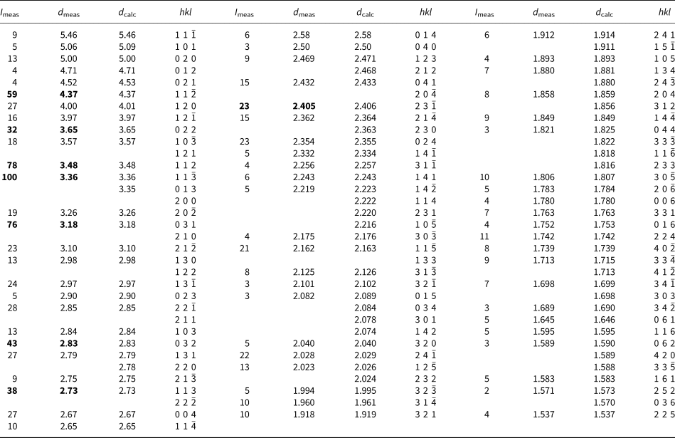
Powder X-ray diffraction data (d in Å) for kristjánite.*

* Intensities of the eight strongest diffraction maxima are in bold.
Single crystal X-ray diffraction
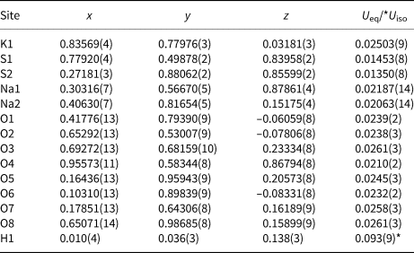
The structure solution (using the program SUPERFLIP based on the charge-flipping method) revealed the positions of all non-hydrogen atoms. The position of hydrogen was obtained from a difference-Fourier map after one isotropic refinement of the model obtained by the structure solution. The last refinement was made with anisotropic displacement parameters for all atomic sites except the H site that was kept isotropic. Fractional atomic coordinates and displacement parameters are in Table 4, other details of structure refinement are in Table 1 and in the crystallographic information file which has been deposited with the Principal Editor of Mineralogical Magazine and is available as Supplementary material (see below).
Fractional atomic coordinates and equivalent isotropic displacement parameters or isotropic displacement parameter (for H) (Å2).

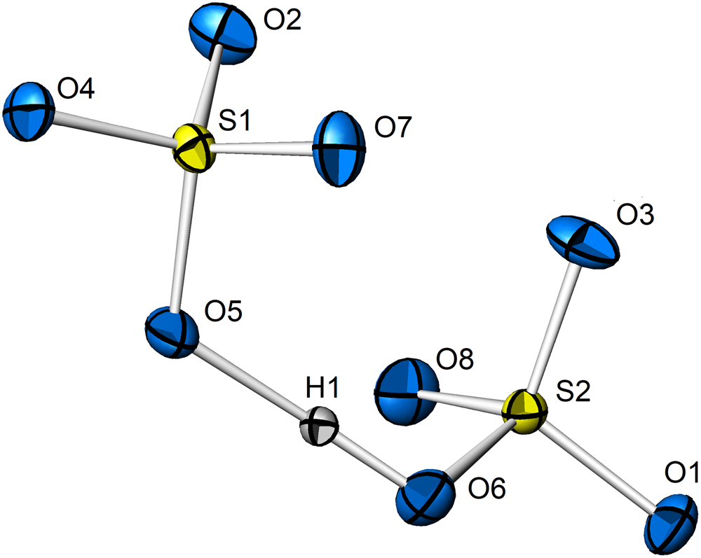
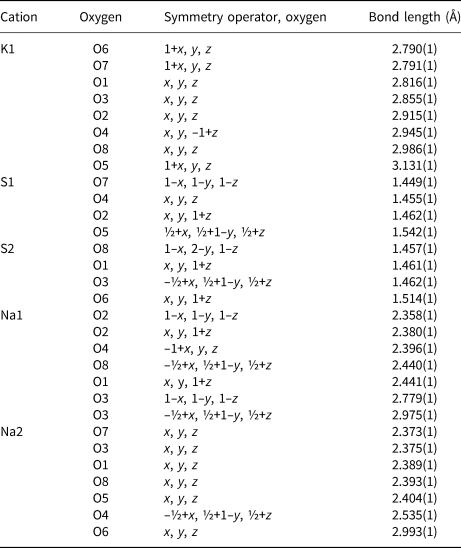
Sulfur atoms at the S1 and S2 sites are in tetrahedral coordination with three bond lengths of 1.46 Å and the fourth over 1.5 Å, which is the distance to the O atom (O5 in S1 or O6 in S2 coordination) involved in the hydrogen bond. H sits approximately on a straight line between them with distances 1.08(3) Å to O6 and 1.37(3) Å to O5, bond angle 176(3)°. Its total valence is 1.103 (from the bond valence sum). The SO4–H–SO4 dimer is shown on Fig. 4.
SO4–H–SO4 dimer. Atomic displacement ellipsoids drawn at the 50% probability level.

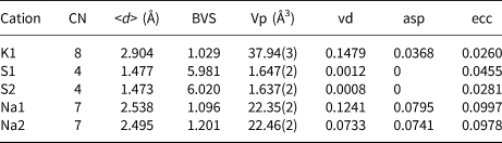
The potassium atom at the K1 site is in a distorted bis-disphenoid [coordination number (CN) 8 with bonds between 2.8 and 3.1 Å].
The sodium atoms at the Na1 and Na2 sites are in a CN 7 coordination best described as a split octahedron (Na1–O bond distances of 2.36 to 2.97 Å, Na2–O 2.37 to 2.99 Å).
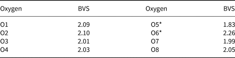
Parameters of the coordination polyhedra are in Table 5, bond valence sums for oxygens are in Table 6 and all bond lengths in Table 7 except those in hydrogen bonds, described earlier.
The parameters of the coordination polyhedra calculated with the program IVTON (Balić Žunić and Vicković, Reference Balić Žunić and Vicković1996).

Notes: CN = coordination number; <d> = average bond length; BVS = bond valence sum, calculated using the exponential function of Brown and Altermatt (Reference Brown and Altermatt1985) with the parameters of Brese and O'Keeffe (Reference Brese and O'Keeffe1991); Vp = polyhedral volume; vd = volume distortion; asp = asphericity; ecc = eccentricity.
Bond valence sums (BVS) for oxygen atoms. The oxygens that participate in the hydrogen bond are marked with a *.

The bond lengths in the coordination polyhedra of cations, hydrogen bond excluded (can be found in the text).

The general appearance of the crystal structure is the following: sulfate tetrahedra and alkali atoms are arranged in (100) parallel layers; the layers are interconnected through K–O, Na–O and hydrogen bonds; and sulfate tetrahedra and alkali atoms also form two-sulfate-tetrahedra thick slabs parallel to (001), interconnected by K–O and Na–O bonds (Fig. 5).
Crystal structure viewed along [010]. Dark violet = K atoms; magenta = Na atoms; yellow = S coordinations; blue = O atoms; grey = H atoms. One unit cell indicated. Atomic displacement ellipsoids drawn at the 50% probability level. Drawn using Atoms (www.shapesoftware.com).

The K1 and Na2 coordinations share two triangular faces and thus form relatively straight chains along [100]. Na2 coordinations additionally share a vertex with one of the neighbouring chains and thus connect chains in (010) wrapped sheets. SO4 tetrahedra interconnect the sheets. Na1 coordinations that, taken alone, form [010] zigzag chains by mutually sharing two of the vertices, additionally connect K–Na2 sheets and SO4 groups through vertex, edge and face sharing. The 3D arrangement of K, Na and S coordination polyhedra forms a tight structure that features narrow [100] channels around inversion centres at (x,0,0) and (x,½,½), lined along the two narrow opposite sides by hydrogen bonds, and along the two longer sides by K and Na2 coordination polyhedra (Fig. 6).
Crystal structure along [100]. Dark violet = K coordinations; magenta = Na coordinations; yellow = S coordinations; blue = O atoms; grey = H atoms. One unit cell indicated. Atomic displacement ellipsoids drawn at the 50% probability level.

Discussion
Kristjánite belongs to the mineral crystal structure group 7AD: anhydrous sulfates with only large cations (Strunz and Nickel, Reference Strunz and Nickel2001). It moreover contains SO4–H–SO4 dimers where a very short hydrogen bond (<2.5 Å) connects two SO4 groups. In letovicite, (NH4)3H(SO4)2 (Leclaire et al., Reference Leclaire, Ledesert, Monier, Daoud and Damak1985), Rb3H(SO4)2 (Swain and Row, Reference Swain and Row2007) and K3H(SO4)2 (Oh et al., Reference Oh, Sohn, Meven and Heger2019) the hydrogen is placed symmetrically, whereas in ivsite, Na3H(SO4)2 (Joswig et al., Reference Joswig, Fuess and Ferraris1982) and kristjánite it is asymmetrical but with a small difference between the donor and acceptor bonds (1.16, 1.28 Å in Na3H(SO4)2 and 1.08, 1.37 Å in kristjánite). The differences in crystal structures between kristjánite and other structures with (SO4)–H–(SO4) dimers are profound and it thus represents a novel structure type.
Conclusions
The new mineral, kristjánite, was found in association with metathénardite, belomarinaite, aphthitalite and ivsite at a high-temperature fumarole (≥600°C) on Fimmvörðuháls, south Iceland. Its chemical formula is KNa2H(SO4)2. It is most probably formed through a reaction between lava minerals (sanidine and augite) and SO2 and probably H2O from volcanic gas. It has a unique crystal structure for which we could not find a closely related counterpart. It contains the SO4–H–SO4 dimers featuring an almost straight linear and very short (<2.5 Å) hydrogen bond also observed in some other A3H(SO4)2 compounds (where A stands for large monovalent cations).
Acknowledgements
We thank Nina Søager from Department of Geosciences and Natural Resource Management, Copenhagen University for help in preparing the polished section and Berit Wenzel from the Danish Technical University for assistance in chemical analysis. The reviewers Peter Leverett and Igor Pekov and an anonymous reviewer gave useful comments, which improved the content. We also thank Principal Editor Stuart Mills and Associate Editor Anthony R Kempf for their professional help. The Royal Danish Library financially supported the open access publishing of the article.
Supplementary material
The supplementary material for this article can be found at https://doi.org/10.1180/mgm.2024.4.
Competing interests
The authors declare none.


 Open access