


Highlights
-
• Portable ultra low field MRI (ULF-MRI) can improve access to neuroimaging and promote equity in diverse settings.
-
• Portable ULF-MRI has shown promise in diagnosing various neurological conditions, such as multiple sclerosis and stroke.
-
• Portable ULF-MRI has great potential for neuroimaging but needs further research to improve its low SNR, which affects image quality.
Introduction
The emergence of portable ultra-low field MRI (ULF-MRI) brain scanners offers the promise of addressing accessibility gaps, affordability and long-standing health inequities in neuroimaging. Reference Deoni, Bruchhage and Beauchemin1,Reference Shen, Wolf and Bhavnani2 ULF-MRI systems typically use lighter permanent magnets, enabling a reduction in cost, size and weight. This allows for easier portability and brings imaging procedures to bedside emergency units, Reference Sheth, Yuen and Mazurek3 intensive care units (ICU) Reference Arnold, Freeman, Litt and Stein4,Reference Deoni, Medeiros and Deoni5 and remote healthcare centres and community settings. Reference Shen, Wolf and Bhavnani2,Reference Mazurek, Cahn and Yuen6 By lowering financial and technical barriers to neuroimaging, such systems open opportunities to reach new populations and expand possibilities for how and where MRI is used and by whom. Reference Arnold, Tu and Okar7,Reference Campbell-Washburn, Keenan and Hu8
Portable ULF-MRI uses magnets with a considerably lower strength than for conventional MRI, typically below 0.1 T, Reference Cawley, Padormo and Cromb9,Reference Hayashi, Watanabe and Masumoto10 which helps overcome some of the challenges associated with high-field MRIs (HF-MRIs) (i.e. 1.5 and 3T) such as immobility, special infrastructural requirements, rigid safety precautions and the need for highly trained technicians. Reference Liu, Leong and Zhao11,Reference Shoghli, Chow, Kuoy and Yaghmai12 It also has no risk of thermal burns and ferrous projectiles. Reference Shoghli, Chow, Kuoy and Yaghmai12
Furthermore, portable ULF-MRI has an affordability advantage over HF-MRI systems. This is because a ULF-MRI system costs much less to buy and to run, takes up less space, does not need a dedicated magnetically shielded room and requires less power and maintenance. Reference Deoni, Bruchhage and Beauchemin1,Reference Cawley, Padormo and Cromb9,Reference Abate, Adu-Amankwah and Ae-Ngibise13 For example, a portable ULF-MRI with a magnet strength of 0.064T could cost only 10% of a conventional MRI scanner (1.5 and 3T), which costs between $1.5 and $3 million. Reference Deoni, Medeiros and Deoni5 This high cost makes conventional MRIs unaffordable for many, resulting in over 90% of them being available only in developed countries, leaving just one MRI for every 1.25 million people in low-resource countries. Reference Liu, Leong and Zhao11,Reference Ogbole, Odo, Efidi, Olatunji and Ogunseyinde14
Existing studies have suggested that portable MRI scanners can produce clinically useful diagnostic images with comparable accuracy to high-field scanners, all the while being more cost-effective and providing greater patient comfort. Reference Savukov, Karaulanov and Castro15 For example, compared to HF-MRI, a portable ULF-MRI (0.064T) scanner was shown to be able to identify multiple sclerosis (MS) lesions with 94% accuracy, Reference Arnold, Tu and Okar7 had 80.4% sensitivity in detecting intracerebral haemorrhage,Reference Mazurek, Cahn and Yuen 6 90% accuracy in detecting ischaemic stroke (IS) Reference Yuen, Prabhat and Mazurek16 and identified midline shift in stroke patients with a sensitivity of 93% and specificity of 96%. Reference Sheth, Yuen and Mazurek3 While these findings demonstrate the clinical potential of ULF-MRI, it is important to note that its lower magnetic field strength may affect image quality, potentially hindering clinicians’ ability to make accurate decisions. Reference Kimberly, Sorby-Adams and Webb17
The major limitation of ULF-MRI is its lower magnetic field, which leads to a reduced signal-to-noise ratio (SNR). This reduction can compromise imaging resolution and cause clinicians to overlook subtle diagnostic details. Reference Altaf, Shakir and Irshad18–Reference Chetcuti, Chilingulo and Goyal20 For example, a study Reference Arnold, Tu and Okar7 compared SNR, accounting for background noise, between ULF-MRI and HF-MRI. The results showed that SNR was significantly higher in 3T images (paired t-test, t = 4.36, p = 0.00184). Besides, ULF scanners tend to have longer acquisition times, as a mechanism to compensate for their low SNR, which can result in greater image degradation due to patient movement. Reference Hayashi, Watanabe and Masumoto10 Researchers argue that despite its limitations, ULF-MRI has the potential to create the next generation of MRI that is affordable, convenient and accessible in any healthcare system. Reference Liu, Leong and Zhao11,Reference Altaf, Shakir and Irshad18
Recently, researchers have become more interested in ULF-MRI, and some studies have compared how well ULF-MRI scanners work for diagnosis, their practicality and the cost savings when compared to high-field scanners. Reference Sheth, Yuen and Mazurek3,Reference Yuen, Prabhat and Mazurek16,Reference DesRoche, Johnson and Hore21 While such a body of work is growing, to our knowledge, this is the first systematic review to establish evidence of the costs, safety and diagnostic capability of ULF-MRI. Therefore, this systematic review (a) explores the diagnostic capacity and cost viabilities of ULF-MRI compared to HF-MRI in brain imaging and (b) examines the feasibility, safety and potential limitations and challenges associated with ULF-MRI.
Methods
Overview
This study followed the Preferred Reporting Items for Systematic Reviews and Meta-Analysis (PRISMA) guidelines. Reference Page, McKenzie and Bossuyt22 A protocol was preregistered on PROSPERO (CRD42024557927). Three reviewers (GB, RT and VG) were involved in the title and abstract screening, full-text review and data extraction, as detailed below. The Rayyan software Reference Ouzzani, Hammady, Fedorowicz and Elmagarmid23 was employed for deduplication and screening based on titles and abstracts. Disagreements at any stage of the review were resolved through discussions and consensus among the three reviewers. The database search was last conducted in May 2025.
Search strategy
A comprehensive search was conducted on four databases (Medline, Embase, Scopus and Global Health) to identify studies comparing portable ULF-MRI with HF-MRI in terms of diagnostic accuracy, costs, safety and feasibility of MRI in brain imaging. The search terms employed a combination of free text and indexing terms. Besides, the reference lists of studies identified through searching the databases were also examined. Detailed search strategies can be found in the supplementary full search strategy.
Selection and eligibility criteria
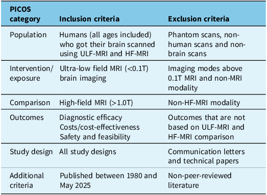
The inclusion and exclusion criteria were established based on the PICOS framework, and the search included all study designs and publications from 1980 to May 2025 (Table 1). Accordingly, articles were required to include study participants who underwent brain imaging with ULF-MRI, used HF-MRI as a comparator and reported cost-effectiveness or cost analysis, diagnostic accuracy or clinical effectiveness and safety as outcome measures. We included all types of study designs except for letters of communication, technical papers and methodological papers. Furthermore, studies involving non-human subjects such as phantoms and animals and comparators that are not HF-MRI were excluded.
Population, intervention, comparison, outcomes and study (PICOS) framework
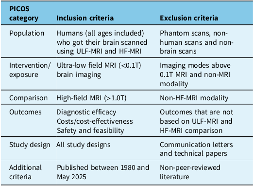
Table 1. Long description
-
• For the population category, studies involving humans of any age who underwent both ultra-low-field MRI (ULF-MRI) and high-field MRI (HF-MRI) brain imaging were included. Phantom scans, non-human scans, and non-brain scans were excluded.
-
• For the intervention or exposure category, studies involving ultra-low-field MRI brain imaging with magnetic field strengths below 0.1 Tesla were included, whereas imaging modalities above 0.1 Tesla and non-MRI modalities were excluded.
-
• For the comparison category, high-field MRI systems with field strengths greater than 1.0 Tesla were included. Studies using non-high-field MRI modalities as comparators were excluded.
-
• The outcomes of interest included diagnostic efficacy, costs or cost-effectiveness, and safety and feasibility. Studies reporting outcomes that did not involve comparisons between ULF-MRI and HF-MRI were excluded.
ULF-MRI = ultra-low field MRI; HF-MRI = high-field MRI.
Data extraction and synthesis
Data extraction was completed using a standardized summary table with the following categories: study design, year of publication, sample size, demographic information of participants, settings where the study took place and modalities of scanning. Outcome-related measures, such as diagnostic capacity (e.g. sensitivity, specificity), adverse events and cost measurements, were also extracted from each study when available. Two researchers (GB and RT) independently extracted the data, and any discrepancies between the two reviewers were resolved through discussion. In case of disagreement, a third reviewer (VG) was brought in to make the final decisions.
Due to the heterogeneity in design, outcome measures and the limited availability of quantitative data, only a narrative (thematic) synthesis approach was utilized. After extracting the data, the studies were categorized according to their main areas of focus, including economic or cost evidence, safety and diagnostic capacity. Researchers (GB, RT and VG) discussed and agreed on these grouping criteria. Subsequently, the findings were summarized descriptively, and comparisons were made within and between the studies to identify patterns, themes and key findings.
A series of discussion sessions were conducted among researchers, particularly to interpret the findings in the context of the research questions and the characteristics of the studies, such as study quality, relevance and appropriateness. This iterative discussion helped the research team shape the emerging narratives based on recurring results, trends, conflicting evidence and gaps in the literature. Disagreements were addressed through discussions or consensus. The full data set is available in the provided supplementary data.
Quality assessment
To assess the risk of bias, we used the Quality Assessment of Diagnostic Accuracy Studies 2 (QUADAS-2) tool recommended by Cochrane for reviews of diagnostic test accuracy (DTA). Reference Whiting, Rutjes and Westwood24 The extension of QUADAS-2, designed to assess risk of bias in primary studies comparing two or more tests, QUADAS-C, was applied to the selected studies, as they compare two or more modalities. The tool has four specific domains that may contribute to risk of bias: patient selection, evaluation of index tests, evaluation of reference standards and flow and timing. Each of the domains was ranked as high risk of bias, low risk of bias or unclear based on rating several specific yes/no questions.
Results
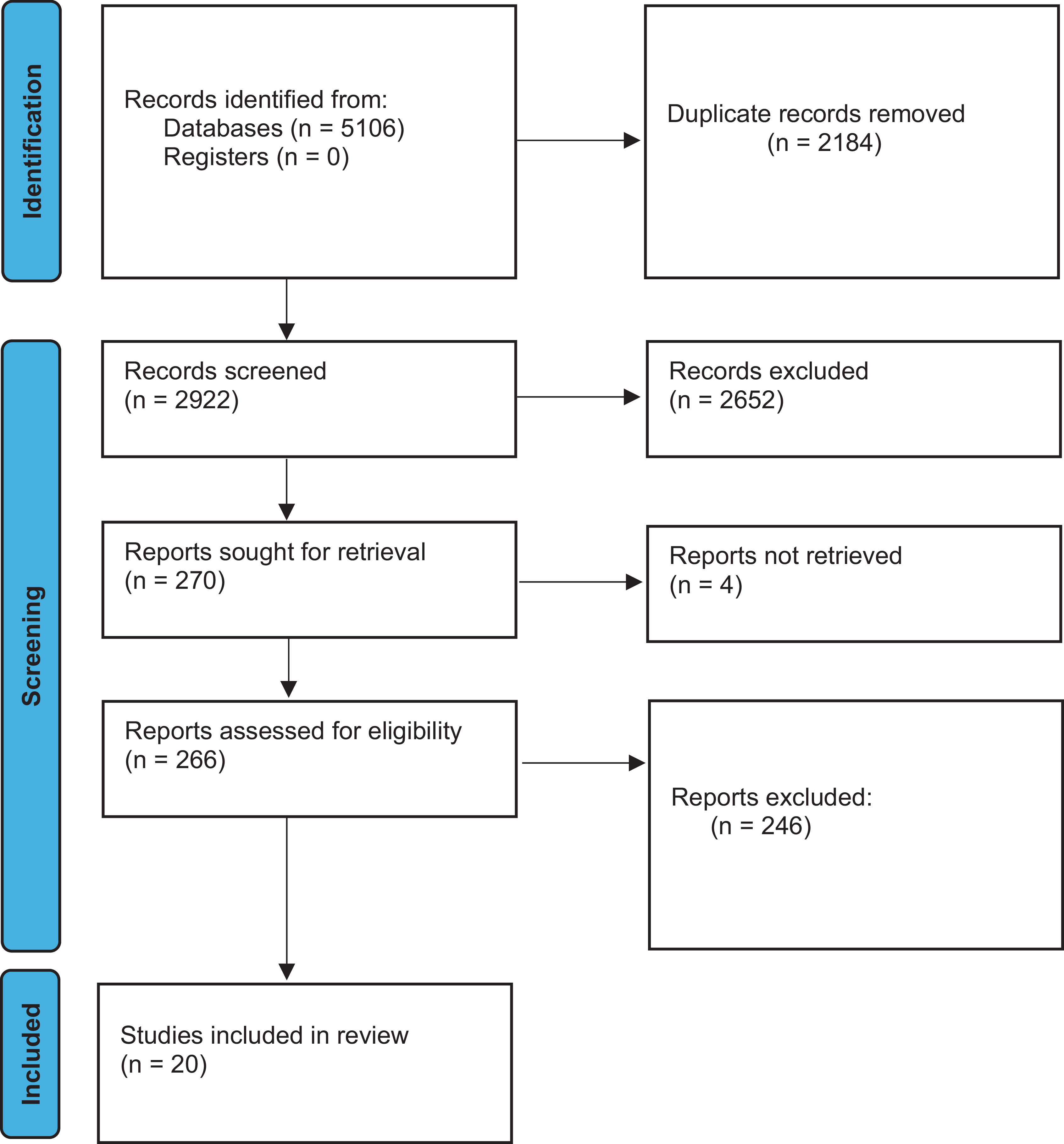
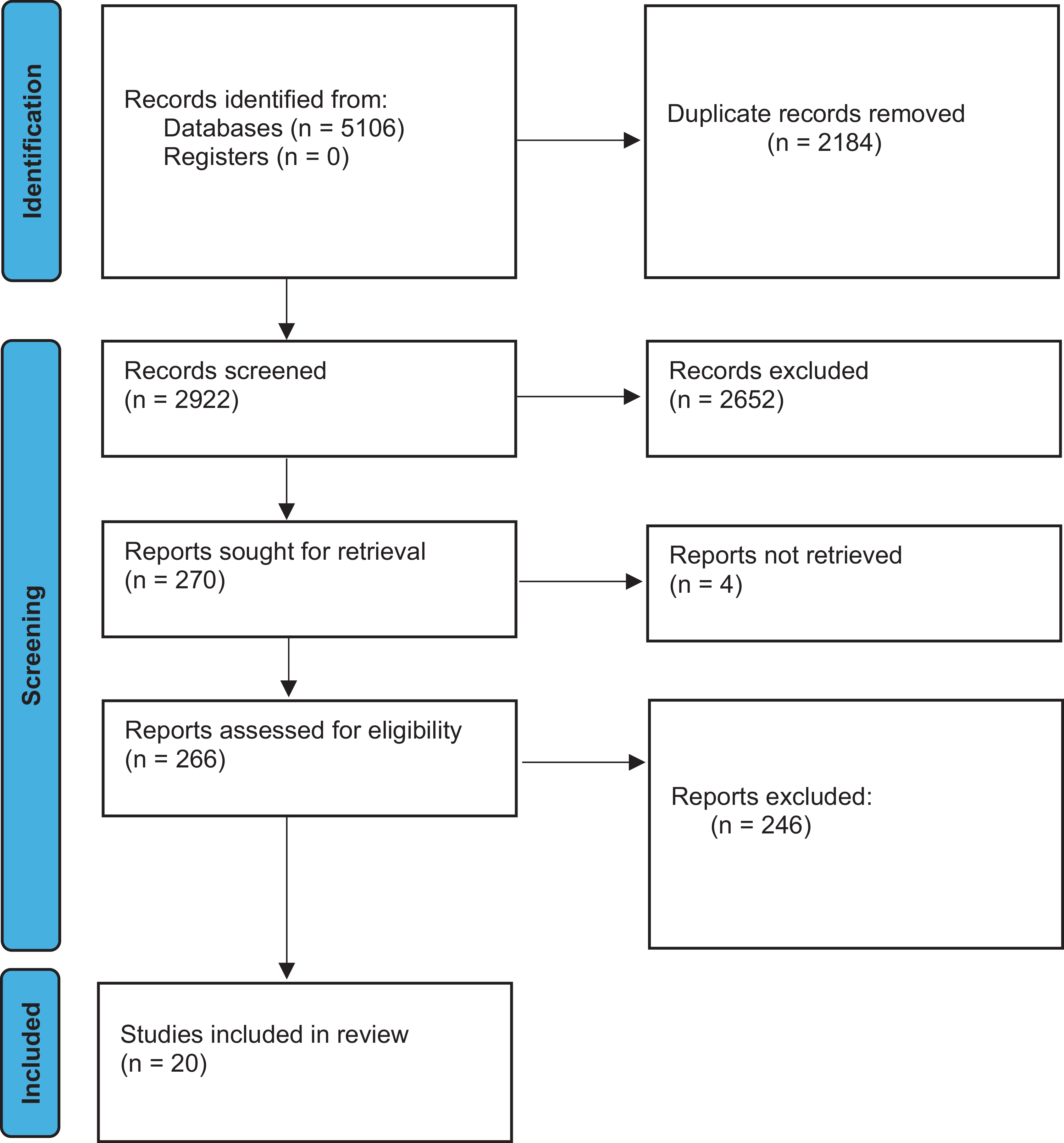
The search across the four databases identified a total of 5106 studies. After removing 2184 duplicate records, 2922 were screened based on their title and abstract (Figure 1). The Rayyan software Reference Ouzzani, Hammady, Fedorowicz and Elmagarmid23 was used to support the deduplication process. Following the screening of titles, abstracts and full texts, 20 primary studies were retained. Nineteen of these studies utilized the 0.064T Hyperfine MRI system, while one study used a 0.055T MRI scanner as the exposure. Conventional 1.5 and 3T HF-MRI systems were used as comparators.
Preferred Reporting Items for Systematic Reviews and Meta-Analysis flow chart outlining the search and review process used to identify and select articles for inclusion in this systematic review.
QUADAS appraisal
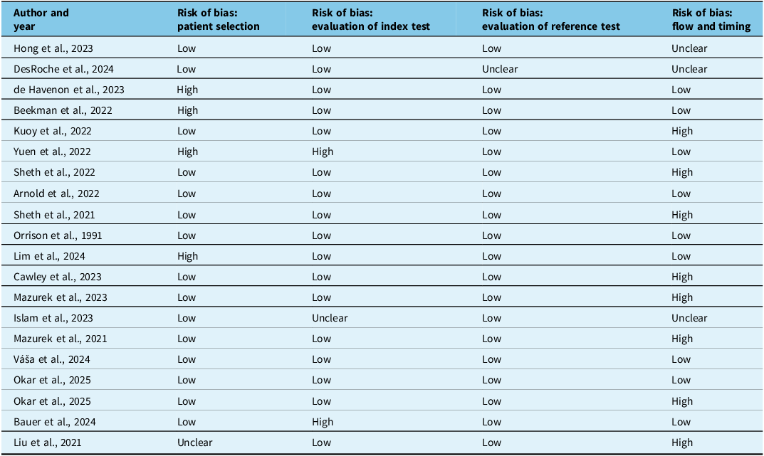
We appraised the quality of the selected studies using the QUADAS tool, and this showed a few methodological limitations. As demonstrated in Table 2, the most notable limitations included the risk of bias related to flow and timing. Most studies did not have an appropriate interval between the index test and the reference standard. In some cases, they compared tests that were months apart. Besides, in most cases, not all selected participants received a reference standard test. Moreover, a few studies showed high-risk in patient selection and evaluation of index tests, which could affect the reliability of reported diagnostic performances. While most studies were methodologically sound, limitations identified in a few studies suggest that the results should be interpreted with caution. Therefore, confidence in the synthesized evidence is moderate. GB, VG and RT were involved in assessing the quality of each study, and disagreements were resolved through consensus.
Summary of QUADAS-2 C tool quality assessment
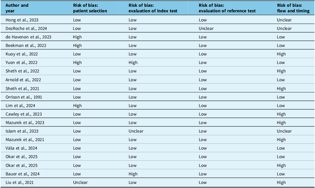
Table 2. Long description
Table 2 summarizes the quality assessment of the included studies using the QUADAS-2C tool. Twenty studies were evaluated across four domains: patient selection, evaluation of the index test, evaluation of the reference test, and flow and timing. Most studies showed a low risk of bias for the evaluation of the index test and the reference test. High risk of bias in patient selection was identified in studies by de Havenon et al. (2023), Beekman et al. (2022), Yuen et al. (2022), and Lim et al. (2024). Yuen et al. (2022) was the only study with a high risk of bias in the index test domain. The reference test domain showed predominantly low risk of bias, except for DesRoche et al. (2024), which had an unclear risk. In the flow and timing domain, high risk of bias was identified in studies by Kuoy et al. (2022), Sheth et al. (2022), and Sheth et al. (2021), while unclear risk was reported for Hong et al. (2023) and DesRoche et al. (2024). Several studies, including Arnold et al. (2022) and Orrison et al. (1991), demonstrated low risk of bias across all four domains.
QUADAS-2 = Quality Assessment of Diagnostic Accuracy Studies 2.
Characteristics of the included studies
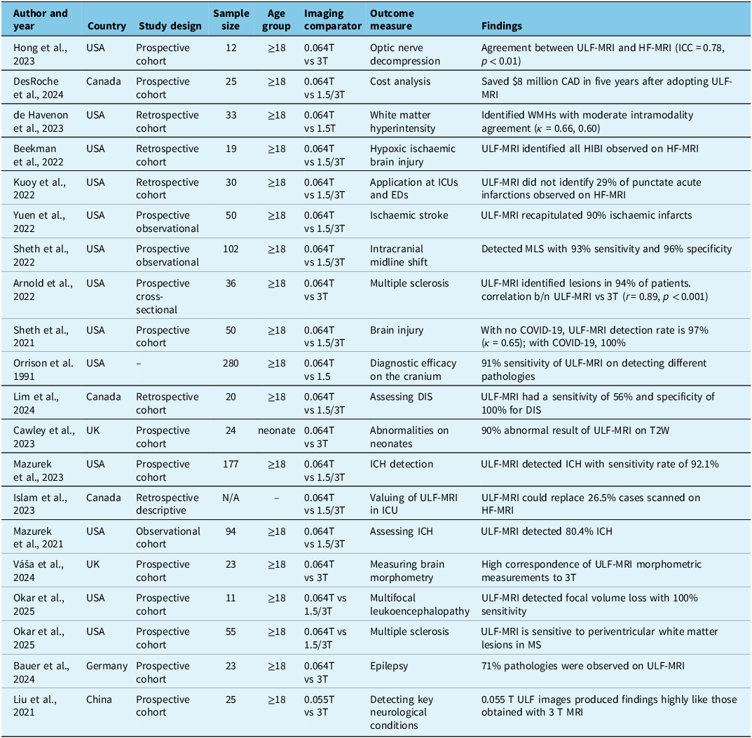
Most of the studies included were conducted in the USA (k = 13), with the rest originating from the UK (k = 2), Germany (k = 1), Canada (k = 3) and China (k = 1). Many studies Reference Sheth, Yuen and Mazurek3,Reference Arnold, Tu and Okar7,Reference Mazurek, Parasuram and Peng25–Reference Lim, Suthiphosuwan and Micieli28 employed a prospective cohort design or a retrospective hospital record review/cohort design. Two studies Reference DesRoche, Johnson and Hore21,Reference Islam, Lin and Bharatha29 used a cost analysis approach (see Table 3).
An overview of studies involving ULF-MRI applications
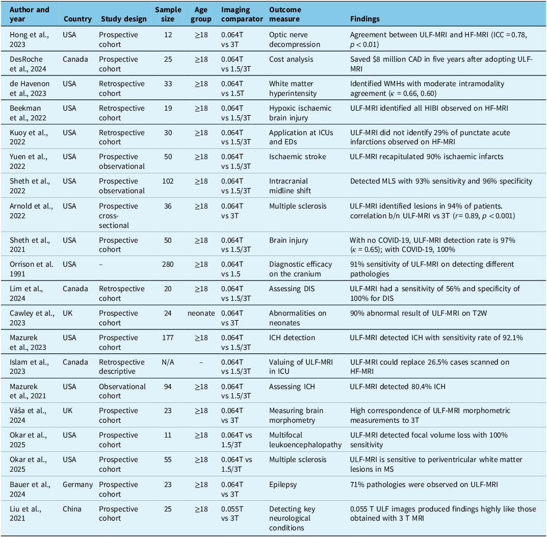
Table 3. Long description
Twenty studies conducted between 1991 and 2025 that evaluated the clinical applications of ultra-low-field MRI (ULF-MRI) has been described. Most studies were primarily in the United States, with additional studies from Canada, the United Kingdom, Germany, and China. Sample sizes ranged from 11 to 280 participants, and most studies involved adults, with one study focusing on neonates. The studies compared ULF-MRI systems with field strengths of 0.055 T or 0.064 T against conventional high-field MRI systems ranging from 1.5 T to 3 T. Outcomes assessed included diagnostic accuracy, cost analysis, feasibility, and the detection of various neurological conditions. Applications investigated included optic nerve decompression, white matter hyperintensities, hypoxic ischemic brain injury, ischemic stroke, intracranial midline shift, multiple sclerosis, intracerebral hemorrhage, brain injury, epilepsy, multifocal leukoencephalopathy, neonatal abnormalities, brain morphometry, and other neurological conditions. One study evaluated the economic impact of ULF-MRI and reported savings of approximately 8 million Canadian dollars over five years. Another study found that ULF-MRI could potentially replace approximately 26.5 percent of intensive care unit scans performed using conventional MRI. Overall, the studies demonstrated moderate to high agreement between ULF-MRI and high-field MRI. Diagnostic performance was generally favourable, with reported sensitivities ranging from approximately 56 percent to 100 percent depending on the condition being assessed. Several studies reported high sensitivity for detecting intracerebral hemorrhage, ischemic infarction, midline shift, multiple sclerosis lesions, and other neurological abnormalities.
ULF-MRI = ultra-low field MRI; HF-MRI = high-field MRI; ICC = intraclass correlation coefficient; CAD = Canadian; WMHs = white matter hyperintensities; HIBI = hypoxic ischaemic brain injury; ICUs = intensive care units; EDs = emergency departments; MLS = midline shift; COVID-19 = coronavirus disease 2019; DIS = Dissemination in Space; ICH = intracranial haemorrhage; T2W = T2-weighted; MS = multiple sclerosis; ULF = ultra-low field.
The analysis of the 20 studies included revealed several distinct themes and patterns, including feasibility and applicability to different neurological conditions and settings, safety, diagnostic capability and costs.
Feasibility and applicability
Several studies have shown that adopting and utilizing portable ULF-MRI is feasible and applicable in a variety of settings, including in emergency departments, acute care units, paediatric centres and less-resourced remote healthcare centres. Reference Yuen, Prabhat and Mazurek16,Reference DesRoche, Johnson and Hore21,Reference Mazurek, Parasuram and Peng25,Reference De Havenon, Parasuram and Crawford27 In these settings, ULF-MRI has been deployed to effectively scan a range of neurological indications, including IS, intracerebral haemorrhage and acute brain injuries.
Portable ULF-MRI has been successfully deployed in various ICUs to conduct scans on critically ill patients, including in inpatient neuro ICUs, coronavirus disease 2019 ICUs and cardiac arrest ICUs. Reference Yuen, Prabhat and Mazurek16,Reference Beekman, Crawford and Mazurek26,Reference Islam, Lin and Bharatha29–Reference Sheth, Mazurek and Yuen31 This demonstrates the applicability and feasibility of ULF-MRI technology across different environments for a wide variety of pathologies.
Moreover, ULF-MRI was deployed at the emergency department of a tertiary hospital in the USA Reference De Havenon, Parasuram and Crawford27 to identify white matter hyperintensity (WMH), a condition associated with stroke. The study showed that a diverse population can be swiftly enrolled using portable ULF-MRI, making it a valuable neuroimaging tool for estimating population-level disease conditions.
Furthermore, portable ULF-MRI has also been deployed and utilized in a paediatric setting. Transporting neonates to conventional MRI rooms with the commonly used neonatal ICU support equipment is not suitable and safe. One study Reference Cawley, Padormo and Cromb9 reported that a 0.064T ULF-MRI successfully produced images with sufficient contrast to differentiate white matter and grey matter in infants.
Finally, portable ULF-MRI technology has been successfully deployed and utilized in a remote healthcare centre where there was no conventional MRI system. A study conducted in Canada Reference DesRoche, Johnson and Hore21 examined the adoption of ULF-MRI in a remote hospital that serves over 12,000 predominantly indigenous people. In this community, patients needing brain imaging were usually transported over 800 km to access an MRI scan. The study reported that out of 25 patients imaged at this centre, more than half (56%) would not require transfer to a facility with HF-MRI capabilities.
Diagnostic capability
Several studies have reported the diagnostic capacity of portable ULF-MRI on various clinical applications, including epilepsy, Reference Bauer, Olbrich and Groteklaes32 MS, Reference Arnold, Tu and Okar7,Reference Okar, Nair and Kawatra33 stroke Reference Yuen, Prabhat and Mazurek16 and acute brain injury. Reference Sheth, Mazurek and Yuen31 It has also been used to monitor and identify further brain infarcts after post-operative and post-cardiac arrest procedures. Reference Beekman, Crawford and Mazurek26,Reference De Havenon, Parasuram and Crawford27
Portable ULF-MRI was able to detect 71% of potentially epileptogenic pathologies known from clinical HF-MRI. Reference Bauer, Olbrich and Groteklaes32 A complete diagnosis was possible in approximately half of the cases (50%), indicating that all relevant information from the clinical HF-MRI reading was also present in the ULF-MRI report for those cases. However, researchers reported that both grey and white matter SNRs, as well as the contrast-to-noise ratio (CNR), were significantly lower on ULF-MRI, which contributes to reduced quality of the images and therefore difficulties with diagnosis.
ULF-MRI successfully detected ischaemic infarcts in 90% of patients with a sequence-specific sensitivity of 98% for T2-weighted (T2W) imaging, 100% for Fluid Attenuated Inversion Recovery (FLAIR) and 86% for diffusion-weighted imaging (DWI). Reference Yuen, Prabhat and Mazurek16 This study also found a strong correlation in stroke volume between ULF-MRI and HF-MRI measurements for T2W (intraclass correlation coefficient [ICC] = 0.994, 95% confidence interval [CI]: 0.986–0.997), FLAIR (ICC = 0.989, 95% CI: 0.976–0.995) and DWI (ICC = 0.940, 95% CI: 0.871–0.972) sequences. Additionally, ULF-MRI was able to identify and quantify intracranial midline shift (MLS) in patients with intracranial haemorrhage (ICH) and IS with a sensitivity of 93% and specificity of 96%. Reference Sheth, Yuen and Mazurek3 This study observed a significant agreement between ULF-MRI and HF-MRI measured MLS assessments (κ = 0.87, p = 1.7 × 10⁻¹² for dichotomous measurements; ICC = 0.94, p = 5.9 × 10⁻²³ for continuous measurements).
Two other studies showed that ULF-MRI could detect intracranial haemorrhage with a sensitivity rate of 80.4% Reference Mazurek, Cahn and Yuen6 and 92.1%. Reference Mazurek, Parasuram and Peng25 The former study demonstrated a strong correlation (ICC = 0.955) between haematoma volumes on ULF-MRI and conventional imaging volumes. In the latter study, raters received ULF-MRI images along with the patients’ clinical information, which improved the accuracy of their assessments. Reference Mazurek, Parasuram and Peng25
A newly developed 0.055T ULF-MRI in China was investigated to assess its ability to diagnose patients with major neurological diseases such as brain tumours, IS and intracerebral haemorrhage. According to the study, the 0.055T ULF-MRI demonstrated excellent sensitivity in detecting brain pathologies and displayed good image correspondence with HF-MRI images. Reference Liu, Leong and Zhao11 For example, in a case of brain tumour, both 0.05T and 3T images revealed a mass in the right parietal cortex that appeared hypointense in T1W images and hyperintense in T2W images.
ULF-MRI has been shown to be capable of identifying moderate to severe WMHs in point-of-care settings. Reference De Havenon, Parasuram and Crawford27 The study reported that ULF-MRI can detect moderate to severe levels of WMH with moderate agreement (κ = 0.66) compared to HF-MRI. In another point-of-care context, ULF-MRI identified 71% of punctate acute infarctions observed on HF-MRI examination. Reference Kuoy, Glavis-Bloom and Hovis30 According to this study, missed punctuate acute infractions were measuring 4 mm or less in the maximal axial dimension, indicating limitations in image resolution and in detecting small lesions and their locations.
Two identical 0.064T ULF-MRI scanners were employed to measure test–retest reliability and the correspondence of brain morphometric measurements with HF-MRI. Reference Váša, Bennallick and Bourke34 This study found high reliability between both 0.064T scanners with ICC values of 0.98–1.0 for global structure/tissue volumes and a median ICC of 0.96 for local structures. It also showed that morphometric measurements extracted from 0.064T scans have excellent correspondence to HF-MRI, with Pearson’s r values ranging from 0.95 to 1.0 for global structures and a median correlation of r = 0.94 for local/individual structures.
Several studies Reference Arnold, Tu and Okar7,Reference Lim, Suthiphosuwan and Micieli28,Reference Okar, Nair and Kawatra33 have examined the performance of ULF-MRI in detecting lesions associated with MS. For example, ULF-MRI successfully detected white matter lesions in 94% of confirmed MS patients, using 3T MRI as ground truth. Reference Arnold, Tu and Okar7 This study revealed a strong correlation in lesion volumes between ULF-MRI and HF-MRI (r = 0.89, p < 0.001). The average size of the smallest manually detected lesions was found to be 5.7 ± 1.3 mm in maximum diameter at 0.064 T compared to 2.1 ± 0.6 mm at 3 T, approaching the spatial resolution of the respective scanner sequences (3 T: 1 mm, 0.064 T: 5 mm slice thickness). Reference Arnold, Tu and Okar7
Furthermore, ULF-MRI has been utilized to diagnose Dissemination in Space (DIS) in an MS clinic. Reference Lim, Suthiphosuwan and Micieli28 The results showed high specificity (100%) but low sensitivity (56%) due to limitations in depicting small lesions. There was also moderate concordance between ULF-MRI and HF-MRI for DIS (80%, k = 0.58). Lastly, a study Reference Okar, Nair and Kawatra33 indicated that ULF-MRI consistently exhibited high sensitivity in detecting at least one white matter lesion, particularly in the periventricular region, in established cases of MS. In this study, two raters showed good case-level sensitivity for detecting at least one white matter lesion (90% for both).
Portable ULF-MRI has also been deployed as a prognostic tool to evaluate optic nerve decompression after endoscopic endonasal surgery Reference Hong, Lamsam and Yadlapalli35 and detect hypoxic ischaemic brain injury (HIBI) after cardiac arrest. Reference Beekman, Crawford and Mazurek26 In the former study, ULF-MRI demonstrated decompression of the optic chiasm following surgery with a strong agreement between ULF-MRI and HF-MRI (ICC = 0.78, p < 0.01). This suggests that ULF-MRI has the potential for broader application, and using such a scanner could be an alternative strategy when intraoperative MRI is not available. In the second study, all patients (63%) who had HF-MRI evidence of HIBI after cardiac arrest were found to have HIBI upon ULF-MRI examination.
The sensitivity and specificity of ULF-MRI were compared with HF-MRI in identifying various cranial central nervous system diseases. The overall sensitivity of ULF-MRI in identifying any abnormality was 91%, with a 64% specificity. Reference Orrison, Stimac and Stevens36 This study reported that the sensitivity of ULF-MRI was statistically indistinguishable from that of HF-MRI in diagnosing white matter disease and neoplasms.
Finally, the capabilities of ULF-MRI have also been tested in neonatal brain imaging. Reference Cawley, Padormo and Cromb9 According to this study, ULF-MRI provided abnormal results in 90% of T2W scans and 70% of T1W scans compared to HF-MRI. In the T1W scans, ULF-MRI failed to detect pathologies, such as periventricular cysts, small subependymal cysts, products of an old germinal matrix haemorrhage and white matter punctate lesions. Reference Cawley, Padormo and Cromb9 The study found that these undetected pathologies on ULF-MRI were typically small or subtle in nature.
Cost implications of ULF-MRI technology
While no studies have conducted a full economic evaluation or cost-effectiveness analysis, a limited number of studies have assessed cost-related issues of portable ULF-MRI. A cost analysis study Reference DesRoche, Johnson and Hore21 conducted in Canada reported that if portable MRI were available for routine clinical use, 56% of patients would not require transfer to a centre with HF-MRI. The deployment of ULF-MRI for one year resulted in a cost saving of $854,841 (in 2022 Canadian dollars) per 50 patients. Additionally, travel costs could be averted, saving $889,500 in transportation expenses from the community centre to the location of the conventional MRI system. Furthermore, a budget impact analysis based on a five-year adoption model projected total savings of $7,835,162 if 100 patients per year could use ULF-MRI by the fifth year (DesRoche et al., 2023).
Another study Reference Islam, Lin and Bharatha29 demonstrated that the adoption of portable ULF-MRI in an ICU setting for select neurological conditions such as encephalopathy, seizures and focal neurological deficits could replace fixed MRIs in 26.5% of cases. Therefore, the utility of ULF-MRI will free up time on HF-MRI, allowing an additional 324 patients to be scanned on it annually.
Safety and adverse events
All studies that examined portable ULF-MRI in different contexts and clinical conditions reported no adverse events or complications associated with portable ULF-MRI scanning. For example, patients were scanned without being disconnected from ICU monitoring equipment, including cooling gel pads and infusion pumps. Reference Mazurek, Cahn and Yuen6,Reference Beekman, Crawford and Mazurek26
Other studies Reference Yuen, Prabhat and Mazurek16,Reference Sheth, Mazurek and Yuen31 reported that ULF-MRI scanning was successfully performed in environments with equipment containing ferromagnetic materials, such as standard monitoring devices, mechanical ventilators and haemodialysis machines, without any adverse events. Clinical staff and ULF-MRI operators were able to freely enter and exit the patient’s room without any risk of projectiles. Reference Mazurek, Cahn and Yuen6,Reference Beekman, Crawford and Mazurek26
In a post-cardiac arrest examination of brain injury using portable ULF-MRI, no adverse events or complications were reported. ULF-MRI scans were completed in all patients without disrupting ICU treatment and monitoring processes. Reference Beekman, Crawford and Mazurek26
Ease of operation
Many of the studies included in this review have highlighted the major advantage of ULF-MRI technology as being user-friendly and patient-centric. For example, setting up a new ULF-MRI in a remote hospital in Canada only required three hours, with no changes to infrastructure needed. Reference DesRoche, Johnson and Hore21 It also did not require specialized technicians to operate the technology, needing only a 45-minute training session for existing staff members.
ULF-MRI power consumption during scanning is very low (<1650 W), Reference Sheth, Yuen and Mazurek3,Reference Liu, Leong and Zhao11 which is significantly lower than the energy consumption of HF-MRI (>19,900 W). Reference DesRoche, Johnson and Hore21 This enables the technology to run using a standard power supply available in any room Reference Liu, Leong and Zhao11,Reference Sheth, Mazurek and Yuen31 and allows the technology to be easily deployed in resource-limited settings, particularly in areas with limited power supply. In addition, images from the ULF-MRI can be easily uploaded to – and automatically analysed in – a cloud platform, allowing radiologists to access data and results from anywhere. Reference Hong, Lamsam and Yadlapalli35
Challenges of imaging with ULF-MRI system
Many studies in this review have highlighted the challenges facing portable ULF-MRI, particularly in relation to its reduced image quality compared to HF-MRI. Reference Arnold, Tu and Okar7,Reference Yuen, Prabhat and Mazurek16,Reference Bauer, Olbrich and Groteklaes32 ULF-MRI images are characterized by lower resolution, reduced contrast between tissue types and significantly lower grey and white matter SNR and CNR due to the lower magnetic field. Reference De Havenon, Parasuram and Crawford27,Reference Bauer, Olbrich and Groteklaes32,Reference Váša, Bennallick and Bourke34 The low SNR and image resolution ultimately affect robust clinical decisions and the ability of ULF-MRI to differentiate various tissues and pathologies. Reference Liu, Leong and Zhao11
A study revealed that the SNR of portable ULF-MRI is not high enough to provide accurate gradation of white matter hyperintensities, especially when trying to determine how severe they are. Reference De Havenon, Parasuram and Crawford27 The reduced SNR issue of portable ULF-MRI also resulted in a low detection rate of small foci Reference Yuen, Prabhat and Mazurek16 and poorer delineation of CSF and brain parenchyma. Reference Okar, Nair and Kawatra33,Reference Dirgahayu, Ilyas and Rahma37 Additionally, based on bias-field magnitude measurement of image quality, a study indicated that ULF-MRI had a larger bias-field magnitude than HF-MRI (mean = 0.97 vs 0.27, t22 = 6.54, p < 0.001). Reference Bauer, Olbrich and Groteklaes32 A larger bias field is typically linked to lower image quality, which may impact the interpretation and analysis of abnormalities. Another study assessed ULF-MRI image quality by calculating lesion conspicuity, SNR and CNR. This assessment indicated that lesion conspicuity was preserved in the images, while SNR and CNR, which account for background noise, were found to be significantly lower in ULF-MRI. This suggests there is similar lesion-to-tissue contrast between the ULF and HF sequences but more noise in low-field images. Reference Arnold, Tu and Okar7
Discussion
This systematic review discusses the diagnostic capability, costs, safety and feasibility of portable ULF-MRI in brain imaging. The review highlights the prospects for broader applications of ULF-MRI in various settings and clinical conditions. In addition, it examines the benefits and limitations of ULF-MRI in real-life clinical applications.
The findings of this review indicate that portable ULF-MRI is applicable and feasible in nontraditional settings, such as emergency rooms, ICUs, paediatric units, interventional surgery and under-resourced remote healthcare centres. Reference Yuen, Prabhat and Mazurek16,Reference Mazurek, Parasuram and Peng25,Reference Sheth, Mazurek and Yuen31 In these contexts, ULF-MRI can be used for diagnosing and monitoring a wide variety of brain-related pathologies, including stroke, MS, trauma and neurodegenerative diseases. Reference Arnold, Tu and Okar7,Reference Cawley, Padormo and Cromb9,Reference Mazurek, Parasuram and Peng25,Reference Sheth, Mazurek and Yuen31 This advancement and clinical applicability have multiple benefits, such as mitigating potential secondary risks during transportation to a conventional MRI, expediting neurological emergencies, screening high-risk individuals and meeting unaddressed neuroimaging needs in resource-limited settings.
ULF-MRI has the potential to expand access to neuroimaging in nontraditional circumstances primarily due to its low risk of adverse events and safety concerns. None of the studies reviewed reported any safety issues, even when imaging was performed in the presence of standard hospital medical equipment and ferromagnetic materials nearby. This enables patients with active implants and life support medical equipment to safely undergo ULF-MRI scans. For example, patients on extracorporeal membrane oxygenation (ECMO), those on ECMO with an intra-aortic balloon pump, and patients with a pressure monitoring intracranial bolt were safely scanned using ULF-MRI. Reference Chinedozi, Boskamp and Darby38–Reference Wilcox, Acton and Rando40 However, a phantom experiment, not included in this review, showed that a HeartMate 3 left ventricular assist device system shut off 40 cm from the centre of the scanner, where the magnetic field increased the workload of the device. Reference Khanduja, Rando and Chinedozi41 This suggests that devices should still be thoroughly tested individually.
Portable ULF-MRI systems require substantially lower installation and maintenance costs, reduced power consumption and smaller footprints and do not need shielding or cryogenic cooling. Reference DesRoche, Johnson and Hore21,Reference Samardzija, Selvaganesan and Zhang42 This offers a promising option to shift from the current high-cost and low-access HF-MRI to high-access and low-cost options. This is particularly significant in developing economies and low-resource settings where access to conventional MRI is limited. Low- and middle-income countries such as Nigeria and Ghana have 0.30 and 0.48 MRI units per million people, while high-income countries such as Japan and the USA have 51.67 and 38.96 units per million, respectively. Reference Ogbole, Odo, Efidi, Olatunji and Ogunseyinde14,Reference Baljer, Zhang and Bourke43 Affordable and less complex ULF-MRI systems can make MRI accessible and economically viable in such environments. Reference Altaf, Shakir and Irshad18 However, an optimal trade-off between the cost and diagnostic capability is crucial for clinical adoption. Reference Liu, Leong and Zhao11 Further optimization in image quality and consideration of contextual, organizational and systemic constraints of underserved regions in the adoption process could facilitate efficient clinical applicability.
Clinical implications
Portable ULF-MRI is increasingly utilized in neurocritical care settings, especially in detecting pathologies related to intracranial haemorrhage, IS Reference Mazurek, Cahn and Yuen6,Reference Yuen, Prabhat and Mazurek16,Reference Mazurek, Parasuram and Peng25,Reference Sheth, Mazurek and Yuen31 and MS, Reference Arnold, Tu and Okar7,Reference Lim, Suthiphosuwan and Micieli28,Reference Okar, Nair and Kawatra33 with reported diagnostic accuracy ranging from 56% to 98%. This suggests that with further optimization and testing, ULF-MRI has the potential to become a valuable diagnostic and monitoring tool that could revolutionize neuroimaging. It can complement HF-MRI or be used in place of it in areas with limited resources where conventional MRI is unavailable.
The clinical applicability of ULF-MRI may be most effective in contexts such as follow-up point-of-care imaging for established or suspected cases, frequent scanning to manage disease progression or evaluating responses to clinical interventions. Reference Arnold, Freeman, Litt and Stein4 In addition, ULF-MRI has considerable potential applications for the subset of patients that face multiple barriers to receiving MRI scans, such as availability, cost, distance, clinical status and inconvenience of traditional MRI scanning. For instance, this technology can be used as a monitoring and prognostic tool in post-surgery, post-cardiac arrest and post-thrombectomy situations, Reference Beekman, Crawford and Mazurek26,Reference Hong, Lamsam and Yadlapalli35,Reference Sujijantarat, Koo and Jambor44 where serial imaging with HF-MRI is not feasible due to cost or safety risks. Moreover, the open design of ULF-MRI can provide scanning access to claustrophobic patients, who make up over 3% of all patients Reference Dewey, Schink and Dewey45,Reference Eshed, Althoff, Hamm and Hermann46 and who may not be able to undergo traditional MRI scans due to claustrophobia.
ULF-MRI can also be used in economically challenged healthcare settings in a similar role as high-field systems. Reference Samardzija, Selvaganesan and Zhang42 Due to limited trained professionals in such settings and the simplicity of operating ULF-MRI, its clinical application would have an immense benefit. Reference Arnold, Tu and Okar7 However, as an evolving technology, ULF-MRI has not yet been rigorously validated for many disease conditions. This limits its current ability to replace the roles of HF-MRI in brain imaging, even if ULF-MRI is accessible and affordable.
ULF-MRI has shortcomings in key aspects of MRI acquisitions, such as low spatial resolution, high levels of noise and artefacts in images and lengthy scan time. Reference Altaf, Shakir and Irshad18 This affects image quality and the diagnostic accuracy of subtle lesions. However, researchers are exploring different techniques to enhance the quality of images. One such technique is super-resolution, which aims to increase the resolution of an imaging system. Reference Kimberly, Sorby-Adams and Webb17,Reference Koonjoo, Zhu, Bagnall, Bhutto and Rosen47 Machine learning techniques are used in this process to predict a high-resolution image from a low-resolution input Reference Kundu, Mohseni Salehi and Cahn48,Reference Zhao, Ding and Lau49 . Besides, a dual acquisition 3D super-resolution model was developed and applied to 0.055T images, significantly enhancing image spatial resolution and suppressing noise/artefacts. Reference Lau, Xiao and Zhao50
Limitations and future work
This systematic review has a few limitations. The studies included are extremely heterogeneous in terms of design, sample size, outcomes measured, clinical conditions examined and methods of analysis. As a result, conducting a meta-analysis was not feasible. Of the five studies initially identified for a potential meta-analysis, only one provided complete diagnostic accuracy data; others had missing specificity values, methodological issues or incomplete statistical reporting, which prevented meaningful statistical pooling.
According to PRISMA-DTA, Reference McInnes, Moher and Thombs51 a minimum requirement for pooling is complete 2 × 2 table data (true positives [TP], false positives [FP], true negatives [TN], false negatives [FN]) along with CIs. Only Lim et al. Reference Lim, Suthiphosuwan and Micieli28 provided such information (TP = 5, FP = 0, TN = 11, FN = 4). The Mazurek studies Reference Mazurek, Cahn and Yuen6,Reference Mazurek, Parasuram and Peng25 cannot contribute specificity data because they only enrolled patients with confirmed disease, creating case-only designs that fundamentally lack TN and FP data. The Okar study Reference Okar, Nair and Kawatra33 presents significant methodological concerns by using interquartile ranges instead of CIs, while Yuen Reference Yuen, Prabhat and Mazurek16 only reported sensitivity values without corresponding specificity, predictive values or CIs. As these missing data mechanisms indicate that the data is Missing Not At Random, it is not possible to use an imputation method to impute missing data, and advanced hierarchical models designed to handle missing diagnostic accuracy data require at least 4–5 studies with complete information for stable estimation. Reference Deeks, Bossuyt, Leeflang and Takwoingi52
There is also excessive clinical heterogeneity in the studies considered here, which undermines any rationale of pooling. The studies target fundamentally different neurological conditions across distinct anatomical regions and pathophysiological processes. This clinical diversity violates core assumptions of diagnostic accuracy meta-analysis, where pooled estimates should represent performance for a clinically coherent diagnostic question.
The other limitation for this study is the inclusion of studies that are assessed as having a high risk of bias in one or more domains. These studies had to be retained because ULF-MRI is an emerging innovation with a limited and heterogeneous evidence base. Some of the studies included are the initial investigations of ULF-MRI that prioritize the validation and feasibility over a rigorous comparative design. Excluding these studies in this review would have further limited the scope of the evidence that would provide critical foundational evidence for further development of the technology. However, the inclusion of these studies may reduce the overall certainty of the evidence, and a cautious approach needs to be taken when interpreting the results.
Future studies should evaluate the diagnostic accuracy of ULF-MRI relative to other disease processes or healthy controls, which is particularly essential to measure specificity. Besides, conducting thorough economic and/or cost-effectiveness studies will be essential in promoting the wider clinical adoption of ULF-MRI. Researchers should also include diagnostic accuracy metrics, including sensitivity, specificity, negative predictive value and positive predictive value, in their reports to facilitate future meta-analysis studies on the clinical efficacy of ULF-MRI.
Conclusion
In conclusion, this review demonstrates that ULF-MRI is feasible and applicable in diverse and nonconventional healthcare settings, including ICUs, emergency departments, paediatric centres and resource-limited settings. It has shown good correspondence with HF-MRI findings in diagnosing various neurological conditions, such as MS, IS and intracranial haemorrhage. ULF-MRI could also help healthcare systems reduce imaging costs, as it does not require special power supply, trained technicians and complex infrastructure. However, it faces limitations, such as a reduced SNR and lower image resolution compared to HF-MRI, which can affect the detection of subtle pathological details. While ULF-MRI holds immense potential to enhance neuroimaging accessibility and address global health inequities, it is important to note that ULF-MRI cannot yet fully replace HF-MRI in brain imaging. Future research should address the potential imaging quality issues of ULF-MRI and further facilitate its clinical adoption.
Supplementary material
The supplementary material for this article can be found at https://doi.org/10.1017/cjn.2026.10632.
Acknowledgements
N/A.
Author contributions
GB, VG and RT developed the search strategy protocol, screened the articles and wrote the manuscript. NB, CB and SW contributed to the study conception and reviewed and edited the manuscript.
Funding statement
No funding was received in support of this study.
Competing interests
The authors report no conflicts of interest relevant to the content of this article.


 Open access

