

The rapid appearance and evolution of marine life, known as ‘the Cambrian Explosion’, has been well documented by numerous diverse fossil lineages. For a long time, it has been convincingly established that the various arthropod fossils in Cambrian sediments are dominated by trilobite exoskeletons, exhibiting a worldwide distribution. However, with the increasing discovery of soft-bodied fossils from many fossil Lagerstätten, such as the Burgess Shale-type deposits (e.g., Briggs et al. Reference Briggs, Erwin and Collier1994; Chen Reference Chen2004; Hou et al. Reference Hou, Siveter, Siveter, Aldridge, Cong, Gabbott, Ma, Purnell and Williams2017), more and more of the hidden majority of early animal life has emerged into view (Ahlberg Reference Ahlberg1990). Even so, among these soft-bodied fossil assemblages, arthropods always constitute the dominant component (Du et al. Reference Du, Ortega-Hernández, Yang, Yang, Guo, Li, He, Li, Du, Hou and Zhang2020, fig. 7). It seems inevitable that none of these fossil Lagerstätten can reflect the original composition of the biota (Hou et al. Reference Hou, Siveter, Siveter, Aldridge, Cong, Gabbott, Ma, Purnell and Williams2017). Nevertheless, the subsequent documentation of exquisite material tends to provide a window into a better understanding of ancestral forms of life.
Numerous diverse metazoans have been globally recognised during the early Cambrian, flourishing in marine environments. Apart from trilobites – the most abundant and biostratigraphically meaningful arthropod clade – there are quite a number of specialised groups of various euarthropods from the Cambrian fossil Lagerstätten that have been comprehensively studied (Daley et al. Reference Daley, Antcliffe, Drage and Pates2018). For example, bradoriid arthropods, representing one of the distinctive arthropod groups and displaying near-simultaneous appearance with the trilobite forerunners, have had the lowest occurrence in the early Cambrian strata (Zhang Reference Zhang2007). Notably, some bradoriids from the Chengjiang biota exhibit rare preservation of soft parts (e.g., Hou et al. Reference Hou, Siveter, Williams, Walossek and Bergstrom1996; Zhai et al. Reference Zhai, Williams, Siveter, Harvey, Sansom, Gabbott, Siveter, Ma, Zhou, Liu and Hou2019), but they were mostly known from their small bivalved carapaces (Zhang Reference Zhang2007). In addition, the soft-bodied fuxianhuiids, which were regarded as upper-stem euarthropods (Yang et al. Reference Yang, Ortega-Hernández, Butterfield and Zhang2013; Ortega-Hernández Reference Ortega-Hernández2016), so far known merely from the Yangtze Platform, display a distinctive body configuration constrained by a set of shared features (Yang et al. Reference Yang, Ortega-Hernández, Legg, Lan, Hou and Zhang2018). As another unique group, pectocaridids with bivalved carapace, stalked eyes, multi-segmented trunk, diversified cephalic appendages and slim trunk limbs were first found from the Cambrian Stage 3 Chengjiang Lagerstätte (Hou & Sun Reference Hou and Sun1988; Hou et al. Reference Hou, Bergström and Xu2004), where from the same horizon a new bivalved arthropod, Jugatacaris (Fu & Zhang Reference Fu and Zhang2011), may also belong to the pectocaridids. Subsequently, some strikingly diversified pectocaridids were reported from the Chengjiang with continuing exploration on the Chengjiang euarthropods (Jin et al. Reference Jin, Mai, Chen, Liu, Hou, Wen and Zhai2021) and, most recently, another new taxon – Pectocaris paraspatiosa Jin et al. Reference Jin, Chen, Mai, Hou, Yang and Zhai2024 – has been unearthed from a younger horizon at the Xiazhuang section.
Here we describe a new bivalved euarthropod based on many specimens collected from the Cambrian Series 2, Stage 3 Xiaoshiba Lagerstätte in Kunming, China. With exceptionally preserved soft parts, our material provides critical morphological evidence for assessing the essential body organisation, appendage specialisation and possible associated function and lifestyle of the new taxon. On this basis, the probable affinity between Cassicaris clarksoni and other stem bivalved euarthropods (e.g., Pectocaris, Chuandianella, Isoxys) has been evaluated.
1. Materials and methods
More than 200 specimens used for this study were collected from the lower part (within the Yunnanocephalus–Chengjiangaspis–Hongshiyanaspis biozone) (Yang et al. Reference Yang, Smith, Lan, Hou and Zhang2014) of the Cambrian (Stage 3) Hongjingshao Formation at the Xiaoshiba section in the eastern suburb of Kunming in southern China (Yang et al. Reference Yang, Ortega-Hernández, Butterfield and Zhang2013, Reference Yang, Smith, Lan, Hou and Zhang2014; Hou et al. Reference Hou, Yang, Zhang, Hughes and Lan2019). These euarthropod fossils are mostly isolated carapaces or trunk and appendage fragments; exceptionally preserved specimens with soft parts represent only a small portion of the fossil assemblage. All of them are deposited in the Collections of the Institute of Palaeontology, Yunnan University (YKLP).
Specimens were prepared by manually removing the overlying matrix under a Leica M125-C stereomicroscope. Photographs were taken using a DFC 500 digital camera mounted on a Leica M205-C stereomicroscope and a Leica DFC 7000 T monochrome digital camera mounted on a Leica M205 FA fluorescence stereomicroscope. Energy-dispersive micro X-ray fluorescence (micro-XRF) was performed on uncoated specimens using a Bruker M4 Tornado (Salge et al. Reference Salge, Tagle, Patzschke and Hecht2011). Digital figures were processed using Adobe Photoshop CS6 (Adobe Inc. 2012) and CorelDRAW 2018. Measurements of the specimens were taken using ImageJ (Schneider et al. Reference Schneider, Rasband and Eliceiri2012).
To resolve the phylogenetic position of C. clarksoni, we conducted maximum parsimony analyses using an updated dataset of 46 taxa and 86 characters modified from Jin et al. (Reference Jin, Mai, Chen, Liu, Hou, Wen and Zhai2021) (see supplementary materials). The dataset was analysed with TNT 1.6 (Goloboff & Morales Reference Goloboff and Morales2023) employing New Technology Search with Parsimony Ratchet, Sectorial Searches, Tree Drifting and Tree Fusing. Searches comprised 100 replicates to find minimum-length trees, collapsing threes after each search under implied weighting (k = 3).
2. Systematic palaeontology
The terms used here usually follow Land (Reference Land and Autrum1981), Briggs (Reference Briggs, Erwin and Collier1994), Fu et al. (Reference Fu, Zhang, Budd, Liu and Pan2014), Yang et al. (Reference Yang, Ortega-Hernández, Lan, Hou and Zhang2016), and Hou et al. (Reference Hou, Siveter, Siveter, Aldridge, Cong, Gabbott, Ma, Purnell and Williams2017). If a term appearing in current literature has more than one linguistic expression, we always followed one and listed others, such as:
-
anterior sclerite = ocular sclerite = ocular somite;
-
cardinal process = anterior spine;
-
antennula (pl antennulae) = first antenna (pl first antennae);
-
antenna (pl antennae) = second antenna (pl second antennae);
-
mesopelagic = area in the water column between 200 and 800 m (Land Reference Land and Autrum1981).
Phylum Euarthropoda Lankester, Reference Lankester1904
Class Pancrustacea Zrzavý & Štys, 1997
Order Odaraiida Simonetta & Delle Cave, Reference Simonetta and Delle Cave1975
Family Pectocarididae Hou, 1999
Genus Cassicaris gen. nov.
Type species. Cassicaris clarksoni sp. nov.
Etymology. Cas (Latin), shield, referring to the shape of the cephalic shell, and cari (Latin), shrimp.
Diagnosis. A pectocaridid with a bivalved carapace carrying an anterior cardinal spine. Multi-segmented trunk consisting of many short segments, each somite bearing a pair of appendages. A pair of stalked eyes protruding from the anterior segment, behind which there are two pairs of uniramous antennae, paired biramous head limbs and thin trunk limbs. All of these biramous limbs bearing multi-segmented endopods and flat exopods. The abdomen strongly sclerotised and the tail bearing a pair of broad rami.
Remarks. Cassicaris gen. nov. is similar to Pectocaris Hou, 1999 and Jugatacaris Fu & Zhang, 2011 in having a bivalved carapace, paired stalked eyes, a multi-segmented body, and each somite bears a pair of appendages. But the former differs from Pectocaris and Jugatacaris in having a curved anterior cardinal spine and a highly armoured abdomen.
Cassicaris clarksoni sp. nov.
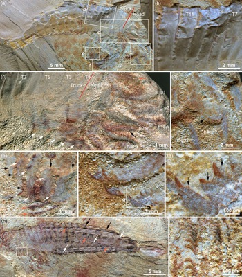
Bivalved euarthropod Cassicaris clarksoni gen. et sp. nov. from the Cambrian Stage 3 of China. (a–f ) YKLP 12493 (holotype), appendage-bearing individual with its tail twisted as showing its ventral side upwards: (a) left view; (b) close-up of area b in (a), showing a stalked eye; (c) close-up of area c in (a), showing fragmentary limbs and possible sternum (arrow); (d) close-up of fragmentary endopod (arrow) in area d in (c); (e) close-up of endopod with feather-like podites in area e in (c); (f ) close-up of tail flukes without setae? in area f in (a). (g) YKLP 12494, empty head shield with cardinal spine (arrow) aside distorted abdomen tergite. (h) Close-up of tiny tubercles and spines situated along posteroventral margin in area h in G. (i) YKLP 12495, details of the posterior trunk with oar-shaped flukes. (j–l) YKLP 12496 (paratype): incomplete individual: (j) right view, showing eye and antenna beneath anterior cardinal spine (white arrowed) and gut fillings (black arrowed); (k, l) close-up of areas k in (j) and l in (k), showing the robust antennula (arrow) and right stalked eye.

Visual system of Cassicaris clarksoni. (a–d) YKLP 12499: (a) right view; (b) close-up of an eye and broken antenna (arrow) in area b in (a); (c) visual surface (orange arrow), antennula (black arrow) and antenna (white arrow) in area c in (b); (d) interpretive drawing of the eye in (b, c). (e–j) YKLP 12500, showing carapace and paired stalked eyes attached to anterior tergite (arrow): (e) YKLP 12500a, left valve (counterpart); (f) YKLP12500b, right valve (part); (g) close-up of area g in (f), showing median eye (arrow); (h) interpretive drawing of (g); (i) close-up of area i in (g), showing the median eye presumably bearing fine lenses (arrows) along round ‘facet’; (j) interpretive drawing of (i); (k) YKLP 12501, articulated head shells with an eye beneath the cardinal spine; (l) close-up of the eye in area l in (k); (m) interpretive drawing of (l). (n) YKLP 12502, fragmentary head shield with an eye preserved; (o), close-up of area o in (n), showing minute lenses (arrows) in the facet; (p) YKLP 12503, head shield with an eye; (q) close-up area q in (p), showing poorly preserved lenses (arrows). Abbreviations: AS, anterior sclerite; B, the base of the eye and the cuticular part of the eye stalk, containing the first optical ganglia; F, globular facets, for field of view (grey area); ME, median eye (presumably a compound eye); VS, visual surface, indicating globular dioptric apparatuses of facets.

Trunk sclerites and appendage microstructures of Cassicaris clarksoni. (a–g) YKLP 12497 (paratype): (a) individual missed head shield, its head and trunk roughly separated along the red line; (b) trunk sclerites (underneath head shield) with smooth surface and serrated posterior margin; (c) close-up of area c in (a) showing possible left antennula (orange arrow), right antennula, antenna, and other head limbs most anteriorly situated, together with incomplete exopod with fine setae (black arrows), and each thoracic segment corresponding to a limb (white arrows); (d) close-up of possible (not in situ) head limbs showing endopods and broad exopod (white arrow) in area d in (a); (e) close-up of possible trunk limbs showing relatively narrow exopods (black arrows) with setae along rear margins (white arrows), and poorly survived endopods (orange arrows) in area e in (a); (f) close-up of area f in (a), showing spinose terminals of endopods; (g) close-up of terminal spine fringed with even fine setules (arrows) in area g in (f). (h) YKLP 12498, left view (head shield removed), showing smooth anterior trunk sclerites and armoured posterior trunk, which displays rows of dorsal spines (black arrows), left lateral spines (white arrows) and depressions of right lateral spines (orange arrows). (i) Close-up of area i in (h), showing densely fringed setules on the endites of endopods underneath anterior trunk. Abbreviations: a1, antennula; a2, antenna; h1en, endopod of first head limb; h4en?, presumed endopod of fourth limb; T1–T7, first to seventh trunk segments.

Appendages, trunk and abdomen of Cassicaris clarksoni. (a–d) YKLP 12504: (a) left view, showing cardinal spine (white arrow) and a possible eye (orange arrow); (b) close-up of multi-segmented trunk limbs with short podomeres in area b in (a); (c) close-up of area c in (a), showing antennula (black arrow), antenna (white arrow) and a possible endopod (orange arrow). (d) Close-up of limb podomeres, each bearing fine setules (arrows) in area d in (b). (e) YKLP 12506, damaged individual, showing anterior trunk segments (arrow) and succeeding spiny abdomen segments. (f) Close-up of area f in (e), showing endopods bearing feather-like podites. (g) YKLP 12505, trunk limbs with feather-like podites. (h) YKLP 12507, individual with laterally buried head shield and vertically compressed tail, showing cardinal spine (white arrow), smooth trunk segments (black arrow) and abdomen sclerites. (i) Close-up of area i in (h) showing details of armoured abdomen, which displays rows of dorsal spines (black arrows), lateral spines (white arrows) and depressions of lateral spines (orange arrows) underneath the visible abdomen sclerites. (j) YKLP 12508, individual showing cardinal spine (arrow), segmented trunk and gut. (k) enlargement of the ‘segmented’ gut deposits in area k in (j).

Etymology. The new species is named in memory of Euan N. K. Clarkson (1937–2024), the British palaeontologist, a forerunner and leading scholar who explored the extraordinary trilobite eyes and vision in million-year-old arthropods.
Diagnosis. As from the genus.
Holotype. YKLP 12493 (Fig. 1a), a slightly distorted individual with the anterior portion of its trunk and some appendages moved out of the carapace.
Other material. Fourteen specimens with soft parts (YKLP 12494–12507).
Description. Carapace is sub-elliptical in lateral outline, weakly convex, displaying a straight dorsal margin and smooth or finely ornamented surface. The length of measured carapaces varies from 25 mm to 35 mm (excluding the anterior cardinal spine, which is curved ventrally, ranging from 3.2 mm to 6.4 mm in length). Underneath the cardinal spine is a pair of bulb-like stalked eyes that are anterolaterally stretching out from a narrow anterior sclerite with a node situated at the middle of the sclerite (Fig. 2e, g–j). Immediately behind the anterior sclerite are two pairs of antenniform appendages: the first pair of antennulae is small, possibly connected to the second head segment, whereas the second pair is robust and longer (Figs. 1k, 2b, 3c, 4c) than any other head ones. The succeeding five pairs of head limbs are elongate, containing at least ten podomeres (Figs. 1k, 3c), each bearing a broad paddle-like exopod. There are 12 to 13 pairs of successive trunk limbs (Fig. 3a) appearing to be reducing in size backwards (Figs. 3a, h, 4e, f). More uniquely, these biramous trunk limbs, which correspond with the trunk segmentation in a one-to-one manner (Fig. 3c), display remarkable differentiation: the exopod is relatively small (Fig. 3d, e), whereas the endopod is with elongated podomere (Figs. 1e, 3i, 4f, g). Each podomere of the endopod carries an elongate claw-shaped endite on its adaxial surface, making the endopod feather-like. This endite is fringed with a series of filaceous setules (Fig. 4d) on the side facing the proximal end of the limb and fine terminal spines (Fig. 3f) on which there are even finer setules (Fig. 3g). The anterior trunk of the multi-segmented body, being covered by the carapace, contains at least 12 segments with a smooth outer surface, and the exposed abdomen, which bears 10 to 13 highly armoured sclerites ornamented with longitudinal rows of ridges and spines (Figs. 1a, g, 3a, h, 4e, h, i). Tail is wide, bearing a pair of articulated furcal rami, each displaying two ridges: the outer one is situated marginally and the inner one, which acts as the inner margin of furcal rami, is fringed with an oval-shaped membrane, instead of marginal setae (Figs. 1a, f, i, 4h).
Remarks. Cassicaris clarksoni is generally similar to the members of the genus Pectocaris Hou, 1999, including the type species P. spatiosa Hou, Reference Hou1999, P. eurypetala (Hou & Sun, Reference Hou and Sun1988), P. inopinata Jin et al., 2021 and P. paraspatiosa Jin et al., 2024, as well as Jugatacaris agilis Fu & Zhang, 2011, in having a bivalved carapace, a pair of stalked eyes, a multi-segmented body, and the slim trunk appendages with many podomeres. However, C. clarksoni is obviously distinguishable from all of these previously known pectocaridids by its anterior cardinal spine of the carapace, multi-segmented body with fewer segments and associated trunk limbs, and the strongly sclerotised abdomen with rows of spines and tail.
Clypecaris serrata Yang et al., Reference Yang, Ortega-Hernández, Lan, Hou and Zhang2016 is another bivalved euarthropod that also came from the Xiaoshiba biota, characterised by a pair of suboval-shaped valves with sawtooth-like spines along both anteroventral margins of the carapace. The trunk bears at least 20 tergites, a pair of stalked eyes, a pair of robust raptorial appendages and a paired series of dorsal spines (Yang et al. Reference Yang, Ortega-Hernández, Lan, Hou and Zhang2016). In comparison, the newly known Cassicaris clarksoni differs from C. serrata in possessing a bivalved carapace with anterior cardinal spines, a heavily segmented trunk, paired bulbous eyes situated on the anterior sclerite and relatively small antennulae and antennae.
Jugatacaris agilis was suspected to be the same species as P. spatiosa (Hou et al. Reference Hou, Siveter, Siveter, Aldridge, Cong, Gabbott, Ma, Purnell and Williams2017), but to clarify the taxonomy additional material is needed. Instead, temporarily regarding J. agilis as a close relative of other pectocaridids (Jin et al. Reference Jin, Mai, Chen, Liu, Hou, Wen and Zhai2021) is accepted here, and all of these described pectocaridids possibly constitute a monophyletic clade (Family Pectocarididae Hou, Reference Hou1999) within the Phylum Euarthropoda.
Chuandianella ovata (Lee, Reference Lee1975), a bivalved euarthropod widely found on the Yangtze Platform, is characterised by its rectangular carapace, elongated antennae, and long abdominal segments (Hou & Bergström Reference Hou and Bergström1997; Liu & Shu Reference Liu and Shu2008; Hou et al. Reference Hou, Siveter, Siveter, Aldridge, Cong, Gabbott, Ma, Purnell and Williams2017). Based on these distinctive features, it could be easily distinguished from the Cassicaris clarksoni.
Each valve of the carapace of Xiazhuangocaris chenggongensis Zeng et al., 2021 possesses a protruded anterior tip, and its tail bears a pair of caudal rami, both of which are somewhat comparable to the bivalved carapace and tail of Cassicaris clarksoni, correspondingly, but differ in detailed morphology and structures. Moreover, the former is of about 13 trunk segments separated by arthrodial membrane, apparently fewer than and different from that of the latter.
Locality and horizon. Xiaoshiba section in the eastern suburb of Kunming, China. Yunnanocephalus–Chengjiangaspis–Hongshiyanaspis biozone of the Cambrian Stage 3 Hongjingshao Formation (Yang et al. Reference Yang, Ortega-Hernández, Butterfield and Zhang2013, Reference Yang, Smith, Lan, Hou and Zhang2014; Hou et al. Reference Hou, Yang, Zhang, Hughes and Lan2019).
3. Discussion
3.1. Appendages, segmentation and body configuration
The whole body of Cassicaris clarksoni, like that of the bivalved arthropod Nereocaris (Legg et al. Reference Legg, Sutton, Edgecombe and Caron2012), can be subdivided into four anatomically distinct parts: the cephalon, thorax (trunk), abdomen and tail. Accordingly, it seems apparent that C. clarksoni also bears a short antennula (Figs. 1k, 4c) and a robust antenna (Figs. 2b, 4c). Although we do not know exactly how many pairs of cephalic limbs exist behind the antenna, as in the well-examined Pectocaris inopinata (Jin et al. Reference Jin, Mai, Chen, Liu, Hou, Wen and Zhai2021), none of the C. clarksoni displays a ‘mandibula’. Instead, all succeeding limbs are certainly biramous (Fig. 3c–e) and the thorax has at least eight pairs of biramous limbs, based on direct observation of sclerites which have been covered by a carapace, displaying a smooth surface, and always situated immediately in front of the armoured abdomen (Figs. 3a, b, h, 4e, h). Like that of the two batches of cephalic and trunk appendages of Isoxys (Zhang et al. Reference Zhang, Liu, Ortega-Hernández, Wolfe, Jin, Mai, Hou, Guo and Zhai2023), C. clarksoni reveals the essential body configuration of the pectocaridid-like euarthropod. The two pectocaridids were initially described as bearing paired antennulae and antennae (Hou et al. Reference Hou, Bergström and Xu2004), and both antennula and antenna were also mentioned in Jugatacaris (Fu & Zhang Reference Fu and Zhang2011) and well illustrated in Pectocaris inopinata, of which the antennula is rather bigger than the antenna (Jin et al. Reference Jin, Mai, Chen, Liu, Hou, Wen and Zhai2021). The size differentiation between antennula and antenna of C. clarksoni is also remarkable (Figs. 1k, 3c, 4c), but curiously its small antennule is similar to that of P. spatiosa (Hou et al. Reference Hou, Bergström and Xu2004), whereas its antenna is rather strong, comparable to the antennule of Pectocaris inopinata (Jin et al. Reference Jin, Mai, Chen, Liu, Hou, Wen and Zhai2021). The size variation among these pectocariids is partially due to incomplete preservation.
Coexisting with Cassicaris clarksoni in the Xiaoshiba biota, some fuxianhuiids display a ‘taphonomic dissection’, which can well expose the head organisation originally concealed by the dorsal shield (Yang et al. Reference Yang, Ortega-Hernández, Butterfield and Zhang2013). In most cases, the head of C. clarksoni is more or less covered by its bivalved shield. Therefore, none of the specimens assigned to the new taxon can clearly show the head organisation, or the attachment between the body and all of its appendages. Most likely, the head shield of C. clarksoni is tightly attached to the trunk so that almost all of the specimens examined here cannot entirely show their cephalic and anterior thoracic portions, which are always tightly covered by their carapaces. Sometimes we have to manually remove the partially survived carapace to see the details of body structures underneath (Fig. 3h). In contrast, the armoured abdomen could be twisted (Figs. 1a, 4h), and single appendages (Figs. 1c, d, 4b, d) or some still articulated ones were shifted from their body to some extent (Figs. 3a, d, 4e), possibly resulting from distortion during the burial process. This makes the anatomical analysis and taxonomical identification for C. clarksoni more complex. Particularly, its carapace with a cardinal spine is anatomically unable to articulate with an anterior sclerite, which, instead, is always found with a pair of stalked eyes beneath the cardinal spine visible from every eye-bearing specimen in our collection (Figs. 1a, j, 2a, e, f, k, n, p). For each individual the eye stalks are obviously connected to the first somite of its soft body. It remains obscure whether or not the actual segmentation of cephalon, thorax and abdomen may vary among them. However, the available material reveals a one-to-one association between limb and tagmosis (e.g., Fig. 3c, h), except for the abdomen, which bears approximately 12 segments and is limbless (Figs. 1a, 3h, 4e, h).
The overall body form of bivalved Tokummia katalepsis from the 508-million-year-old Marble Canyon fossil deposit (Burgess Shale), whose trunk is also heavily segmented, and each bearing a pair of slim biramous limbs (Aria & Caron Reference Aria and Caron2017), is somewhat comparable to that of pectocaridids, as well as C. clarksoni. However, the former is characterised by a pair of chelate maxillipeds, as well as other complex cephalic appendages and feeding structures. Therefore, the unique body organisation of T. katalepsis enhances its assignment to Hymenocarina – a distinct stem-group of Euarthropoda.
In comparison to Cassicaris clarksoni, like the bivalved Nereocaris exilis Legg et al., Reference Legg, Sutton, Edgecombe and Caron2012, possesses a strongly sclerotised, elongate abdomen and tail, but differs remarkably in morphology of appendages, abdomen and tail, especially the latter which has relatively narrower thoracic segments with filamentous appendages, which were assumed to be unsuitable for walking (Legg et al. Reference Legg, Sutton, Edgecombe and Caron2012).
The bivalved arthropod Isoxys, which adapted to a pelagic lifestyle, is a cosmopolitan genus recorded in several Cambrian Lagerstätten (Legg & Vannier Reference Legg and Vannier2013; Fu et al. Reference Fu, Zhang, Budd, Liu and Pan2014). With a thin carapace, prominent eyes, slender endopods and broad tail flukes, Pectocaris has been suggested to be a nektonic swimmer (Hou et al. Reference Hou, Bergström and Xu2004, Reference Hou, Siveter, Siveter, Aldridge, Cong, Gabbott, Ma, Purnell and Williams2017). These key characters are also shared by C. clarksoni, which may also support a nektonic lifestyle. However, to date, the new taxon has only been unearthed from the Xiaoshiba biota – a single fossil site. Without solid evidence derived from additional material from elsewhere, we are unable to conclude that C. clarksoni also adopted a pelagic life mode.
3.2. Visual system of Cassicaris clarksoni
The stalked eyes of C. clarksoni appear in a club-like shape protruding from the carapace, where the rest of the stalk is hidden, similar to many other, especially bivalved, arthropods from the early Cambrian, such as Waptia (Vannier et al. Reference Vannier, Aria, Taylor and Caron2018), Isoxys (Schoenemann & Clarkson Reference Schoenemann and Clarkson2011) and others. The part of the stalked eye visible here consists of a conical element tapering proximally, and the almost hemispherical visual surface covers its top, appearing hat-shaped. Some relics of the former compound eye’s facets are visible, increasing in size from the base of the eye towards the top. The largest preserved facets measure approximately 80–100 µm, similar to those of Isoxys auritus (Schoenemann & Clarkson, Reference Schoenemann and Clarkson2011). The field of view is about 110 °, but is significantly enlarged by the mobility of the eye stalks. The optical performance of this eye can be estimated if it is assumed that an arc of the visual surface at its summit can be covered by about 25 facets, resulting in an angle of light acceptance of ∼4.4 ° for each facet, and an eye parameter of about 7.7 [µm rad]. The eye parameter is a measure developed by Snyder and colleagues in the 1970s, describing the adaptations of eye morphology to light ecological constraints (Snyder, Reference Snyder1977, Reference Snyder and Autrum1979; Snyder et al. Reference Snyder, Stavenga and Laughlin1977a, Reference Snyder, Laughlin and Stavenga1977b). The theory assumes that the eye operates at the lower threshold of perceivable light intensity and that, typical for most compound eyes of day-active arthropods today, each facet perceives only one pixel of the overall mosaic-like image (apposition compound eye). An optimal apposition compound eye establishes a compromise between accuracy (the smallest angle of light acceptance possible to establish as many facets as possible and thus achieve the highest possible resolving power) and light-gathering capacity (a lens diameter as large as possible). The light parameter p describes this compromise: p = D • Δφ [µm rad], with D meaning lens- (facet-) diameter and Δφ the angle of light acceptance. This construction achieves the highest possible acuity with an aperture as small as possible that still allows the eye to function, thus establishing as many facets as possible within the limited space of a compound eye. The relationship between p and light intensity is not linear. The eye parameter of 7.7 [µm rad] resulting for C. clarksoni is close to that of Isoxys auritus (Jiang, Reference Jiang, Luo, Jiang, Wu, Song and Ou1982) from Chengjiang with 9.9 [µm rad] (Schoenemann & Clarkson Reference Schoenemann and Clarkson2011). Horridge (Reference Horridge1977, Reference Horridge1978) demonstrated that indeed the eye parameter correlates strongly with the ecological lifestyle of many arthropods. He found that organisms with values lower than 2 [µm rad] include diurnal terrestrial insects, such as values of 2 ≤ p < 4 [µm rad], for example, that characterise diurnal subaqueous crustaceans, and at the range of 2 ≤ p < 4 [µm rad] and higher there are twilight and nocturnal arthropods such as the larvae of the dragonfly Cordulegastre annulatus hunting at twilight (Land Reference Land and Autrum1981, p. 551).
There is limited research on aquatic arthropods in this regard. To date, the most crucial study is that by Mike Land (Reference Land1989), which compares the eyes of hyperiid amphipods inhabiting different depths in a clear ocean and describes the relationships between optical structure and depth. Here surface amphipodes (<200 m) show eye parameters <4.4 [µm rad], for mid-water crustaceans (<200–800 m) p ranges from 6.9 to 7.9 [µm rad] in the relevant area of the internally differentiated compound eyes, and for deeper living amphipodes he finds an eye parameter >11 [µm rad] in the relevant area of the internally differentiated compound eye. So, the eye parameter of C. clarksoni (7.7 [µm rad]) matches perfectly here the parameter of mid-water, mesopelagial environments.
The eye parameter was also frequently used for fossilised arthropods, such as trilobites (e.g., Fordyce & Cronin Reference Fordyce and Cronin1989; McCormick & Fortey Reference McCormick and Fortey1998; Schoenemann et al. Reference Schoenemann, Poschmann and Clarkson2019). McCormick and Fortey (Reference McCormick and Fortey1998) assign the trilobite Pricyclopyge bindosa (Salter in Murchison Reference Murchison1859) with an eye parameter of p vertical rows of facets 4.23–4.98 [μm rad] and p horizontal rows of facets 7.06–8.31 [μm rad] to a mesopelagic lifestyle in conditions of dim illumination during the day. This may have been similar to C. clarksoni and I. auritus from the Chengjiang Formation. Land (Reference Land and Autrum1981, p. 556; Land & Nilsson, Reference Land and Nilsson2012, p. 27) also correlates the eye parameter with the depth at which aquatic organisms can live under clear water conditions. Here this eye parameter indicates a depth of the environment not deeper than ∼350 m under clear ocean water conditions, or less if associated with suspended sediment.
The acuity of scanning the surroundings, and thus the fineness of vision, can be estimated in this specimen as well. It depends on the angle of light acceptance for the individual facet, Δφ – the smaller the angle, the finer the resolution of vision. A measure of this is often given as the inverse, ν s = 1/Δφ [rad–1], because this measure increases with the increase of acuity (Snyder Reference Snyder1977). For C. clarksoni, νs = 1/Δφ [rad–1] is ∼13 [rad–1], and thus C. clarksoni looks about twice as fine as Isoxys auritus (∼6 [rad–1]) (Schoenemann & Clarkson Reference Schoenemann and Clarkson2011). The fineness of vision of C. clarksoni is similar to that of the shore crab Leptograpsus with 19 rad–1 (compare the high resolving compound eye of the dragonfly Aeshna with 115 [rad–1] while an eagle reaches 8022 [rad–1] (Land & Nilsson Reference Land and Nilsson2012, p. 52). (For detailed development and background of the theory please see Snyder Reference Snyder1977, Reference Snyder and Autrum1979; Snyder et al. Reference Snyder, Stavenga and Laughlin1977a, Reference Snyder, Laughlin and Stavenga1977b; Land Reference Land and Autrum1981.) C. clarksoni probably had a similar lifestyle to Isoxys auritus (Jiang, Reference Jiang, Luo, Jiang, Wu, Song and Ou1982) from Chengjiang. The eye parameter is consistent with adaptation to moderate light conditions, either for a lifestyle at depths below 350 m (mesopelagic) or for an epibenthic lifestyle in more turbid water. As the Chengjiang biota lived in a deltaic environment, characterised by flash floods and high sedimentation rates, the latter seems more probable. The acuity (∼13 [rad–1]) is relatively high compared to I. auritus (∼6 [rad–1]), much better than the horseshoe crab Limulus (4.8 [rad–1]) and similar to the shore crab Leptograpsus (19 [rad–1]) (Land & Nilsson Reference Land and Nilsson2012). So surely C. clarksoni could recognise predators and mates visually.
C. clarksoni possesses a second visual system – a so-called ‘median eye’. It is located underneath the cardinal spine, situated in the middle of the anterior sclerite (Fig. 2e, g–j) from which the stalked eyes originate. This is the typical position for median eyes. Median eyes are plesiomorphic to panarthropods, and aside from the compound eyes, their second eye system. Median eyes are ocellar systems, often tiny cups floored by a small retina and of diverse function among their bearers (Land & Nilsson Reference Land and Nilsson2012) – here in C. clarksoni we find, however, in this position presumably a compound eye. The dioptric apparatus of some facets, visible as spherical elements (Fig. 2 i, j), are comparable to other compound eyes of the Cambrian (e.g., Schoenemann & Clarkson Reference Schoenemann and Clarkson2011; Strausfeld et al. Reference Strausfeld, Ma, Edgecombe, Fortey, Land, Liu, Cong and Hou2016). The regular spherical shape of all elements, their homogeneous structure, their regular order on the surface of the eye, and their typical size of about 100 µm, clearly indicate that they are biological structures rather than diagenetic artefacts. This is the only median compound eye known so far and, if the observations of this singular find can be stabilised by further research, will give rise to unique insights into the evolution of compound eyes, younger than median eyes, as discussed later.
3.3. Body configuration and phylogenetics
Head segmentation and the nature and arrangement of head appendages have long been regarded as bearing critical information for assessing the early evolutionary radiation and phylogenetics of euarthropods (Scholtz & Edgecombe Reference Scholtz and Edgecombe2006; Yang et al. Reference Yang, Ortega-Hernández, Butterfield and Zhang2013; Ortega-Hernández Reference Ortega-Hernández, Janssen and Budd2017). The Burgess Shale-type deposit, as one of the irreplaceable data sources, has made profound contributions to our understanding of arthropod evolution (Budd & Telford, Reference Budd and Telford2009; Ortega-Hernández, Reference Ortega-Hernández2016; Aria & Caron, Reference Aria and Caron2017). With Burgess Shale-type preservation, Cassicaris clarksoni provides substantial anatomical information for the evaluation of its own phylogenetic position, as well as its relationship with other Cambrian bivalved euarthropods (Fig. 5). First, its paired stalked eyes protrude from the anterior sclerite (Figs. 1a, j, 2a, e, f, k, n, p) – the first cephalic tergite that may define the protocerebral origin (Ma et al. Reference Ma, Hou, Edgecombe and Strausfeld2012; Yang et al. Reference Yang, Ortega-Hernández, Butterfield and Zhang2013). Secondly, the same combination of anterior structure has also been recognised from some bivalved taxa closely aligned with C. clarksoni, such as Pectocaris inopinata (Jin et al. Reference Jin, Mai, Chen, Liu, Hou, Wen and Zhai2021) and Jugatacaris agilis, of which the node seen at the middle of the anterior sclerite was described as the middle eye (Fu & Zhang Reference Fu and Zhang2011). It should be noted that the paired stalked eyes have also been determined from other bivalved euarthropods, such as Isoxys acutangulus (Legg & Vannier Reference Legg and Vannier2013) and I. auritus (Hou et al. Reference Hou, Siveter, Siveter, Aldridge, Cong, Gabbott, Ma, Purnell and Williams2017). However, with a pair of ‘great appendages’, Isoxys represents a lineage separated from the Cassicaris. Thirdly, as a conspicuous group of upper-stem euarthropods (Ortega-Hernández Reference Ortega-Hernández2016), fuxianhuiids, such as Fuxianhuia, Chengjiangocaris, Shankouia, Guangweicaris and Alacaria (Chen et al. Reference Chen, Zhou, Edgecombe and Ramsköld1995; Hou & Bergström Reference Hou and Bergström1997; Waloszek et al. Reference Waloszek, Chen, Maas and Wang2005, Reference Waloszek, Maas, Chen and Stein2007; Yang et al. Reference Yang, Ortega-Hernández, Butterfield and Zhang2013, Reference Yang, Ortega-Hernández, Legg, Lan, Hou and Zhang2018), with a pair of specialised post-antennal appendages and decoupled body segments and appendages (e.g., one tergite may bear up to four limbs), are noticeably distinguished from any bivalved euarthropods, despite all of them having a pair of stalked eyes projecting from the anterior sclerite. The morphologically similar integration of eye structures, which occurs independently in phylogenetically separated euarthropod groups, may imply a deep origin of the vision system in early euarthropod evolution.
Parsimony phylogenetic analysis of Cassicaris clarksoni. Strict consensus under implied weights k = 3 (36 MPTs, 398.86 steps, CI = 0.572, RI = 0.696).

Based on the new soft-bodied material of Cassicaris clarksoni, especially the fresh information about the general features of its appendages, stalked eyes, trunk segmentation and body organisation, our phylogenetic analysis resolves C. clarksoni and Pectocaris inopinata as a sister group, and both are closely related to P. eurypetala, P. spatiiosa and Jugatacaris agilis. Overall, all of these taxa constitute the Family Pectocarididae. Meanwhile our analysis also indicates that the co-occurring Isoxys, Nereocaris and Chuandianella are notable stem lineages of bivalved euarthropods. These contemporaneous bivalved arthropods that once lived on the Yangtze Platform may infer the stepwise divergence which reflects progressive morphological innovations in carapace structure, trunk segmentation, abdominal specialisation and appendage differentiation across this Cambrian clade. It is worth noting that pectocaridids exhibit a low degree of appendage differentiation and the endopods have a large number of podomeres, in contrast to Tokummia which is characterised by chelate maxillipeds and complex cephalic structures. The question of potential convergence in key features like the bivalved carapace and multi-segmented body remains unresolved. Although Jugatacaris and Pectocaris have been referred to Hymenocarina by Vannier et al. (Reference Vannier, Aria, Taylor and Caron2018), and only Pectocaris by Izquierdo-López & Caron (Reference Izquierdo-López and Caron2024), even more bivalved euarthropods with soft parts were regarded as hymenocarines (Zeng et al. Reference Zeng, Zhao, Yin and Zhu2020). Yet based on the diagnosis of Hymenocarina (Aria and Caron Reference Aria and Caron2017), many bivalved euarthropods should be included into this group. However, as demonstrated here, with a unique body configuration Cassicaris clarksoni, as well as other pectocaridids, are notably different from hymenocarines, and our parsimonious tree clearly shows they belong to a separate group (Fig. 5). Bearing no key appendage, mandibles and the antennae of Pectocaris, which were anatomically argued to be a crustacean synapomorphy (Legg et al. Reference Legg, Sutton, Edgecombe and Caron2012), gives strong support to our phylogenetic analysis.
With the application of micro-computed tomography and three-dimensional rendering, more detailed information about the diversified euarthropod appendages has been well documented (Liu et al. Reference Liu, Edgecombe, Schmidt, Bond, Melzer, Zhai, Mai, Zhang and Hou2021). In addition to the head appendages, the discovery of feather-like rachises developed by the endopods of Chuandianella reveals a complex limb organisation in Cambrian bivalved euarthropods (Zhai et al. Reference Zhai, Williams, Siveter, Siveter, Harvey, Sansom, Mai, Zhou and Hou2022), which is special and has never been known before. As reported here, the feather-like rachises of endopods corresponding to trunk segments have also been present in C. clarksoni. The discovery provides new data for assessing the appendage disparity, as well as the related lifestyles evolved by Cambrian bivalved euarthropods.
C. clarksoni, on its anterior sclerite, possesses one median eye (Fig. 2e–j). Median eyes generally are not compound eyes, but typically small ocellar, cup-like organs equipped with a tiny retina and a small lens. Some of them may have the capacity for rough image formation, but the function is diverse and often not really understood (Land & Nilsson, Reference Land and Nilsson2012). While median eyes are plesiomorphic to all panarthropods, they are homologous to the ocelli of lobopodians and onychophorans (Mayer Reference Mayer2006; Mayer et al. Reference Mayer, Hering, Stosch, Stevenson and Dircksen2015), which does not contradict molecular findings, such as those of Fleming and colleagues (Fleming et al. Reference Fleming, Kristensen, Sørensen, Park, Arakawa, Blaxter, Rebecchi, Guidetti, Williams, Roberts, Vinther and Pisani2018). Thus, their original number is two, as still can be found, for example, among Xiphosurans, eurypterids and scorpions, and typically in all other extant chelicerates, where they often function as the ‘main’ eyes (e.g., Harzsch et al. Reference Harzsch, Vilpoux, Blackburn, Platchetzki, Brown, Melzer, Kempler and Battelle2006; Poschmann Reference Poschmann2020; Giribet et al. Reference Giribet, Edgecombe, Wheeler and Babbitt2002). Multiples of two exist in many spiders and the nauplius eyes of the basal subclass of crustaceans, Phyllopoda (Elofsson Reference Elofsson1969; Land Reference Land1985; Land & Nilsson Reference Land and Nilsson2012). Median eyes are evolutionarily older than the compound eyes (Schoenemann & Clarkson Reference Schoenemann and Clarkson2023) and are innervated by neuropiles separate from those of the compound eyes. Their neural centres lie within the protocerebrum, not positioned serially according to the tagmata. While the compound eyes are consistently innervated by the lateral part of the protocerebrum, the median eyes are innervated by the central parts (Mayer Reference Mayer2006; Strausfeld et al. Reference Strausfeld, Mok-Strausfeld, Loesel, Rowell and Stowe2006a, Reference Strausfeld, Strausfeld, Stowe, Rowell and Loesel2006b; Homberg Reference Homberg2008). There is a remarkable plasticity in the final conformation of median eyes, and sometimes the position of both types, median as compound eyes, in the cephalon can be very variable. A comprehensive review of the current discussion on cephalisation in arthropods is given by Chipman et al. (Reference Chipman and Edgecombe2019) and Lev et al. (Reference Lev, Edgecombe and Chipman2022). Probably by duplication, we find four median eyes in larval Phyllopoda and chelicerates, among fossils in Cinderella eucalla (Schoenemann & Clarkson Reference Schoenemann and Clarkson2023). By fusion of the median pair, three median eyes (Paulus Reference Paulus and Grypta1979; Schoenemann & Clarkson Reference Schoenemann and Clarkson2012, Reference Schoenemann and Clarkson2023), as typical for evolutionarily advanced eurarthopods such as trilobites, is considered an autapomorphy of Tetraconata/Pancrustacea (Bitsch & Bitsch Reference Bitsch, Bitsch and Koenemann2005; Dohle Reference Dohle2001; Mayer Reference Mayer2006; for further overview and references see Schoenemann & Clarkson Reference Schoenemann and Clarkson2023). A loss of median eyes must be considered, consequently, as secondary (e.g., many crustaceans, where the nauplius eyes are still just larval retained).
In C. clarksoni, we find an exception: the median eye is ‘in the right position’, in the middle of the anterior sclerite, but it is not an ocellar system, and it is not paired – it clearly is a single stalked compound eye with clearly discernible units that appear as spheres from the outside (Fig. 2g, i). To possess a stalk (Fig. 2g–j) is untypical for median eyes, but probably realised in Opabinia regalis also. It is a common misconception to assume that all compound eyes are like those of modern bees, equipped with densely packed and hexagonal facets (visual units). In the early compound eyes of the Cambrian, the dioptric apparatuses of the visual units are often still round. Hexagonality arises later from dense packing of as many lenses as possible in the limited space of the visual surface, such as in anomalocarids (Paterson et al. Reference Paterson, García-Bellido, Lee, Brock, Jago and Edgecombe2011) or certain trilobites (Schoenemann Reference Schoenemann2021), as during the ontogeny of modern compound eyes). So, this raises the question of whether we stay here as witnesses to the evolution of compound eyes, which emerge from median eyes. This is to investigate and clarify the object of current research.
The bizarre-looking radiodont Stanleycaris hirpex from the Burgess Shale, which displays a large median eye situated behind the preocular sclerite but in front of the paired lateral eyes, as is typical for median eyes (Moysiuk & Caron Reference Moysiuk and Caron2022), we interpret as fused (three) median eyes. The five eyes of Opabinia, generally considered as compound eyes (e.g., Fleming et al. Reference Fleming, Kristensen, Sørensen, Park, Arakawa, Blaxter, Rebecchi, Guidetti, Williams, Roberts, Vinther and Pisani2018), in the phylogenetic context surely have to be interpreted as two lateral compound eyes and three median (ocellar) eyes. To prove this, closer investigations would be necessary.
3.4. Lifestyle and ecosystem
Details of appendage features and body configuration known from well-preserved fossils, especially from soft-bodied material, are critical and valuable in evaluating the lifestyle of the fossilised animals and the palaeoenvironment involved.
Pectocaris spatiosa and the coeval P. eurypetala from the Chengjiang biota (Hou et al. Reference Hou, Bergström and Xu2004) clearly show stalked eyes, stout first appendages equipped with spines, strong abdomen and broad tail fans; both pectocaridids were regarded as active swimmers, adopting to filter-feeding habits (Hou et al. Reference Hou, Bergström and Xu2004). With broad furcal rami and oar-like exopods, Jugatacaris was regarded as an efficient swimmer; together with its U-shaped food groove and filter limbs this pectocaridid may adopt to a typical filter-feeding habitat (Fu & Zhang Reference Fu and Zhang2011).
The widespread geographical occurrence of the Cambrian Age 3 Chuandianella ovata suggests enhanced dispersal capability. Its well-developed stalked eyes would have provided multi-directional vision for various uses including detection of predators.
Moreover C. ovata occurs in supposed coprolites in the Chengjiang biota which also indicates that it was a prey or carrion item. In comparison, the occurrence of Cassicaris clarksoni is limited to its type locality, and it remains uncertain if it is an endemic or cosmopolitan lineage. However, a recent study reveals that C. ovata possesses a set of biramous limbs, each comprising a short, paddle-shaped exopod and a feather-like endopod (Zhai et al. Reference Zhai, Williams, Siveter, Siveter, Harvey, Sansom, Mai, Zhou and Hou2022). Such unusual post-antennal appendages have also been adopted by C. clarksoni (Figs. 1e, 3i, 4g). The shared fine appendage feature appears most likely to be in association with the feeding habits of the two euarthropod taxa as epibenthic animals lived in a similar ecological niche.
With a heavily segmented trunk, Nereocaris exilis from the Cambrian Miaolingian Stage 5 is quite unlike other bivalved arthropods. Still, its possession of ‘a strongly sclerotised, elongate abdomen and telson’ (Legg et al. Reference Legg, Sutton, Edgecombe and Caron2012) is comparable to that of C. clarksoni, whose abdomen and tail bear longitudinal rows of ridges and spines that are obviously different from those of N. exilis. Functionally, the strongly armoured abdomen of C. clarksoni is unlikely to be an adaptive feature for high-speed movement, but its antennulae and antennae are suitable for performing raptorial functions as compared to typical grasping frontal appendages seen from some assumed Cambrian predators, and may act efficiently when coworking with the post-antennal limbs to serve both for feeding and locomotion. The combination of these features supports the reconstruction of C. clarksoni (Fig. 6).
Reconstruction of the bivalved euarthropod Cassicaris clarksoni from the Cambrian Stage 3 Xiaoshiba biota of China.

4. Conclusions
The discovery of Cassicaris clarksoni reveals not only its detailed body configuration, but also the probable lifestyle it adopted in the associated marine ecosystem.
-
1. During the early Cambrian ‘Evolutionary Radiation’, the rapid emergence of various life forms is well recorded by exceptionally preserved Burgess Shale-type material. As demonstrated here, the bivalved C. clarksoni from the Xiaoshiba biota, together with other pectocaridids, constitute a small but distinctive group belonging to Euarthropoda, because they share some unique features while displaying prominent variations in body organisation.
-
2. The stalked eyes of C. clarksoni, like those of Pectocaris and Jugatacaris, are located on the anteriormost segment – the anterior sclerite, which is well known from many Cambrian stem-group arthropods with remote affinities to each other, such as the phosphatocopine crustacean (Zhang & Pratt Reference Zhang and Pratt2012), the monophyletic clade Isoxys (Legg et al. Reference Legg and Vannier2013) and the stem-group euarthropod fuxianhuiids (Yang et al. Reference Yang, Ortega-Hernández, Butterfield and Zhang2013, Reference Yang, Ortega-Hernández, Legg, Lan, Hou and Zhang2018). The visual system, widespread among the early diversified arthropod groups, opens a potentially critical window for assessing the essential ocular forms and evolution of early arthropods. In particular, as demonstrated here, the dedicated microstructures of the stalked eyes of C. clarksoni can provide further unexpected information.
-
3. On the anterior sclerite we find a node which can be identified by its position as a single, stalked median eye. Most remarkably, it is presumably, but very clearly, a compound eye, which is unique so far because median eyes typically are ocellar systems. More specimens, however, are needed to verify this intriguing observation and its reality.
-
4. Characterised by having a robust cardinal spine and highly sclerotised abdomen and tail, the body organisation of C. clarksoni infers a defensive life mode. Moreover, its multi-segmented trunk with many slim limbs, each of which bears minute spiny structures, may imply an efficient feeding strategy resulting from the increasingly complex ecosystem. With such a specialised body configuration, it seems likely that the new taxon adapted to a unique lifestyle which differs from that exhibited by other stem-group pectocaridid crustaceans.
Supplementary material
The supplementary material for this article can be found https://doi.org/10.1017/S1755691025101011.
Acknowledgements
We thank Stephen Pates and an anonymous referee for their constructive and valuable comments, and Tian Lan and Jin-bo Hou for assistance with field work.
Funding statement
This work was supported by a National Natural Science Foundation of China grant (no. 42162002) to J.Y. and a research grant from the Yunnan Province (K204202250035) to X.Z.
Competing interests
The authors declare none.


 Open access