

Introduction
Funerary practice in Iron Age Britain (c. 800 BC–AD 43) is difficult to discern archaeologically. With a few notable exceptions, such as the Arras Culture inhumation cemeteries in East Yorkshire (e.g. Halkon Reference Halkon2013; Lamb Reference Lamb2020), cemeteries displaying apparently ‘formal’ funerary rites appear only towards the end of the period and even then, are geographically restricted (Harding Reference Harding2016). There are, in contrast, abundant examples of human remains found in contexts that would not traditionally be regarded as funerary, such as house floors, grain pits, middens, settlement boundaries and entranceways. This has led to suggestions that the remains of the dead were particularly potent within Iron Age communities, and highly visible within the world of the living (Armit Reference Armit, Bradbury and Scarre2017).
Evidence for the circulation and deposition of human remains is particularly prominent in north-west Scotland, including the Northern and Western Isles, where environmental conditions are often highly conducive to the survival of bone (Armit & Ginn Reference Armit and Ginn2007). Here, we see some of the strongest evidence for the postmortem curation and manipulation of human remains, including mummification (Parker Pearson et al. Reference Parker Pearson2021) and the modification of bones into tools or decorative objects (Shapland & Armit Reference Shapland and Armit2012).
It is within this context that we must consider two individuals buried within a low stone cairn at Loch Borralie, Sutherland, close to the north-west extremity of the Scottish mainland (MacGregor Reference MacGregor2003; Figure 1). As part of the European Research Council-funded COMMIOS project, which examines the demography and funerary archaeology of Iron Age Britain (Armit Reference Armit and Fernández-Götz2022), we conducted osteoarchaeological reanalysis, multi-isotope analysis, ancient DNA (aDNA) analysis and radiocarbon dating of the human remains. This article reports the results of these analyses and considers their significance for wider understandings of funerary practice, mobility and social networks in Iron Age Britain.
Map showing the location of the Loch Borralie cairn and other Iron Age sites mentioned in the text (map by Helen Goodchild. Produced using Copernicus data and information funded by the European Union - EU-DEM layers).

The Loch Borralie burial cairn
The Loch Borralie cairn survives as a low, disturbed spread of stones set on a shallow slope on the Durness Peninsula (NGR NC 3790 6761; Canmore ID 184571 at https://canmore.org.uk/), on the exposed north coast of Scotland. Although remote and sparsely populated in modern times, the area has a dense distribution of later prehistoric settlement, including the remains of numerous Bronze and Iron Age (dating to the second and first millennia BC) roundhouses (Reid et al. Reference Reid1968: 28–32). Although the cairn was robbed-out to provide stone for nearby field walls, it might have never been substantial; its surviving maximum measurements of approximately 7m north–south by 3.5m east–west and around 1.2m high may approximate its original extent (MacGregor Reference MacGregor2003).
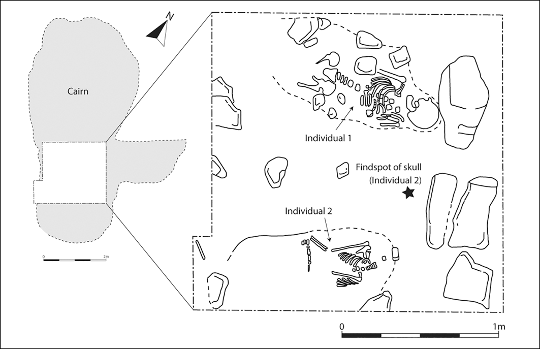
Excavation of part of the cairn was conducted in 2000 in response to the discovery of a human cranium that had eroded out from the feature (MacGregor Reference MacGregor2003, Figure 2). The work revealed two inhumations: an adult (Individual 1) and a juvenile (Individual 2, to whom the displaced cranium belonged). Both had been laid in an extended supine position with their heads to the east (Figure 3). Individual 1 was associated with a ring-headed pin, broadly dateable to the Iron Age, that may have been used to fasten clothing or a shroud (although it was recovered a short distance from the body), and two pieces of crudely struck quartz. Individual 1 appeared to have been placed on a small, primary mound of stones, while Individual 2 had been buried in a more readily identifiable grave cut that appeared to be stratigraphically slightly later than the interment of Individual 1; both burials pre-date the main body of the cairn (MacGregor Reference MacGregor2003: 12).
Excavation photograph of Individual 1 (SC 1061426 © Crown Copyright: HES. Excavation photograph by GUARD).

Plan of the original excavations at Loch Borralie (drawn by Lindsey Büster after MacGregor Reference MacGregor2003; illustration 4).

Chronology
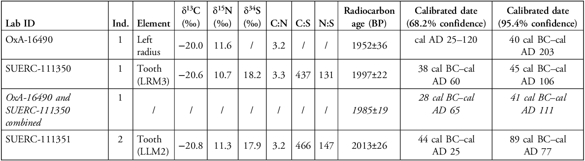
Four radiocarbon determinations have been obtained from the human skeletal remains from the Loch Borralie cairn. An initial determination from the left humerus of Individual 1 (OxA-10253; MacGregor Reference MacGregor2003: 11) was withdrawn following the identification of technical issues with the pretreatment of bone samples analysed between 2000 and 2002 at the Oxford Radiocarbon Accelerator Unit (Sheridan & Higham Reference Sheridan and Higham2006) and replaced with a determination obtained from the left radius of the same individual (OxA-16490; Table 1). More recently, radiocarbon dates for both individuals were obtained from molar teeth (SUERC-111350/1; Table 1). The results form a consistent series, placing the deaths of both individuals within the first centuries BC/AD, most likely between c. 50 BC and AD 70, and almost certainly prior to the initial Roman incursions into southern and eastern Scotland that began in AD 79.
AMS determinations and associated isotope measurements from Loch Borralie. The original, withdrawn date (OxA-10253) is omitted. The radiocarbon ages have been calibrated using the IntCal20 calibration curve (Reimer et al. Reference Reimer2020) and OxCal v.4.4 (Bronk Ramsey Reference Bronk Ramsey2009).

LRM3: lower right third molar; LLM2: lower left second molar.
Reanalysis
The following presents a summary of the results from the osteoarchaeological, ancient DNA and stable isotope analysis of Individuals 1 and 2. Full analysis and methods are available in the online supplementary material (OSM).
Osteological analysis of Individual 1
Individual 1 is a probable female aged over 30 years at death. Much of the lower part of the skeleton is missing, as is part of the left upper limb (Figure 4). No complete long bones are available for stature estimation. Observable non-metric traits include bilateral supraorbital foramina, bilateral mastoid foramina and complete bridging of the sella turcica of the sphenoid bone. Palaeopathological assessment revealed osteoarthritis of the cervical vertebrae and a range of traumatic lesions and bone modifications in three main anatomical regions: cranium, scapulae and long bones.
Schematic representation of the surviving skeletal elements of Individual 1 (figure by authors).

Traumatic lesions
The base of the cranium displays an unusual break where both the condylar and basilar parts of the occipital bone, and a portion of the sphenoid, have become detached (Figure 5a). The characteristics, texture and colour of the break surfaces and the presence of three radiating fractures on the right side of the occipital bone (Figure S2) suggest that the break probably occurred at or around the time of death, when the bone was still wet. Cranial base fractures generally result from high-velocity impacts, which today are associated with motor vehicle collisions, sport accidents, falls or assaults (Bobinski et al. Reference Bobinski2016). They can also result from long-drop hanging (Waldron Reference Waldron1996) and falls from a significant height, where impact forces are transmitted from the feet or buttocks through the spine to the cranium (Lovell Reference Lovell1997). In all cases, the resultant fractures display relatively consistent patterns (Figure S3), which differ from the pattern observed in Individual 1 suggesting that the fracture resulted from an intentional targeted impact.
Perimortem lesions observed in Individual 1: a) endocranial (i.e. interior) view of the cranial base fragment formed by the fracturing of the lateral and basilar portions of the occipital and the body and left lesser wing of the sphenoid from the rest of the cranium (schematic shows location of the fragment within the cranium); b) bilateral and almost symmetrical perimortem fractures at the base of the left (left image) and right (right image) scapular spines (photographs by Rebecca Ellis-Haken; figure by authors).

Bilateral fractures were identified on both scapulae, at the base of the scapular spine (Figure 5b). The fracture surfaces are smooth and show areas of splintering but as there are no signs of healing, these fractures are classified as perimortem.
Postmortem modifications
Examination of the endocranial surface revealed a series of straight and parallel striations on the frontal bone (Figure 6a). These could have resulted from taphonomic processes, such as trampling, fungal activity and root etching, or could represent intentional cut marks. Trampling marks are typically superficial, with a symmetrical U-shaped cross-section and irregular groove trajectory, they also vary in size and direction (Madgwick Reference Madgwick2014; Courtenay et al. Reference Courtenay2020). Microbial bioerosion is only visible under electron microscopy (Jans et al. Reference Jans2004) and root marks are rarely straight and most often imbricated (Fernández–Jalvo & Andrews Reference Fernández-Jalvo and Andrews2016). As none of these characteristics fit the observations, it is more likely that the incisions resulted from deliberate scratching or cutting into the endocranial surface of the cranium with a sharp implement. Taken together, breakage of the cranial base and internal cutmarks are suggestive of deliberate removal of the brain soon after the death of this individual.
Evidence of intentional postmortem manipulation observed in Individual 1: a) endocranial (i.e. interior) surface of the frontal bone (inset: 20× magnification), black arrows indicate incisions, brown arrows show possible root etching; b) the preserved long bones (right and left humeri, left ulna and left femur); c) Reflectance Transformation Imaging (RTI) representation of modification to the left femoral diaphysis (note the flattening of the distal edge); d) RTI representation of modification to the diaphysis of the right humerus (note the whittling marks arrow) (photograph by Rebecca Ellis-Haken; figure by authors).

At least four of the long bones belonging to Individual 1 had been modified prior to deposition. Both humeri and the left ulna and femur were present in the grave as fragments (comprising around 50% of the original bone elements). Although the original report suggests that these bones may have been gnawed by rodents (Roberts Reference Roberts and MacGregor2003), they lack the characteristic striations that would be expected from this process. Instead, the humeri and ulna taper towards the end of the fragment; the outer layers of cortical bone have been removed and the internal layers have been whittled/worked to a sharp edge and a singular pointed end (Figure 6b). The left humerus also displays a U-shaped scar on the whittled surface, which suggests that a sharp implement, such as a knife, was used to work the bone to its final shape (Figure 6c). The femur, in contrast, does not have a sharp edge but the fragment terminates in a flat and smooth margin, as if the sharp edge was worked down against another, possibly softer, surface (Figure 6d) after having been whittled.
Despite clear evidence for the working of these long bones and (in the case of the left femur) the accrual of use-wear, all four had been placed in the grave in their correct anatomical position.
Osteological analysis of Individual 2
Individual 2 is a juvenile, aged between 14.5 and 15.5 years at death. Estimation of osteological sex was not attempted due to the young age of the individual. Only around 25% of the skeleton survives, and the displaced cranium was found on the modern ground surface between the two burials (Figure 2). The presence of linear enamel hypoplasia on the teeth suggests that this individual may have suffered periods of growth disruption and malnutrition, and there are also indications that they may have suffered from vitamin C deficiency. The fourth and fifth cervical vertebrae are fused, most likely due to a congenital failure of segmentation (see OSM). None of the complex patterns of trauma observed in Individual 1 are displayed in the skeleton of Individual 2.
Ancient DNA analysis
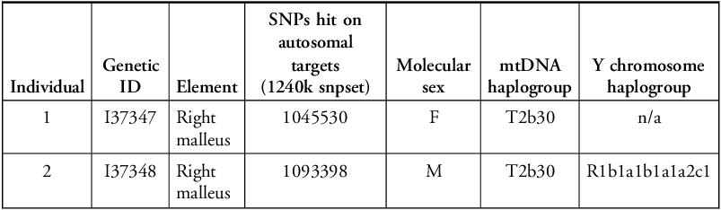
We successfully analysed aDNA from inner ear ossicles belonging to both individuals (Table 2; see OSM). Individual 1 is determined to be genetically female while Individual 2 is male. Principal component analysis of the genomic data for both individuals suggests that they are genetically typical of Scottish Iron Age populations (see Figure S6).
Summary genetic data from Loch Borralie.

The Y chromosome haplogroup for Individual 2 (R1b1a1b1a1a2c1) is common in Iron Age Britain and largely uninformative regarding ancestry. Both individuals, however, share mitochondrial haplogroup T2b30, which is otherwise unique among published ancient individuals from Britain, suggesting that they are maternally related. Although we were not able to detect a close biological relationship between the two using READv2 (Monroy Kuhn et al. Reference Monroy Kuhn2018; Alaçamlı et al. Reference Alaçamli2024), identity by descent (IBD) analysis identified that they share four DNA fragments longer than 8 centimorgans (cM) (43.345cM in total), with the longest segment of 12.144cM suggesting a biological relationship of the fifth degree or more distant. Given the similar dating of the two individuals and the sharing of a highly unusual mitochondrial haplogroup, they are most probably maternally related possibly at the level of second cousins sharing a pair of great-grandparents (corresponding to fifth-degree relationships).
IBD analysis further revealed that both individuals have distant biological relationships (likely eighth degree or more) with individuals found in Orkney (see OSM). Individual 1 is related to an adult male from the Atlantic roundhouse site of Bu, dated to 399–207 cal BC (2271±33 BP; SUERC-69075). This time gap of at least 150 years (perhaps five–six generations) suggests that the latter could have been a direct ancestor of Individual 1, or a collateral relative of a direct ancestor. Individual 2 is biologically related to an individual from Knowe of Skea with a slightly later date of cal AD 25–215 (1915±35 BP; SUERC-8410). Both Orcadian individuals also have distant biological relationships with an adult male (Individual 3) buried in an informal stone cairn at Applecross, on the west mainland of Scotland, around 140km south-west of Loch Borralie (Sheridan et al. Reference Sheridan2019). The Applecross individual, dating to 42 cal BC–cal AD 119 (1980±28 BP; SUERC-73992), is broadly contemporaneous with the Loch Borralie burials.
Isotope analysis
Multi-isotope analysis was conducted to assess diet and mobility. Tooth enamel from both individuals was sampled for strontium (87Sr/86Sr), oxygen (δ18Ocarb) and carbon (δ13Ccarb) stable isotope analysis, while radiocarbon dating of dentine from the same teeth yielded carbon (δ13Ccoll), nitrogen (δ15N) and sulphur (δ34S) isotope measurements. Tooth enamel isotope signatures relate to the period of enamel formation. For this study, a permanent second and third molar were sampled, reflecting enamel formation between approximately 2.5 and 8.5 years (Individual 2) and between 7.5 and 15.5 years (Individual 1), respectively (AlQahtani et al. Reference AlQahtani2010; Hillson Reference Hillson2024).
Both individuals have δ13Ccoll values of around −21‰ (Table 1), indicative of a C3-based diet, which is supported by the enamel δ13Ccarb values (Table 3). The δ15N value is slightly higher for Individual 2, which could suggest that they consumed more animal protein than Individual 1 (although the data reflect the diet of the two individuals at different ages). The combined δ13C and δ15N results from the dentine do not suggest that either individual consumed a significant amount of marine protein during childhood.
Summary isotope results from Loch Borralie. *Approximate estimated age of individual captured by enamel sampling.

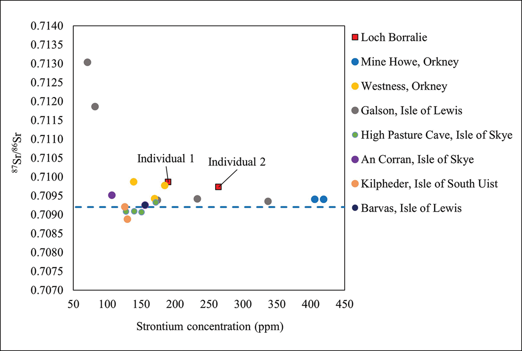
The strontium concentrations (Sr ppm) for both Individual 1 (190ppm) and Individual 2 (264ppm) (Table 3) are higher than for most individuals from mainland Britain, which normally lie within the range 20–180ppm (Evans et al. Reference Evans2012). These high concentrations are, however, closely comparable to Iron Age communities in coastal island environments such as Orkney and the Western Isles of Scotland (Figure 6) (Evans et al. Reference Evans2012; Montgomery et al. Reference Montgomery2014). The 87Sr/86Sr ratios also resemble those from coastal sites, which tend to have isotope ratios closer to seawater (0.7092) due to factors including sea spray, seaweed fertiliser and high rainfall, which can overwrite the biosphere signature (Figure 7). The δ34S values of around 18‰ (Table 1) are also more typical of those associated with coastal communities (>15‰; Richards et al. Reference Richards2001). Overall, the isotope data suggest that both individuals spent the early part of their lives in a coastal environment.
87Sr/86Sr and Sr ppm of human remains from Loch Borralie and other Iron Age sites (data from Evans et al. Reference Evans2012); the Westness data include only ‘Pictish’ (Late Iron Age) individuals (Montgomery et al. Reference Montgomery2014). The87Sr/86Sr value for seawater (0.7092) is shown by the dashed blue line (figure by authors).

The British Geological Survey (BGS) Biosphere Isotope Domains map website (Evans et al. Reference Evans2022) was used to restrict the possible childhood residential origins of the two individuals based on their combined 87Sr/86Sr and δ18Ocarb composition (Figure 8a). The geology of the site and surroundings is well characterised, and full details can be found in the OSM. The 87Sr/86Sr ratios for both individuals (Individual 1: 0.70986; Individual 2: 0.70973) fall outside the 50% central range anticipated for the immediate area around Loch Borralie (0.7094–0.7096) but are common across several areas of Britain and are therefore unhelpful in constraining possible childhood residence. The δ18Ocarb values of Individuals 1 and 2 (25.7‰ and 26.2‰, respectively), however, are not consistent with western Britain and exclude areas of northern Scotland. In terms of regions in reasonable geographical proximity to Loch Borralie, a stretch of the east coast of Sutherland, extending broadly between the modern towns of Helmsdale and Golspie, could accommodate these results and is an area of dense Iron Age settlement (Figure 8b). Other potential locations all lie further to the south and appear considerably less likely, particularly in view of the genetic evidence for biological relationships with individuals in Orkney.
Maps of combined 87Sr/86Sr and δ18Ocarb (‰) (VSMOW) for Individuals 1 and 2. a) Areas in orange have a data range compatible with these results. b) Detail showing the stretch of east Sutherland coastline that represents the most likely area where the Loch Borralie individuals spent their childhoods (figure by authors).

Discussion
Multi-method analysis has successfully reconstructed aspects of the life histories and mortuary treatments of the two individuals buried at Loch Borralie. The pair are shown, through aDNA analysis, to be close biological relatives, possibly as close as maternal second cousins. Isotope analysis demonstrates that they moved to the area around Loch Borralie sometime after childhood and, for Individual 1 at least, probably after adolescence. The most likely area where they would have spent their childhoods is the east coast of Sutherland, around 80km to the south-east. The journey could have been made on foot over a few days, or else by sea around the challenging waters of the Pentland Firth. Given the closeness of the radiocarbon dates, their similar isotopic profiles and their biological relatedness, it is possible that these individuals travelled together, perhaps as part of a larger group. Nevertheless, the stratigraphy of the cairn suggests that they may not have been buried at the same time, while osteological analysis reveals very different postmortem treatments of their bodies.
The biological relationships identified through IBD analysis add a further layer to our understanding of social networks in Iron Age Britain, beyond Loch Borralie itself. While genome-wide aDNA analysis has been successful in identifying the biological relationships of individuals up to the second or third degree, IBD offers the possibility of reconstructing more distant relationships, potentially far beyond the genealogical knowledge of the individuals concerned. These more distant biological relationships, rather than necessarily offering insights into prehistoric kinship practices, act as a proxy for understanding mobility and connectivity between groups on a broader chronological and geographical scale. In this case, IBD analysis reveals a network of biologically related individuals extending over around 265km from Applecross in the south to Knowe of Skea in the north, while isotope analysis extends this to the east coast of Sutherland (Figure 9). While there is perhaps little likelihood that even the broadly contemporaneous individuals were known to each other, these connections attest to the mobility and interconnectedness of communities over several centuries. The movement of people between coastal communities provides a human dimension to the marked similarities in material culture across Iron Age Atlantic Scotland, most evident in the construction of the monumental broch towers that characterise the region (Armit Reference Armit2003), and reminds us that seaways were the highways of prehistoric Britain.
Schematic representation of the network of Iron Age connections identified in northern Scotland (map by Helen Goodchild. Produced using Copernicus data and information funded by the European Union - EU-DEM layers).

Relatively few Iron Age inhumations have been identified in north-west Scotland, yet parallels for the Loch Borralie burials can be discerned. The closest geographical parallel is Sangobeg, around 5km to the east, where the flexed skeleton of a child (around 8–10 years at death) was found on a platform of quartzite pebbles under a low cairn of sand, capped by larger quartzite stones (Brady et al. Reference Brady2007). This inhumation yielded a radiocarbon determination of 166 cal BC–cal AD 59 (2050±35 BP; SUERC-4527), placing it chronologically close to the Loch Borralie burials. Although the Sangobeg bones are poorly preserved, the ends of the surviving long bones are suggested to display indications of ‘rodent gnawing’ (Roberts Reference Roberts2007: 67). This observation might now warrant reanalysis.
Structural parallels can also be drawn between the Loch Borralie burials and a series of Iron Age inhumations in Orkney, which were placed into rubble deposits within earlier buildings. The best understood examples come from the ruins of Atlantic roundhouses such as Howe of Howe (Ballin Smith Reference Ballin Smith1994) and Bu (Hedges Reference Hedges1987), the latter of which includes an individual identified as a distant biological relative of Loch Borralie Individual 1. The largest burial population yet found in Atlantic Scotland was recovered from the rubble infill of collapsed buildings at Knowe of Skea, Orkney. This comprised bones (many disarticulated) relating to at least 214 individuals, including a substantial proportion of infants (Gooney Reference Gooney2015: 122). Again, one of these individuals has now been shown to be biologically related to Individual 2 at Loch Borralie.
Perhaps the closest structural parallel is a low cairn of rounded cobbles found on a storm beach at Applecross on the west mainland of Scotland (Dagg Reference Dagg2015; Canmore ID 378165 at https://canmore.org.uk/), around 140km south-west of Loch Borralie, within which were the remains of at least six adult males. The burials ranged from the second century BC to the third century AD, encompassing the date range of the Loch Borralie individuals. This pattern of periodic burial within an informal stone mound echoes the sequence at Loch Borralie and, as previously noted, Individual 3 from Applecross is biologically related to the same two Orcadian individuals who have biological links to the individuals from Loch Borralie.
The genetic links now evident between individuals buried in rubble cairns at Loch Borralie and Applecross, and inhumations placed into accumulating rubble at Bu, Howe of Howe and Knowe of Skea, suggest that, rather than expedient and informal deposition of the dead, we may be seeing the archaeological manifestation of a previously unrecognised funerary tradition (Figure 9).
Despite the close similarities in burial form, none of these sites provides close parallels for the complex sequence of postmortem manipulation incurred by Individual 1 at Loch Borralie. Elucidation of the sequence of traumatic lesions—cranial base blunt-force trauma, possible brain removal, probable perimortem scapular fracture—is not possible, but taken in combination with the bone modifications—working of the long bones to form points, use of at least one worked femur as an implement of some kind—it is possible to suggest that the heavily-processed remains of this female were curated for a period of time before being reassembled for deposition within the cairn.
While these modifications might represent deliberate and precise interactions with the remains, it is, however, difficult to reconstruct the motivations behind these complex mortuary treatments. Removal of the brain could, for example, relate to cannibalism, although there is no corroborating evidence such as the processing of long bones for marrow extraction, or it could have been part of an attempt to clean and preserve the skull, for curation and/or display. The long bone modifications are equally hard to explain; potentially the bones may have been used as implements of some kind, as suggested by use-wear on the modified femur, although no signs of use are evident on the other three modified bones. The closest parallel for this modification comes from the site of Wag of Forse, Caithness, where a human femur, worked to a point and showing extensive wear, polish and red staining on one end, had been placed beneath the entrance of an Iron Age roundhouse (Shapland & Armit Reference Shapland and Armit2012). This latter bone has been directly dated to cal AD 247–404 (SUERC-24238; 1735±30 BP), placing it rather later than the Loch Borralie example. Further afield, the only other broadly comparable Iron Age example is the broken shaft of a femur found in a ditch at Fairfield Park, Bedfordshire, that had become polished through use as a tool of some form (Webley et al. Reference Webley2007).
From the analysis of trauma patterns alone, it is not possible to distinguish whether these modifications represent curation of a respected member of the in-group, or purposefully abusive treatment of the body of an outsider or low status individual. The final, careful reassembly and deposition of the bones within the cairn, however, implies not only a high degree of anatomical knowledge but also a level of care that suggests reverence rather than denigration.
Although the specific treatment of Individual 1 at Loch Borralie cannot be paralleled in detail, there is substantial evidence for the curation, modification and circulation of human remains in Late Bronze and Iron Age Britain (e.g. Armit Reference Armit, Bradbury and Scarre2017). Within Atlantic Scotland itself, the long-term curation of human bodies has been identified elsewhere, for example at Cladh Hallan, South Uist (Parker Pearson et al. Reference Parker Pearson2021), the Sculptor’s Cave, Moray (Armit & Büster Reference Armit and Büster2020), and Crosskirk, Caithness (Fairhurst Reference Fairhurst1984; Armit Reference Armit, Bradbury and Scarre2017). Although lacking specific evidence for curation, the body of a juvenile male from Hornish Point, South Uist, had also evidently spent some time above ground before being divided between four pits under the floor of an Iron Age wheelhouse (Barber et al. Reference Barber1989; Armit Reference Armit2012). Modified body parts, including perforated cranial fragments, are also found widely across the region (Shapland & Armit Reference Shapland and Armit2012). The evidence from Loch Borralie further emphasises the active role of the dead within Iron Age communities across Britain.
Conclusion
The work at Loch Borralie demonstrates the potential of a multi-method approach, incorporating osteoarchaeology, multi-isotope and aDNA analysis, to deepen our understanding of mortuary behaviour, mobility and connectivity within prehistoric societies. The genetic and isotopic evidence highlights long-term interconnectedness between maritime communities around the north coast and Northern Isles of Scotland, where individuals and small groups periodically moved across wide areas, facilitating the maintenance and spread of cultural ideas and practices. Meanwhile, the complex and protracted mortuary treatment afforded to the female identified as Individual 1 at Loch Borralie demonstrates that, although sparse in terms of their archaeological survival, the Iron Age dead held a strong and compelling presence in the world of the living.
Acknowledgements
The authors would like to thank Professor Keith Manchester, Dr Laura Evis, Malin Holst, Dr Mik Lisowski and Dr Barbara Veselka for advice on the palaeopathological and taphonomic aspects of the research, and Professor Melanie Giles, Aristotelis Palyvos, Dr Alison Sheridan and Jen Valentine for their help in organising and facilitating the work. We also thank the wet laboratory and bioinformatics teams at Harvard Medical School. The authors also thank the three anonymous reviewers and Antiquity’s editorial team for improving the manuscript.
Funding statement
This research received funding from the European Research Council (ERC) under the European Union’s Horizon 2020 research and innovation programme under grant agreement no. 834087 (COMMIOS Project). The ancient DNA work was supported by National Institutes of Health grant GM100233, John Templeton Foundation grant 61220, the Howard Hughes Medical Institute, and the Allen Discovery Center program, a Paul G. Allen Frontiers Group advised program of the Paul G. Allen Family Foundation.
Online supplementary material (OSM)
To view supplementary material for this article, please visit https://doi.org/10.15184/aqy.2026.10353 and select the supplementary materials tab.
Author contributions: CRediT categories
Laura Castells Navarro: Conceptualization-Equal, Formal analysis-Equal, Investigation-Lead, Methodology-Equal, Visualization-Equal, Writing - original draft-Equal, Writing - review & editing-Lead. Sebastian Metz: Formal analysis-Equal, Investigation-Equal, Visualization-Equal, Writing - original draft-Equal, Writing - review & editing-Equal. Madeleine Bleasdale: Formal analysis-Equal, Investigation-Equal, Visualization-Equal, Writing - original draft-Equal, Writing - review & editing-Equal. Jane Evans: Methodology-Equal, Supervision-Supporting. Michael Legge: Investigation-Equal, Writing - review & editing-Equal. Lindsey Büster: Investigation-Equal, Visualization-Equal, Writing - review & editing-Equal. David Reich: Methodology-Equal, Supervision-Supporting, Writing - review & editing-Supporting. Ian Armit: Conceptualization-Equal, Funding acquisition-Lead, Methodology-Lead, Project administration-Lead, Supervision-Lead, Visualization-Equal, Writing - original draft-Lead, Writing - review & editing-Equal.







 Open access

