


Introduction
Myelin oligodendrocyte glycoprotein antibody-associated disease (MOGAD) is a neuroinflammatory condition that comprises approximately 35%–40% of cases of acquired central nervous system (CNS) demyelinating syndrome in the pediatric population. MOGAD may have diverse clinical manifestations including encephalitis, acute disseminated encephalomyelitis (ADEM), optic neuritis and/or myelitis. Reference Hor and Fujihara1 The pathogenic process in MOGAD involves both humoral and cellular immune mechanisms, and histopathological studies have demonstrated the involvement of CD4+ T cells, B cells, macrophages and, in some cases, complement activation. Reference Corbali and Chitnis2 In 2023, diagnostic criteria for MOGAD were published with the express purpose of identifying distinct clinical and MRI characteristics associated with the presence of serum MOG-IgG antibody. Reference Banwell, Bennett and Marignier3 Central to the consensus definition is the stipulation that the presenting condition is not explained by other etiologies. This approach assumes that the MOG-IgG antibody exists alone. However, based on previous reports of children with features that are typical for both MOGAD and other processes such as Anti-NMDA receptor (NMDAR) encephalitis- and Autoimmune glial fibrillary acidic protein (GFAP) astrocytopathy, coexistence with other phenomena may occur. Reference Yan, Tian, Zhang and Wang4,Reference Jia and Graham5
Hemophagocytic lymphohistiocytosis (HLH) is an inflammatory syndrome characterized by dysregulated immune activation, elevated cytokine levels and pathological infiltration of nonmalignant lymphocytes and histiocytes. HLH may be classified as either primary (familial), resulting from genetic mutations affecting cytotoxic lymphocyte function, or secondary, triggered by infections, malignancies, immune dysregulation or autoinflammatory processes. Severe HLH can lead to cytokine storm, multiorgan dysfunction and death. Reference Janka6 CNS involvement has been reported in 30%–73% of HLH patients and, when present, is an important negative predictor of outcome. Reference Schmid, Côte and Ménager7 Neurological manifestations include seizures, altered consciousness, focal deficits and meningismus and may occur at disease onset or emerge during the disease course.
In rare instances, HLH may manifest exclusively in the CNS, a presentation referred to as isolated CNS-HLH. Reference Goo and Weon8 This form poses significant diagnostic and therapeutic challenges due to its rarity and clinical overlap with other neuroinflammatory disorders.
Herein, we report two children meeting the diagnostic criteria for MOGAD, who also fulfill diagnostic criteria for HLH, one of whom was positive for a gene mutation associated with primary HLH.
Case description
Patient #1
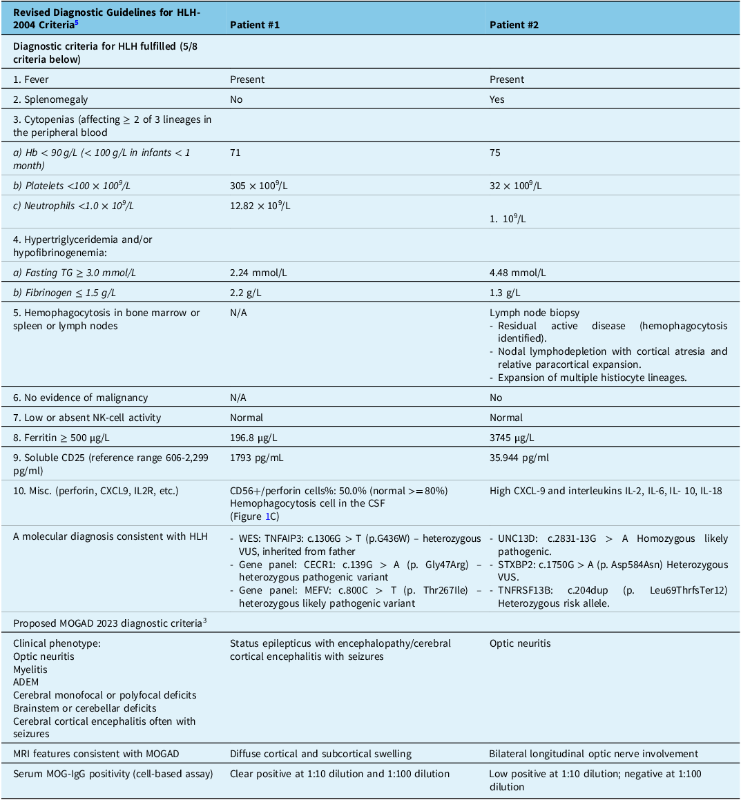
A previously healthy and developmentally appropriate 4-year-old girl presented with focal seizures that evolved to intractable status epilepticus. This was preceded by a 1-week history of fever, headache, neck pain and upper respiratory tract symptoms. Workup at presentation was remarkable for Hb 71, CSF pleocytosis (WBC 19 × 10^6/L,71% lymphocytes), elevated CSF glucose (4.6 mmol/L), elevated CSF protein (0.55 g/L) and MOG-IgG antibody serum positivity (fixed cell-based assay , London Health Sciences Centre) (Table 1). Comprehensive infectious workup and autoimmune encephalitis antibody panel were negative. Brain MRI demonstrated extensive cortical swelling with abnormal T2-FLAIR hyperintensities involving both cerebral hemispheres, with patchy diffusion restriction in the cortical and subcortical white matter (Figure 1A and 1B). She received a 2-day course of Intravenous Immunoglobulin (IVIG), a 5-day course of pulse methylprednisolone (30 mg/kg/day), plasmapheresis and anakinra, and was started on a ketogenic diet with no seizure improvement and an electroencephalogram (EEG) showing continued encephalopathy. Initial flow cytometry revealed low levels of CD56+/perforin+ cells (50%, normal>80%) and impaired NK degranulation assay. Both were later repeated and were found to be in normal range (Figure 1C) (Table 1). These findings, including CSF hemophagocytosis, supported the diagnosis of HLH, and she was started on treatment with ruxolitinib (5 mg BID), dexamethasone (5 mg/m2 BID) and intrathecal methotrexate (12 mg OD). She continued to demonstrate seizure activity and was treated with a course of etoposide (150 mg/m2/dose) twice weekly for 2 weeks, four doses in total. Seizure control was achieved, and the patient was discharged on a dexamethasone taper. Ruxolitinib was not continued as the patient developed neutropenia. One month later, she returned with recurrent seizures following steroid tapering, and a repeat brain MRI showed increased T2-FLAIR hyperintensities in the deep and periventricular white matter with some areas of enhancement. Repeat CSF studies showed normal White Blood Cell count (WBC)(2 × 10⁶/L) and elevated protein (0.61 g/L). She received a 5-day course of pulse methylprednisolone. Whole exome sequencing revealed three variants of unknown significance (Table 1) though none aligned with her clinical phenotype. Nine months post-onset, she demonstrated nearly complete recovery. Follow-up EEG at 7 months after presentation demonstrated multifocal interictal discharges. She is still on three anti-seizure medications with her last documented seizure 12 months after her initial presentation. Follow-up brain MRI 9 months after presentation demonstrated complete resolution of lesions. Serum MOG-IgG antibody remained positive 11 months after onset.
Neuroimaging and CSF findings in two patients with overlapping features of MOGAD and CNS-HLH. Panels A–C for Patient #1: (A) Axial FLAIR MRI shows diffuse cortical swelling and hyperintensities involving the subcortical white matter and cerebral cortex. (B) Axial diffusion-weighted imaging demonstrates patchy diffusion restriction in the same regions. (C) CSF cytology (Wright–Giemsa stain, original magnification 1000×) reveals a phagocytic histiocyte engulfing red blood cells, consistent with hemophagocytosis. Panel D for Patient #2: (D) Axial T2-weighted MRI reveals bilateral, symmetric, long-segment hyperintense swelling of the optic nerves. MOGAD = myelin oligodendrocyte glycoprotein antibody-associated disease; CNS-HLH = central nervous system-hemophagocytic lymphohistiocytosis.

Summary of HLH and MOGAD diagnostic criteria in both patients

WES = Whole-exome sequencing; VUS = Variant of Uncertain Significance. ADEM = acute disseminated encephalomyelitis; HLH = hemophagocytic lymphohistiocytosis; MOGAD = myelin oligodendrocyte glycoprotein antibody-associated disease.
*The degranulation assay measures surface exposure of CD107a NK cells by flow cytometry as per the published protocol (Bryceson et al., Blood119: 2754-2763, 2012). The results are expressed as the net percentage of cells expressing CD107a for resting and/or activated NK cells (CD3-CD56+) after exposure to K562 target cells.
Patient #2
A previously healthy 8-year-old girl born to consanguineous parents presented with persistent fevers, lymphadenopathy and massive splenomegaly. Laboratory investigations (Table 1) revealed severe pancytopenia, hyperferritinemia (3745.9 mg/L), hypofibrinogenemia (1.3 g/L) and positive Epstein–Barr virus (EBV) serology suggestive of reactivation (EBNA IgG+, EA IgG+, VCA IgG+). She was diagnosed with HLH, suspected to be secondary to EBV infection. Ruxolitinib was started at 10 mg BID. She demonstrated significant improvement both clinically, with resolution of her fevers, and biochemically, with improvement in HLH markers. Ruxolitinib was discontinued after 1 week of treatment, and the patient was discharged home. Two days following discharge, she re-presented with fever and abdominal pain. She was admitted to the hospital, and ruxolitinib was restarted. Her soluble CD25 level was significantly elevated, out of proportion to the ferritin level, raising concern for lymphoma. An excisional biopsy of an enlarged axillary lymph node was performed, showing normal histology. Bone marrow biopsy demonstrated no evidence for malignancy but showed evidence for hemophagocytosis. She developed blurred vision, and an ophthalmology assessment revealed bilateral disc edema and retinal hemorrhages. Given the worsening of symptoms, ruxolitinib was discontinued (after 14 days of treatment), and dexamethasone was started (5 mg/m2 BID).
MRI showed bilateral optic nerve involvement, a focal T2/FLAIR hyperintensity in the lentiform nucleus, dural thickening along the tentorium and enhancement along perineural regions of the cranial nerves (Figure 1D). Lumbar puncture revealed WBC 18 (93% lymphocytes), low glucose, normal protein and normal opening pressure; CSF culture was negative. Serum MOG-IgG antibody returned weakly positive (positive at 1:10 dilution and negative at 1:100 dilution). She was treated with 5 days of IV pulse methylprednisolone, IVIG 2 g/kg and seven cycles of PLEX, with marked improvement in visual acuity (20/25 bilaterally). Genetic testing revealed a homozygous variant in the UNC13D (c2831-13G > A), confirming familial HLH. She received bone marrow transplantation following this.
Discussion
Although MOGAD and CNS-HLH are pathologically distinct entities, they share notable immunopathogenic and clinical features. Both are characterized by immune dysregulation and are often triggered by infections, and their clinical manifestations may overlap. The exaggerated immune response seen in HLH can resemble the pathogenic mechanisms observed in MOGAD, with both conditions demonstrating prominent involvement of pro-inflammatory cytokines – potentially driven by similar cytotoxic activation pathways. The contribution of MOG-IgG antibodies to these overlapping presentations remains uncertain; however, our cases highlight the importance of investigating hyperinflammatory syndromes when encountering acute neuroinflammatory presentations.
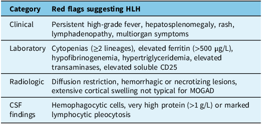
CNS-HLH is not a single disease, but a syndromal entity that identifies a hyper-immune syndrome that may be either genetically mediated or acquired. Reference Schmid, Côte and Ménager7 MRI abnormalities can include multifocal or symmetric lesions in the leptomeninges, periventricular, juxtacortical, cortical or cerebellar regions. These lesions are often poorly defined, with associated perilesional edema, and may show nodular or ring enhancement. Thalamic and basal ganglia involvement is rare, while hemorrhagic transformation may occur due to perivascular infiltration and ischemia. Diffusion restriction may be present. Reference Ma, Zhou and Li9 CSF analysis frequently shows pleocytosis (≥10 cells/μL), elevated protein (≥0.5 g/L) and, in some cases, direct evidence of hemophagocytosis. Reference Shyu, Luca, VandenBussche and Ho10 Elevated cytokines such as IFN-γ, sCD25, IL-10, IL-18, CXCL9 and neopterin are also observed but are nonspecific. Reference Zhao, Ou and Wei11 Table 2 outlines the key red flags that should prompt consideration of HLH. In contrast, in MOGAD, diffusion imaging is infrequently positive, and MRI findings often involve the optic nerves, spinal cord and brainstem. Lesions in MOGAD tend to resolve and lack the hemorrhagic or necrotizing components seen in HLH. CSF in MOGAD may show pleocytosis and elevated protein. Reference Banwell, Bennett and Marignier3
Key red flags that should prompt consideration of HLH

HLH = hemophagocytic lymphohistiocytosis; MOGAD = myelin oligodendrocyte glycoprotein antibody-associated disease.
The discovery of the MOG-IgG antibody and the establishment of diagnostic criteria have led to the recognition of MOGAD as a distinct clinical entity. Increasing use of antibody testing has raised concerns about false positives, particularly in individuals with a low pretest probability or in the absence of supportive clinical findings. Several case reports have described MOG-IgG antibody positivity in patients with other neuroinflammatory disorders, Reference Yan, Tian, Zhang and Wang4,Reference Jia and Graham5 similar to our cases, raising the question of how these diagnostic entities relate to one another. The relevance of MOG-IgG in our patients is unclear. While both have possible genetic mechanisms that could explain HLH, these entities may coexist, or the MOG-IgG antibody may have occurred in the context of an infection, triggering the hyperinflammatory syndrome, thus playing an essential role in the presentation. At present, it is unclear whether low MOG-IgG titers using the fixed cell-based assay clearly differentiate “true” positives from “false” positives in the pediatric population. Reference Budhram, Rotstein, Yang and Yeh12
Our cases illustrate the diagnostic complexity that can arise when overlapping features of MOGAD and CNS-HLH are present. These findings underscore the importance of broadening the differential diagnosis in patients presenting with demyelinating syndromes. Our approach is to perform screening investigations in children presenting with a first-time neuroinflammatory event including Erythrocyte Sedimentation Rate (ESR), C-reactive protein (CRP), complete blood count, coagulation profile, ferritin and liver enzymes, with the later addition of HLH-specific markers if these are abnormal. These tests are not intended to universally screen for HLH but can serve as important markers of systemic inflammation that may prompt further evaluation when abnormal. While a positive antibody result may suggest an antibody-mediated process, it must be interpreted with caution and understood in the context of the overall clinical and radiological picture. Careful clinical assessment and consideration of hyperinflammatory syndromes are essential to ensure accurate diagnosis and appropriate treatment.
Acknowledgments
We would like to thank Dr. Mohamed Abdelhaleem for kindly providing the photomicrograph illustrating hemophagocytosis.
Author contributions
L.A., R.S. and A.M. contributed to the writing of the first draft., H.B. and A.N. contributed to the concept of the manuscript and review. H.B. contributed to the review of imaging. EAY: conceptualization of manuscript, supervision, drafting, revision of manuscript. All authors reviewed and approved the final draft.
Funding statement
EAY’s work is supported in part by the Garry Hurvitz Chair in Neurology. .
Competing interests
EAY has received research support in the last 3 years from the National Multiple Sclerosis Society, Canadian Institutes of Health Research, Stem Cell Network, SickKids Foundation, Peterson Foundation, Multiple Sclerosis Society of Canada, Leong Center, Guthy Jackson Foundation, OMS Life, Canada’s Drug Agency, Garry Hurvitz Chair in Neurology and the Multiple Sclerosis Scientific Research Foundation. She has served on scientific advisory boards for Biogen, Alexion, and Hoffman‐LaRoche. DSMB: WCG, IQVA. Co-chief Editor: MS and Related Disorders. Speaker/other Honoraria/Support for Travel: SOPNIA Chile, University of Chile, ECTRIMS, ACTRIMS, Johns Hopkins University, New Brunswick Neurological Society, American Academy of Neurology, Consortium of MS Centers, University of Ottawa, Canadian Institutes of Health Research, Michael Smith Health Research Organization, Medlink. Clinical trials: Alexion, Novartis, Hoffman-LaRoche. Governing Council/Steering Committee: Stem Cell Network, Rare Kids CAN, Cantrain.


 Open access

