


Introduction
The fossil record not only documents the appearance of lineages through time. Morphological details may also allow inferences to be made regarding ecology and behavior of extinct animal species. Here we describe a new genus of typhlocybine leafhopper (Hemiptera, Cicadellidae) from the Eocene (Rovno amber) representing the smallest known fossil insect (~2.5 mm) with false eyespots, a presumed defensive strategy against visual predators. We also describe another new genus from the same fauna that represents an apparent morphological transition between the extinct typhlocybine tribe Protodikraneurini and the modern tribe Dikraneurini.
Typhlocybinae is the second-largest subfamily of leafhoppers, with >6,000 described extant species placed in ~300 genera and five tribes. Although the oldest fossil Cicadellidae are known from the Early Cretaceous (Aptian; Hamilton, Reference Hamilton and Grimaldi1990, Reference Hamilton1992; Martill et al., Reference Martill, Brito and Donovan2021) and recent molecular time trees place the origin of this family in the Cretaceous (~112–138 Ma; Dietrich et al., Reference Dietrich, Allen, Lemmon, Moriarty Lemmon, Takiya, Evangelista, Walden, Grady and Johnson2017; Johnson et al., Reference Johnson2018), the oldest typhlocybine fossils known so far are from Eocene Baltic amber (36.4–36.8 Ma; Iakovleva, Reference Iakovleva2017; Iakovleva et al., Reference Iakovleva, Mychko, Aleksandrova, Golubev and Nazarov2021).
Typhlocybinae from Baltic amber previously described with sufficient detail belong to an extinct tribe, Protodikraneurini, that has a unique hind-wing venational pattern not known to occur in modern typhlocybines. Three genera comprising five species of this tribe have been described from this fauna so far (Gębicki and Szwedo, Reference Gębicki and Szwedo2006; Szwedo and Gębicki, Reference Szwedo and Gębicki2008; Szwedo et al., Reference Szwedo, Gębicki and Kowalewska2010). Type specimens for three species of Typhlocybinae described earlier from Baltic amber (Germar and Berendt, Reference Germar, Berendt and Berendt1856; Bervoets, Reference Bervoets1910) have not been located, and the original descriptions and illustrations lack details necessary for definite tribal placement, but they appear to be correctly assigned to Typhlocybinae. The oldest previously reported fossil belonging to a modern typhlocybine tribe is an undescribed species reported from Miocene Dominican amber by Dietrich and Vega (Reference Dietrich and Vega1995) and tentatively assigned to the tribe Dikraneurini.
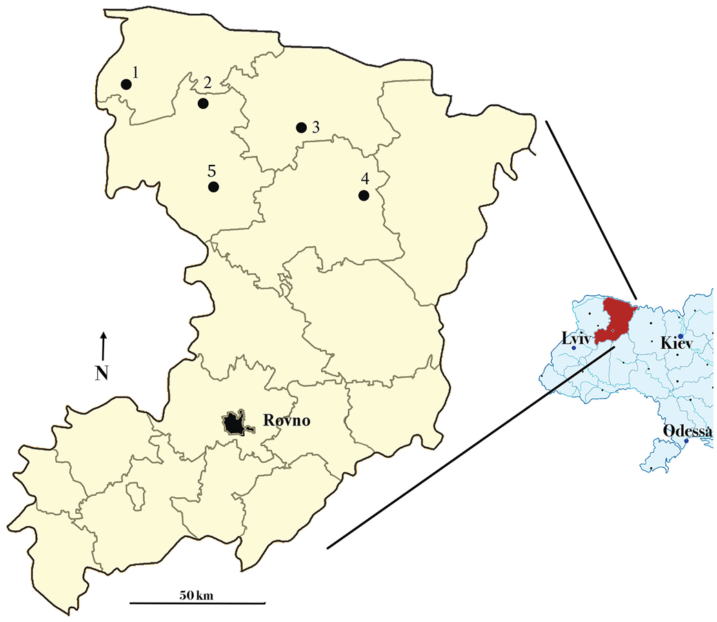
Most known genera and species of Eocene-age Cicadellidae have been described from Baltic amber from northern Europe (reviewed by Szwedo, Reference Szwedo and Holzinger2002; Dietrich and Gonçalves, Reference Dietrich and Gonçalves2014), but, so far, only two cicadellids have been described from Rovno amber, which is similar in age (35–37 Ma; Mänd et al., Reference Mänd, Muehlenbachs, McKellar, Wolfe and Konhauser2018) but collected farther south, mostly in Ukraine (Dietrich and Perkovsky, Reference Dietrich and Perkovsky2020; Dietrich et al., Reference Dietrich, Dmitriev and Perkovsky2021), especially in Rovno Oblast. In recent years, Varash District localities yielded dozens of taxa unknown from the better-studied Klesov deposit (Gilka et al., Reference Giłka, Harbach and Perkovsky2021; Matalin et al., Reference Matalin, Perkovsky and Vasilenko2021; Melnitsky et al., Reference Melnitsky, Ivanov and Perkovsky2021; Kazantsev and Perkovsky, Reference Kazantsev and Perkovsky2022; Legalov et al., Reference Legalov, Nazarenko, Vasilenko and Perkovsky2022a [and references therein], Reference Legalov, Vasilenko and Perkovsky2022b; Olmi et al., Reference Olmi, Guglielmino, Vasilenko and Perkovsky2022; Vitali and Perkovsky, Reference Vitali and Perkovsky2022; Yamamoto et al., Reference Yamamoto, Nazarenko, Vasilenko and Perkovsky2022). From Zhovkini, a new encyrtid genus (Simutnik et al., Reference Simutnik, Perkovsky and Vasilenko2022) and the first Rovno amber ptylodactilid male (Telnov et al., Reference Telnov, Perkovsky, Kundrata, Kairišs, Vasilenko and Bukejs2022) were recently discovered. A map of these and other localities for Rovno amber is shown in Figure 1.
Map showing Rovno amber collection localities (Rovno Region, Ukraine) (modified from Martynova et al., Reference Martynova, Perkovsky, Olmi and Vasilenko2019): 1. vicinity of Kukhotskaya Volya village (basin of Stokhod River); 2. vicinity of Voronki village (basin of Styr River); 3. vicinity of Volnoe village (Dubrovitsa); 4. vicinity of Klesov settlement; 5. vicinity of Zhovkini village.
The fossils described herein from both Klesov and Varash District are the first known Typhlocybinae from Eocene Rovno amber. Three of them clearly belong in Protodikraneurini based on the hind wing with a complete submarginal vein and radius posterior (RP) and media anterior (MA) veins separate from each other and connected by a crossvein. They appear to be related to, but distinct from, the species so far reported from Baltic amber due to their different forewing venation. An additional new species has hind-wing venation that appears to be transitional between Protodikraneurini and Dikraneurini; veins RP and MA are united for a short distance but separate and diverge preapically. Another specimen appears to be the first representative of Dikraneurini reported from the Eocene, the oldest known representative of a modern typhlocybine tribe reported thus far.
Materials and methods
The studied Protodikraneurini leafhoppers were mined at Klesov (51.323°N, 26.897°E, Pugach quarry; Mitov et al., Reference Mitov, Perkovsky and Dunlop2021), and Dikraneurini were found 1 km west of Zhovkini (51.355°N, 26.132°E, Varash District, Rovno Oblast). Morphological terminology follows Dietrich et al. (Reference Dietrich, Dmitriev, Takiya, Thomas, Webb, Zahniser and Zhang2022). Setal rows on the tibia are abbreviated as AD (anterodorsal), PD (posterodorsal), AV (anteroventral), and PV (posteroventral). Diagnosis and classification of Typhlocybinae tribes follow Dietrich (Reference Dietrich2013). Photographs were taken using Leica M16 and Leica Z16 APO microscopes equipped with a Leica DFC 450 camera and processed by LAS Core software.
Repository and institutional abbreviation
Examined specimens are deposited in the Schmalhausen Institute of Zoology of the National Academy of Sciences of Ukraine, Kiev (SIZK), amber collection.
Systematic paleontology
Order Hemiptera Linnaeus, Reference Linnaeus1758
Family Cicadellidae Latreille, Reference Latreille1825
Subfamily Typhlocybinae Kirschbaum, Reference Kirschbaum1868
Tribe Protodikraneurini Gębicki and Szwedo, Reference Gębicki and Szwedo2006
Diagnosis
Forewing without appendix. Hind wing submarginal vein complete, extended around wing apex onto costal area and confluent with vein RA; veins RP and MA separate and connected by radial-medial (r-m) crossvein.
Remarks
This tribe was previously known from three genera and five species, all described from Eocene Baltic amber (Gębicki and Szwedo, Reference Gębicki and Szwedo2006; Szwedo and Gębicki, Reference Szwedo and Gębicki2008; Szwedo et al., Reference Szwedo, Gębicki and Kowalewska2010). Typhlocybinae inclusions from Rovno amber studied so far represent at least two additional genera, described in the following.
Genus Retrorsotettix new genus
Type species
Retrorsotettix vlaskini n. sp., by monotypy.
Diagnosis
As for type species by monotypy.
Etymology
The genus name, a masculine noun, combines the Latin retrorsum (backward) with tettix, a common suffix used for leafhopper names that refers to the false eyespot and leglike markings near the apex of the forewing, which give the insect the appearance of having a head at the posterior end of the body.
Remarks
This genus resembles Microelectrona Szwedo and Gębicki in Szwedo et al., Reference Szwedo, Gębicki and Kowalewska2010, described from Eocene Baltic amber, in having the base of the forewing third apical cell angulate, formed by veins RP and MA converging to meet for a short distance, then diverging toward the wing apex. Other previously described genera of the tribe have these veins separate and connected by a crossvein. Other characters separating the new genus from Microelectrona include the presence of a false eyespot and oblique false veins on the forewing and a relatively long, narrow male subgenital plate with macrosetae restricted to the distal half. The forewing venation of Stareono Gębicki and Szwedo, Reference Gębicki and Szwedo2006 remains unknown, but that genus has the male pygofer with crossed distal spines, which are absent in Retrorsotettix.
Holotype
SIZK K-30290, Klesov, Rovno amber, late Eocene. The specimen, a male, is intact, complete, and in good condition, embedded in a pale-yellow piece of amber with fore- and hind wings spread. Details of the facial sclerites and antennal bases are obscured by a white opaque material. Syninclusions: stellate hairs. Weight of piece before primary treatment: 2 g.
Retrorsotettix vlaskini n. gen. n. sp., holotype: (1) habitus, slight ventrolateral view; (2) habitus, dorsal view; (3) left hind femur, tibia, and tarsus, ventral view; (4) genital capsule, slight ventrolateral view.

Diagnosis
This species differs from other Protodikraneurini in having the following combination of traits: overall body and wing coloration pale; forewing veins RP and MA converging to meet for short distance then diverging toward wing apex, color pattern including two oblique black lines extended to costal margin in apical half and black false eyespot in outer apical cell; hind-wing vein MP confluent with cubitus anterior (CuA) for short distance preapically then diverging toward submarginal vein; male subgenital plate broadest near base with macrosetae situated beyond midlength; pygofer without distal process.
Description
Length of body (without wings): 2.14 mm; forewing: 2.02 mm; length including wing: ~2.5 mm. Dorsal coloration pale, forewing with two broad, reflexed dark false veins along costal margin, one near midlength, and one near distal third; dark elliptical false eyespot in middle of outer apical cell; venter and legs pale. Head slightly wider than pronotum; crown between eyes 1.8× wider than median length, anterior margin parabolically rounded, slightly longer medially than next to eyes; face convex, relatively broad, gena shallowly concave below eye, rostrum not reaching middle trochanter. Pronotum 2.25× longer than crown, anterior margin strongly produced, posterior margin slightly concave. Exposed part of mesonotum and scutellum 0.71× as long as pronotum. Forewing inner apical cell trapezoidal, CuA extended obliquely toward MP then bent at obtuse angle toward inner margin at medial-cubital (m-cu) crossvein and reaching margin well before wing apex; second apical cell with narrow but truncate base, broadened distally; third apical cell with base acutely angulate, formed by meeting of RP and MA at single point; clavus 0.63× total length of forewing. Hind wing with veins RP and MA narrowly separate and nearly parallel preapically, connected by short r-m crossvein; m-cu crossvein relatively long and nearly perpendicular to MP and CuA, CuA branched well distad of crossvein. Front femur with pair of dorsoapical macrosetae. Hind femur macrosetal formula 2 + 1 + 1; tibia row AD with approximately eight long, evenly spaced macrosetae, PD with ~10 macrosetae shorter than those in AD, AV with four long macrosetae in distal half; tarsus elongate and slender, 0.4× as long as tibia. Male with valve concave; subgenital plates in ventral view completely exposed, broad at base, lateral margins roundly tapered distally, each with two macrosetae sublaterally near midlength, apices tapered, compressed, and upturned, extended slightly beyond apex of pygofer.
Etymology
This species is named in honor of Anatoly P. Vlaskin (SIZK), who found, cut, and polished the holotype and other specimens examined for this paper.
Remarks
This species is the first known fossil leafhopper with false eye and leg markings on the forewing. Such markings are present in some modern leafhoppers and presumably function to confuse visual predators such as birds.
Genus Protoparallaxis new genus
Type species
Protoparallaxis clavata n. sp., by monotypy.
Diagnosis
As for type species by monotypy.
Etymology
The genus name, a feminine noun, combines the Greek protos (first) with Parallaxis, the name of a modern genus of Dikraneurini with similar forewing venation.
Remarks
The forewing venation of this genus is very unlike that of other Protodikraneurini for which the forewing venation has been described. The inner apical cell is very short and broad, as in modern Typhlocybini and a few Neotropical Dikraneurini, e.g., Parallaxis McAtee, Reference McAtee1926.
Protoparallaxis clavata new species
Figures 3.6–3.8, 4
(1–5) Retrorsotettix vlaskini: (1) head pronotum, mesonotum and scutellum, dorsal view; (2) forewing; (3) hind wing; (4) valve and subgenital plates, ventral view; (5) left hind femur, tibia, and tarsus, anteroventral view. (6–8) Protoparallaxis clavata n. gen. n. sp: (6) forewing; (7) hind wing apex; (8) genital capsule, ventral view. ac = apical cell; CuA = cubitus anterior; CuP = cubitus posterior; MA = media anterior; MP = media posterior; m-c = media-cubitus crossvein; RP = radius posterior.

Protoparallaxis clavata n. gen. n. sp., holotype: (1) habitus, ventral view; (2) hind wing; (3) genital capsule, ventral view.

Holotype
SIZK K-545, Klesov, Rovno amber, late Eocene. The specimen, a male, lacking the head, is embedded in a medium-yellow piece of amber with wings spread but the dorsum and anterior part of the body poorly visible and venter partly obscured by air bubbles. Syninclusion: SIZK K-546, female of Ceratopogon (Ceratopogonidae). Weight of piece after primary treatment: 1.3 g.
Diagnosis
This species differs from other known Protodikraneurini in having the following combination of traits: forewing with inner apical cell very short and broad, m-cu crossvein connected to vein RP + MA, second apical cell encompassing entire apical margin of wing; hind-wing veins MP and CuA joined by short, oblique m-cu crossvein; male subgenital plate with row of three sublateral macrosetae near midlength, apex slightly expanded in ventral view.
Description
Body length not including wings: 2.8 mm. Forewing inner (first) apical cell unevenly pentagonal, short, and wide; m-cu crossvein forming nearly right angle with distal segment of CuA, joining RP + MA, RP and MA diverging at right angle distally to apparently encompass entire wing apex. Hind wing with CuA branched well basad of m-cu crossvein, which is longer and more basad than r-m crossvein. Hind tibia row AD with macrosetae slightly longer and less numerous than row PD; row AV of hind tibia with four macrosetae in distal half; tarsus relatively short and broad. Male pregenital sternite short, broad, broadly concave posteriorly, partly retracted into segment VII. Subgenital plates slightly shorter than pygofer, separate but closely appressed throughout length, narrow at base then abruptly broadened to widest point in basal fourth, then gradually narrowed toward apex, with slight preapical expansion in ventral view; each with row of three submarginal macrosetae near midlength.
Etymology
The species name refers to the slightly clavate apices of the male subgenital plates in ventral view.
Remarks
This species apparently represents a transitional stage showing considerable consolidation of the forewing venation preapically compared with other Typhlocybinae known from the Eocene. Such consolidation is similar to that found in many modern genera of the tribes Dikraneurini and Typhlocybini. It is somewhat similar to the venation of Eupteryx minuta Bervoets (Reference Bervoets1910) from Baltic amber. However, Bervoets's figure of the forewing (Bervoets, Reference Bervoets1910, fig. 6) shows a sharp bend in vein CuA, suggesting that he omitted from his drawing the distal segment of this vein that terminates at the wing margin; thus, the inner apical cell is, apparently erroneously, shown as open rather than closed, and the second apical cell does not extend to the wing apex. The second apical cell of P. clavata differs in encompassing the entire forewing apex.
Protodikraneura (?) sp.
Figure 5
Description
Body length including hind wings: 2.5 mm. Head with anterior margin bluntly angulate, frontoclypeus relatively short and broad, strongly convex; anteclypeus strongly tapered and relatively short; rostrum not extended to middle trochanter. Hind wing veins RP and MA narrowly separated and slightly divergent toward apex, connected by short r-m crossvein; m-cu crossvein much longer and oblique, CuA branched slightly distad of crossvein, branches unusually long. Front tibia with ~10 AV macrosetae. Middle femur with pair of dorsoapical setae. Hind tibia row AD with 10 macrosetae; PD with ~24 macrosetae; AV with four macrosetae in distal half; tarsus relatively short, 0.32× as long as tibia. Male valve concave posteriorly, subgenital plates separated to base, broad at base, lateral margins roundly tapered distally, apex upturned and compressed, lateral margin with seven to eight short, stout setae in row basad of midlength progressively smaller distally.
Protodikraneura (?) sp., specimen SIZK K-728a: (1) ventrolateral habitus; (2) right hind femur (apex), tibia, and tarsus, anteroventral view.

Materials
SIZK K-728a, Klesov, Rovno amber, late Eocene. The male specimen is complete and in good condition, embedded in a pale-yellow piece of amber with wings spread and hind wings well visible but forewings and dorsum obscured by fractures. Syninclusions: SIZK K-728: Germaraphis dryoides (Germar and Berendt, Reference Germar, Berendt and Berendt1856) (Eriosomatidae), Cicadellidae; SIZK K-729: Drepanosiphidae; SIZK K-730: male of Chironomidae; SIZK K-731: Chironomidae; SIZK K-732: male and female of Orchestina sp. (Oonopidae); SIZK K-733: Chironomidae. Weight of piece after primary treatment: 9.1 g.
Remarks
The hind wing with complete submarginal vein, and veins RP and MA separate and connected by a crossvein, unequivocally place this fossil in Protodikraneurini. The specimen appears to closely resemble Protodikraneura cephalica Gębicki and Szwedo (Reference Gębicki and Szwedo2006), described from Baltic amber, in head structure, leg chaetotaxy, hind wing venation, and shape of the male pygofer and subgenital plates. One possible difference between this species and species of Protodikraneura described from Baltic amber is the presence of a row of small, stout setae on the lateral margin of the subgenital plate near the base. Such setae are apparently absent in P. cephalica and other described species of Protodikraneura. Stareono mirabilis Gębicki and Szwedo (Reference Gębicki and Szwedo2006) has a row of four to five enlarged setae near the midlength of the subgenital plate and extended onto the distal half. Unfortunately, no details of the forewing venation are visible in our specimen. Thus, we are unable to confirm the placement of this species in Protodikraneura, although it appears to be distinct from previously described Protodikraneurini.
Key to genera of Protodikraneurini
1 Hind-wing vein MP connected to CuA at single point (Gębicki and Szwedo, Reference Gębicki and Szwedo2006, fig. 101) or via short m-cu crossvein (Fig. 3.7) … 2
1' Hind-wing vein MP fused to CuA for considerable distance preapically (Fig. 3.3) … 3
2 Male pygofer lobes with paired distal spines crossing each other at midline (forewing venation unknown) … Stareono Gębicki and Szwedo, Reference Gębicki and Szwedo2006
2' Male pygofer lobes without paired distal spines; forewing inner apical cell no longer than maximum width, not extended to apical margin of wing (Fig. 3.6) … Protoparallaxis n. gen.
3 Forewing with third apical cell quadrate at base, r-m crossvein present … Protodikraneura Gębicki and Szwedo, Reference Gębicki and Szwedo2006
3' Forewing with third apical cell narrowly angulate at base, RP confluent with MA for short distance (Fig. 3.2) … 4
4 Forewing with false eyespot in apical cell four and reflexed false vein along costal margin (Fig. 3.2); male subgenital plates narrower than valve, compressed, without macrosetae near base (Fig. 3.4)… Retrorsotettix n. gen.
4' Forewing without false eyespot or false reflexed vein; male subgenital plates short, broader than valve, strongly depressed, with lateral row of macrosetae near base … Microelectrona Szwedo and Gębicki in Szwedo et al., Reference Szwedo, Gębicki and Kowalewska2010.
Tribe Dikraneurini McAtee, Reference McAtee1926
Diagnosis
Forewing without appendix. Hind-wing submarginal vein complete, extended around apex and along costal margin basad of RP + MA (submarginal vein obsolete in Typhlocybella); veins RP and MA completely confluent distally (except Eodikraneura n. gen., described in the following with RP and MA confluent for short distance then divergent and reaching submarginal vein separately). Male subgenital plates usually with macrosetae reduced in number (usually only three to five present) or absent.
Remarks
This modern tribe currently includes >500 described species placed in >70 genera and is distributed worldwide, inhabiting temperate and tropical forests, grasslands, and savannas. Only one fossil definitely attributable to this tribe has been reported previously, an undescribed genus and species from Miocene Dominican amber (Dietrich and Vega, Reference Dietrich and Vega1995).
Genus Eodikraneura new genus
Type species
Eodikraneura obscura n. sp., by monotypy.
Diagnosis
As for type species by monotypy.
Etymology
The genus name, a feminine noun, combines the Greek eos (dawn) with Dikraneura, the name of the type genus of Dikraneurini, and is meant to suggest that this species may be closer to the common ancestor of modern Dikraneurini than any member of Protodikraneurini due to the transitional state of the hind-wing venation.
Remarks
Eodikraneura exhibits a unique pattern in the hind-wing venation that appears to represent a transitional form between the extinct tribe Protodikraneurini and the extant tribe Dikraneurini. Instead of having veins RP and MA separate from each other and connected by a crossvein, as in Protodikraneurini, this genus has the two veins confluent for a short distance preapically before diverging and extending to the submarginal vein. All modern Dikraneurini have hind-wing veins RP and MA completely confluent distally and extended to the submarginal vein as a single vein. Among modern Typhlocybinae, some Alebrini (e.g., Rabela Young, Reference Young1952; Rhabdotalebra Young, Reference Young1952) also have hind-wing veins RP and MA partially confluent and then divergent preapically, but they lack a submarginal vein and have an appendix on the forewing. The appendix is a presumably plesiomorphic trait shared by Alebrini with most non-typhlocybine leafhoppers. Alebrini are mostly restricted to the Neotropical region and remain unknown from the fossil record. Few details of the forewing venation are visible on the holotype of Eodikraneura, but the inner apical cell appears to be long and nearly parallel-sided, the usual condition in modern Dikraneurini, rather than distinctly tapered, as in known Protodikraneurini. The genus is placed in Dikraneurini on the basis of the retention in the hind wing of a complete submarginal vein and the partial confluence of hind-wing veins RP and MA.
Eodikraneura obscura new species
Figure 6
Holotype
SIZK ZH-64 Zhovkini, Rovno amber, late Eocene. The specimen, a male, is intact, complete, and in good condition, embedded in a piece of pale-yellow amber with wings folded over the body in normal rest position and only parts of the face and hind legs obscured by a milky veil. Syninclusions: stellate hairs, SIZK ZH-63: Cicadellidae; SIZK ZH-65: Anystidae; SIZK ZH-65: female of Chironomidae. Weight of piece after primary treatment: 20 g.
Eodikraneura obscura n. gen. n. sp., holotype: (1) habitus, dorsolateral view; (2) habitus, ventrolateral view; (3) enlarged ventrolateral view showing male genital capsule and portions of hind wings; (4) partial reconstruction of hind wing venation showing partial anastomosis of veins RP and MA; (5) head, ventrolateral view; (6) left front femur, tibia, and tarsus, anteroventral view; (7) exposed parts of right hind tibia and tarsus (ventral surface of tibia not visible except near apex). CuA = cubitus anterior; MA = media anterior; MP = media posterior; RP = radius posterior.

Diagnosis
This species may be readily distinguished from other Dikraneurini by the partial confluence of hind-wing veins RP and MA. Other distinguishing traits include head narrower than pronotum, face elongated, ocelli absent; forewing inner apical cell elongate and parallel-sided; male subgenital plates partially concealed by pregenital sternite.
Description
Length: 3.28 mm. Dorsum with coloration apparently uniformly dark, venter and legs pale. Head slightly narrower than pronotum; crown 1.54× wider between eyes than median length, slightly longer medially than next to eye; anterior margin rounded in dorsal view; ocelli not visible, apparently absent; face with frontoclypeus slightly convex, elongate, more than twice as long as greatest width; anteclypeus elliptical, extended well beyond lower margin of gena, medial margin extended less than half distance to base of lorum; lorum flat; rostrum extended to middle trochanter. Pronotum 1.73× longer than crown, anterior margin strongly produced, posterior margin nearly straight. Exposed part of mesonotum and scutellum 0.8× length of pronotum. Forewing clavus 0.67× total length of wing; inner apical cell parallel-sided. Hind wing with veins RP and MA confluent for a short distance then divergent and reaching submarginal vein separately. Front tibia with six AV macrosetae. Middle femur with single dorsoapical seta, tibia with few widely spaced short setae. Hind femur with 2 + 1 + 1 dorsoapical macrosetae; tibial row AD with 10 macrosetae, PD with ~15 macrosetae; tarsus elongate, 0.42× as long as tibia. Male pregenital sternite trapezoidal, overlapping, and obscuring base of subgenital plates; subgenital plates triangular in ventral view, compressed and slightly upturned distally, lateral margins roundly tapered, with three macrosetae near midlength laterally.
Etymology
The species name refers to the dark overall coloration.
Remarks
This species apparently represents a transitional stage in the evolution of hind-wing venation between the extinct tribe Protodikraneurini and the modern tribe Dikraneurini. This and the following species representing the latter tribe are the only typhlocybines recorded so far from Zhovkini, a more western locality than the better-studied Klesov fauna, which so far includes only Protodikraneurini.
Genus Rovnodikra new genus
Type species
Rovnodikra longipes n. sp., by monotypy.
Diagnosis
As for type species by monotypy.
Etymology
The genus name refers to Rovno, the Ukrainian oblast in which the fossil was collected.
Remarks
This appears to be the oldest known representative of the crown group of modern typhlocybine tribe Dikraneurini. Visible parts of the hind-wing venation are indistinguishable from those of the modern genus Dikraneura, which has veins RP and MA completely confluent distally and CuA joining MP for a very short distance and then diverging distally. Other visible aspects of the morphology of the fossil also resemble those of Dikraneura although the male subgenital plate apparently lacks a row of three to four enlarged setae near the midlength. Unfortunately, the only available specimen is poorly preserved, and few characters are visible that would help elucidate its relationship to other known genera of the tribe. The visible parts of the head and wing venation appear to be essentially identical to those of Dikraneura. The elongate hind tarsi distinguish Rovnodikra from other genera of Dikraneurini.
Rovnodikra longipes new species
Figure 7
Holotype
SIZK ZH-162 Zhovkini, Rovno amber, late Eocene. The specimen is an intact male embedded in a piece of pale-yellow amber, with hind wings partly spread to sides and forewings extended dorsally but distorted with details of the venation not visible. The head, pronotum and mesonotum, and all of the ventral surface are covered with a milky veil that conceals many structural details. Syninclusions: stellate hairs, SIZK ZH-160: two Cicadellidae; SIZK ZH-161: female of Chironomidae; SIZK ZH-162: Tipuloidea; SIZK ZH-163: Phoridae; SIZK ZH-164:
Rovnodikra longipes n. gen. n. sp., holotype: (1) habitus, ventral view; (2) head, pronotum, mesonotum, and scutellum, dorsal view; (3) right hind wing, ventral view; (4) partial reconstruction of hind wing venation; (5) left hind wing, ventral view. CuA = cubitus anterior; MA = media anterior; MP = media posterior; RP = radius posterior.

Aphidinea, Latridiidae, Mymarommatidae. Weight of piece after primary treatment: 10 g.
Diagnosis
This species may be distinguished from other Dikraneurini by the following combination of traits: head in dorsal view acutely produced, face with width across eyes approximately equal to length along midline; hind-wing veins RP and MA completely confluent distally, veins MP and CuA confluent for short distance then divergent; hind tarsus very long and slender, nearly half as long as tibia; male subgenital plates slightly longer than pygofer.
Description
Body length without wings: 2.1 mm. Head slightly narrower than pronotum, anterior margin parabolic in dorsal view, crown much shorter than pronotum, face with width across eyes approximately equal to length along midline, rostrum very short, not extended beyond front trochanters; exposed part of mesonotum and scutellum slightly shorter than pronotum. Hind-wing veins RP and MA completely confluent at point well distad of oblique m-cu crossvein; m-cu crossvein connected to CuA at branching point. Hind tarsus elongate and slender, nearly half as long as hind tibia. Male valve concave posteriorly, subgenital plates tapered distally in ventral view, slightly longer than pygofer.
Etymology
The species name combines the Latin longus (long) with pes (foot) and refers to the elongate hind tarsus.
Remarks
The presence of this species with hind-wing venation apparently identical to that of modern Dikraneurini suggests that the typhlocybine leafhopper fauna of Zhovkini is more modern than that of the more eastern Klesov site, at which only Protodikraneurini have been found so far.
Discussion
Studies of leafhopper inclusions from Ukrainian Rovno amber have begun relatively recently, and the fauna remains poorly known compared with that of the much better studied Baltic amber. Some similarities between the two faunas are evident, including the presence of the modern subfamilies Aphrodinae, Bathysmatophorinae, and Typhlocybinae (Szwedo, Reference Szwedo and Holzinger2002; Dietrich and Gonçalves, Reference Dietrich and Gonçalves2014; Dietrich and Perkovsky, Reference Dietrich and Perkovsky2020; Dietrich et al., Reference Dietrich, Dmitriev and Perkovsky2021; unpublished data). Other modern cicadellid subfamilies present in Baltic amber but not yet discovered in Rovno amber include Eurymelinae, Ledrinae, Megophthalminae, and Mileewinae (Szwedo, Reference Szwedo and Holzinger2002; Dietrich and Gonçalves, Reference Dietrich and Gonçalves2014; Dietrich and Thomas, Reference Dietrich and Thomas2018). Both faunas also include cicadellid species with brachypterous adults (Dietrich and Gonçalves, Reference Dietrich and Gonçalves2014; Dietrich et al., Reference Dietrich, Dmitriev and Perkovsky2021) analogous to those inhabiting open grassy habitats in the modern fauna.
Unlike the typhlocybine fauna of Baltic amber, from which only the extinct tribe Protodikraneurini is definitely recorded, Rovno amber apparently includes members of the modern tribe Dikraneurini in addition to Protodikraneurini, and some genera of the latter tribe are distinct from those described from Baltic amber. The presence of one species with hind-wing venation apparently transitional between Protodikraneurini and Dikraneurini suggests that Rovno amber documents an important stage in the evolution of the modern leafhopper fauna.
Dikraneurini are presently the dominant group of Typhlocybinae in lowland South American rainforests but elsewhere constitute a ubiquitous but relatively minor component of the typhlocybine fauna compared with representatives of the tribes Erythroneurini, Empoascini, and Typhlocybini. Recent phylogenetic analyses indicate that Dikraneurini is a monophyletic group and is sister to Erythroneurini (Lu et al., Reference Lu, Dietrich, Cao and Zhang2021; Yan et al., Reference Yan, Dietrich, Yu, Jiao, Yang and Dai2022). Protodikraneurini have not yet been incorporated into formal phylogenetic analyses, and their relationship to modern typhlocybine tribes is uncertain. Nevertheless, the hind-wing venation, in which a complete submarginal vein is retained and veins RP and MA remain separate, suggests that this group could be ancestral to modern Dikraneurini, Erythroneurini, and Typhlocybini. The condition of the hind wing in Eodikraneura, in which veins RP and MA are partly confluent preapically, suggests that the completely confluent condition of these veins found in modern Dikraneurini, Erythroneurini, and most Typhlocybini occurred before the reduction and loss of the submarginal vein characteristic of the latter two tribes.
The newly described protodikraneurine, Retrorsotettix vlaskini, is the first known fossil cicadellid to exhibit a false eyespot and false leg markings (oblique black lines along the costal margin) on the forewing. Such markings are relatively uncommon among modern leafhoppers but have apparently been acquired independently in genera belonging to several different extant cicadellid subfamilies, including Cicadellinae (e.g., Diedrocephala Spinola, Reference Spinola1850), Coelidiinae (e.g., Boulardus Nielson, Reference Nielson1983), Deltocephalinae (e.g., Scaphomonus Viraktamath in Dai et al., Reference Dai, Viraktamath, Zhang and Webb2009; Fig. 8.2), Evacanthinae (e.g., Sophonia Walker, Reference Walker1870; Fig. 8.1), Neocoelidiinae (e.g., Retrolidia Dietrich, Reference Dietrich2003; Fig. 8.4), and Typhlocybinae (e.g., Alconeura Ball and DeLong, Reference Ball and DeLong1925). The markings presumably function to confuse visual predators such as birds and lizards, who may mistakenly attack the leafhopper's “false head,” allowing the leafhopper to escape by leaping in the opposite direction. Various authors have attributed similar markings in insects to spider mimicry, although modern insects exhibiting this syndrome often enhance the effect with spiderlike movements of the body (reviewed by Shcherbakov, Reference Shcherbakov2007). Modern leafhoppers with false eyespots have not been observed engaging in spider-mimicry behavior but, rather, sit motionless until approached, then suddenly leap away.
Modern Cicadellidae with false eyespot and false leg markings on the forewings: (1) Sophonia sp. (Evacanthinae, Nirvanini; China); (2) Scaphomonus sp. (Deltocephalinae, Scaphoideini; Vietnam); (3) Tahurella katharinae Dietrich, Reference Dietrich2013 (Typhlocybinae, Typhlocybini; Ecuador); (4) Retrolidia bimaculata Dietrich, Reference Dietrich2003 (Neocoelidiinae, Krocodonini; Ecuador). Scale bars = 1 mm.

Modern leafhoppers with false eye and leg markings on the forewings are most frequently found on trees in tropical forests, perhaps reflecting the high overall diversity of the cicadellid faunas of such habitats. Nevertheless, among modern typhlocybines, species of the temperate North American dikraneurine genus Alconeura have a prominent false eyespot, although it is present on the third apical cell of the forewing (Young, Reference Young1952), compared with the fourth in Retrorsotettix. Some species of Columbonirvana Linnavuori, Reference Linnavuori1959 and Tahurella Dietrich, Reference Dietrich2013 (Typhlocybini), which inhabit Neotropical rainforests, also have a false eyespot in the third apical cell of the forewing (Fig. 8.3; Dietrich, Reference Dietrich2013, fig. 16), providing further evidence that such spots have been derived independently in various typhlocybinae as well as in members of other cicadellid subfamilies. Apparently, the largest extant leafhoppers with false eye and leg markings on the forewings belong to the modern Neotropical genus Diedrocephala, species of which reach a maximum length of approximately 10 mm. Other modern genera of leafhoppers exhibiting such color patterns are generally much smaller, 2–6 mm long. Like modern Typhlocybinae, Retrorsotettix vlaskini, with total length ~2.5 mm, is smaller than modern leafhoppers belonging to other subfamilies that exhibit similar forewing color patterns.
Among Cretaceous insects, false eyespots on fossilized wings have been reported mainly in very large species of Neuroptera and in a few groups of Hemiptera (Jiang et al., Reference Jiang, Chen and Szwedo2022 and references therein), with the smallest being 12 mm (Shcherbakov, Reference Shcherbakov2007). The apparent absence of small insects (less than 9 mm) with false eyespots in the Mesozoic fossil record may reflect the absence from middle and low latitudes of the most important extant visual predators, crown ornithuromorph birds (Perkovsky et al., Reference Perkovsky, Sukhomlin and Zelenkov2018). Enantiornithine birds (“opposite birds”) exhibited “cranioinertial” feeding behavior: once the food is taken with the beak, the head and neck are rapidly rotated backward, and the beak is simultaneously opened, releasing the prey (Perkovsky et al., Reference Perkovsky, Zhang, Mita, Olmi, Zheng, Martynova, Gantier and Perrichot2020 and references therein). Cranioinertial feeding would have been an obstacle for consuming small (less than 10 mm) arboreal insects (most opposite birds were arboreal) (Perkovsky and Vasilenko, Reference Perkovsky and Vasilenko2020; Perkovsky et al., Reference Perkovsky, Zhang, Mita, Olmi, Zheng, Martynova, Gantier and Perrichot2020; Anisyutkin and Perkovsky, Reference Anisyutkin and Perkovsky2022). Crown ornithuromorph insectivorous birds were numerous and diverse in the Eocene (Mayr, Reference Mayr2022), so their predation pressure on cicadellids may have been comparable to present-day conditions, in which cicadellid species in various lineages have acquired false eyespots and other adaptations for confusing visual predators (Fig. 8).
Most extant leafhoppers with false eyespots occur in tropical rainforests on trees (our data). Thus, it is somewhat surprising that the first fossil leafhopper with such markings is from the Rovno amber forest, which may have been more open than the Baltic amber forest (Legalov et al., Reference Legalov, Nazarenko, Vasilenko and Perkovsky2022a and references therein; but see also Sadowski et al., Reference Sadowski, Schmidt and Kunzmann2022). Nevertheless, wing coloration is often not well preserved or visible even in leafhoppers preserved in amber, particularly when the wings are held in normal resting position at the sides of the body, as in the holotype of Eodikraneura obscura (Fig. 6.1) and many other amber leafhoppers described previously. Type specimens of most previously described Protodikraneurini from Baltic amber have the wings spread, but their coloration appears to be uniform, without distinct spots or pigment lines (Gębicki and Szwedo, Reference Gębicki and Szwedo2006). Such inclusions with spread wings are invaluable not only because they appear to preserve the color pattern of the living insect but also because characters of the venation of both fore- and hind wing, often crucial for correct tribal placement, are most easily visible.
Acknowledgments
We are grateful to M. Kirichenko-Babko (SIZK) for photographing the specimen of Retrorsotettix, to A.P. Rasnitsyn (A.A. Borissiak Paleontological Institute, Moscow, Russia) for discussion, and to N.R. Khomich (Rovno, Ukraine) for help obtaining the specimens from Zhovkini. C.H.D. was supported in part by U.S. National Science Foundation Grant DEB-1639601.
Declaration of competing interests
The authors declare no competing interests.


 Open access