




Non-technical Summary
Venus clams are a well-known and extremely species-rich group of bivalves in modern seas. Their phylogenetic history is well documented from the Jurassic onward, but little has been known about their pre-Jurassic roots. This study proposes that they evolved soon after the greatest known mass extinction at the end of the Permian. These early ancestors of modern Venus clams were small and thin-shelled and remained species-poor until the Jurassic and the Cretaceous, when they evolved siphons (snorkel-like structures) that allowed them to colonize soft sediments well below the seafloor.
Introduction
The impressive Mesozoic–Cenozoic increase in the biodiversity of marine bivalves is to a large extent due to the radiation of the Heterodonta (clades Archiheterodonta and Imparidentia; Stanley, Reference Stanley1968; Bieler et al., Reference Bieler, Mikkelsen, Collins, Glover and González2014). The taxonomy of this superorder is based mainly on the arrangement of its hinge elements (cardinal and lateral teeth), which are less prone to the notorious tendency of bivalves for convergent evolution than are most other shell characters (but see Mikkelsen et al., Reference Mikkelsen, Bieler, Kappner and Rawlings2006). The seminal work of Bernard (Reference Bernard1895) on homologies in the heterodont hinge dentition has revealed two main types: the lucinoid (Fig. 1.3) and the veneroid (Fig. 1.1; formerly referred to as cyrenoid or corbiculoid; Gardner, Reference Gardner2005). As suggested by the differences in complexity, the lucinoid hinge dentition is the ancestral state that evolved from more primitive hinge types during the Paleozoic, whereas heterodonts with a veneroid dentition are unknown before the Mesozoic (Taylor et al., Reference Taylor, Williams, Glover and Dyal2007). Morphologically intermediate between the lucinoid and veneroid hinge type is the arcticoid (formerly cyprinoid; Casey, Reference Casey1952) hinge type (Fig. 1.2), which possibly evolved twice. Gardner (Reference Gardner2005) introduced the term “isocyprinoid” for the geologically older linage. Early Jurassic Eotrapezium Douvillé, Reference Douvillé1913, often held as a subgenus of Isocyprina Roeder, Reference Roeder1882, is generally regarded as the earliest representative of this intermediate state of hinge evolution (Douvillé, Reference Douvillé1913; Cox, Reference Cox1947; Márquez-Aliaga et al., Reference Márquez-Aliaga, Damborenea, Gómez and Goy2010). However, the phylogeny of heterodont bivalves in the Triassic has been poorly studied with regard to a potentially earlier start of the veneroid hinge evolution.
Main types of heterodont hinges (from Bernard, Reference Bernard1895). (1) Veneroid hinge, left and right valve, exemplified by “Cyrena” (= Corbicula) sp. (2) Arcticoid hinge, right (above) and left (below) valve, exemplified by “Cyprina” (= Arctica) islandica (Linnaeus, Reference Linnaeus1767); note the swelling at the posterior end of lateral tooth LAI, which is generally interpreted as a progenitor structure of cardinal tooth 1. (3) Lucinoid hinge, left and right valve, exemplified by Lucina neglecta (de Basterot, Reference de Basterot1825). LA = anterior lateral tooth; LP = posterior lateral tooth; N = nymph; L = ligament. Numbers refer to Bernard’s (Reference Bernard1895) system of hinge teeth indication. Some original lettering has been replaced for better readability.

The lucinoid–veneroid hinge transition
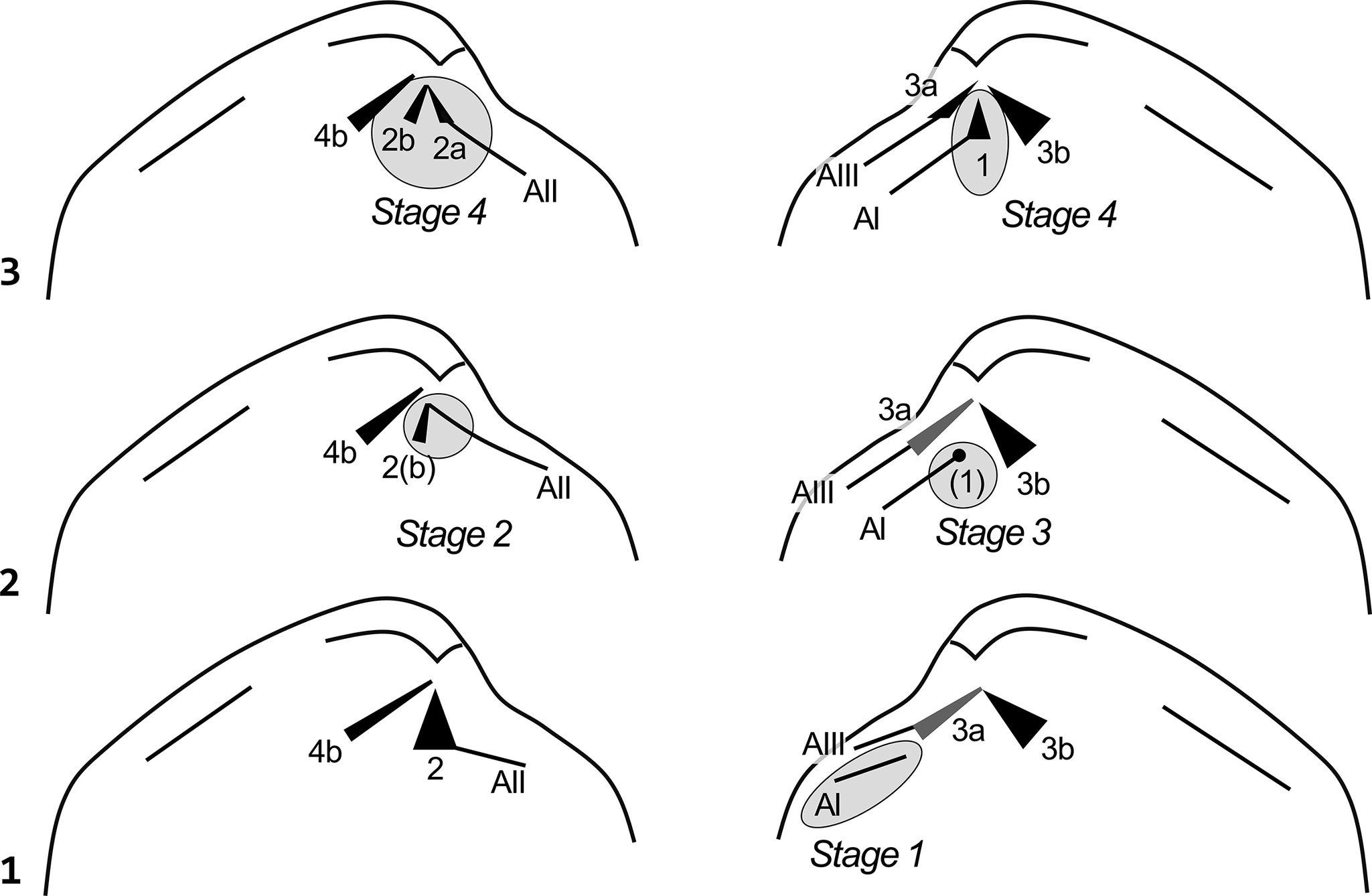
The main difference between the veneroid and lucinoid hinges is that the central, pivotal cardinal tooth is located in the left valve in the case of the lucinoid hinge (cardinal tooth 2 in Bernard’s [1895] system) but in the right valve in the veneroid hinge (cardinal tooth 1 in Bernard’s [1895] system; see Cox, Reference Cox and Moore1969 for a summary of Bernard’s hinge teeth notations). The lucinoid hinge type occurred already in the Paleozoic and is morphologically more primitive than the veneroid hinge type, which evolved from the former through the addition of cardinal tooth 1 in the right valve and the corresponding differentiation of cardinal tooth 2 in the left valve into 2a and 2b. The consensus about the details of the evolutionary transition between these two main hinge types is as follows (Fig. 2; Bernard, Reference Bernard1895; Douvillé, Reference Douvillé1913; Cox, Reference Cox1947, Reference Cox and Moore1969; Casey, Reference Casey1952; Gardner, Reference Gardner2005):
-
(1) Right valve: formation of lateral tooth AI (Fig. 2.1)
-
(2) Left valve: bending upward of the posterior end of lateral tooth AII below the umbo to form an obtuse chevron with cardinal tooth 2(b) (Figs. 2.2, left, 3.1; “Eotrapezium stage” of Cox, Reference Cox1947, p. 142)
-
(3) Right valve: formation of a small tubercle (progenitor of cardinal tooth 1) at the posterior end of lateral tooth AI that fits into the space below the AII–2b complex of the left valve (arcticoid hinge; Fig. 2.2, right)
-
(4) Right valve: offset of this tubercle from AI and its migration to the pivotal position below the umbo where it forms cardinal tooth 1; left valve: differentiation of teeth 2a and 2b to create space for insertion of cardinal tooth 1 (veneroid hinge; Fig. 2.3)
Figure 2.Schematic illustration of the evolutionary transition from the lucinid to the veneroid hinge in four main stages; teeth are illustrated in black; gray ellipses/circles highlight changes in the hinge dentition compared with the previous stage. Note that the shell outline is indicated for general orientation only; the actual position of hinge elements may vary among genera. Also note that for clarity the lateral teeth are shown distant from the shell margin, although some of them may coincide with it. (1) Stage 1: addition of anterior lateral tooth AI; note that the ancestral tooth 3a (indicated in gray) is reduced in Pseudocorbula. (2) Stage 2: formation of AII–2(b) chevron; stage 3: formation of progenitor of cardinal tooth (1). (3) Stage 4: formation of cardinal tooth 1 and differentiation of cardinal teeth 2a and 2b.
 Figure 3.
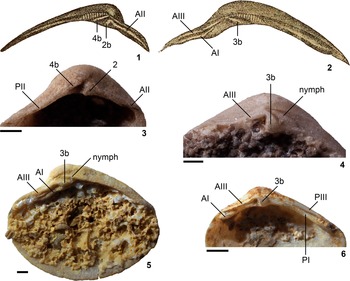
(1, 2) Hinge of Mesodesma germari Dunker, Reference Dunker1844, the type species of Eotrapezium, taken from Böhm (Reference Böhm1901, figs. 22, 23; scale unknown). (1) Left valve. (2) Right valve. (3, 4) Hinge of Pseudocorbula sandbergeri Philippi, Reference Philippi1898. (3) GPIT-PV-75728, left valve, original specimen of Hohenstein (Reference Hohenstein1913, pl. 2, fig. 17). (4) GPIT-PV-124266, right valve, original specimen of Hohenstein (Reference Hohenstein1913, pl. 3, fig. 8). (5) Pseudocorbula sp., MHI 2244, right valve. (6) Pseudocorbula nuculiformis (Zenker, Reference Zenker1836), GPIT-PV-75739, right valve, original specimen of Hohenstein (Reference Hohenstein1913, pl. 3, fig. 3). Hinge teeth notations according to Bernard (Reference Bernard1895), where A indicates anterior lateral teeth, P indicates posterior lateral teeth, and arabic numbers indicate cardinal teeth; even numbers (roman/arabic) refer to teeth of the left valve and uneven numbers to those of the right valve. See text for further details. Scale bars = 1 mm.

Stages (2)–(4) of this evolutionary transition are well documented in the literature (Cox, Reference Cox1947, Reference Cox and Moore1969; Casey, Reference Casey1952; Gardner, Reference Gardner2005). Eotrapezium, the eponymous genus of stage 2, has an Early Jurassic type species; reports of this genus from the Late Triassic (e.g., Golebiowski, Reference Golebiowski1989; Ivimey-Cook et al., Reference Ivimey-Cook, Hodges, Swift, Radley, Swift and Martill1999; Hodges, Reference Hodges2024) have been based on the external shell morphology and are thus uncertain (Nützel et al., Reference Nützel, Nose and Hautmannin press). Accordingly, the initial, pre-Jurassic phase of the hinge evolution of venerids is basically unknown. By comparison with Eotrapezium, I here suggest that Pseudocorbula Philippi, Reference Philippi1898 is a candidate representative of the initial stage of veneroid hinge evolution.
Materials and methods
Material
This study is based on observations of an exceptionally well-preserved sample of silicified shells from the Illyrian (Anisian, Middle Triassic) of southern Germany, previously described by Hohenstein (Reference Hohenstein1913). The Hohenstein collection stems from oolithic, partly silicified limestone of the eastern margin of the Black Forest (Germany). Hohenstein (Reference Hohenstein1913) attributed this fauna to the higher part of the middle Muschelkalk, but a stratigraphic revision by Urlichs (Reference Urlichs1992) suggests an assignment to the lowermost upper Muschelkalk (Trochitenkalk Formation). Hohenstein (Reference Hohenstein1913) regarded Pseudocorbula as a subgenus of Myophoriopis von Wöhrmann, Reference von Wöhrmann1889, but the current consensus is that these two genera are phylogenetically not closely related (see Systematic paleontology). Hohenstein (Reference Hohenstein1913) assigned his material of Pseudocorbula to four different species that he distinguished chiefly by differences in the shape of the valves. Because the collection available to me is too small to allow for an assessment of intraspecific variation, Hohenstein’s (Reference Hohenstein1913) assignments are maintained, but it should be noted that a population-based approach might reveal the synonymy of some of these species.
In addition to the Hohenstein (Reference Hohenstein1913) collection, I studied material from the lower Muschelkalk (Terebratelbank, Pelsonian, Anisian) of Kaltensundheim (Thuringia) and the upper Muschelkalk (Trochitenkalk, Illyrian, Anisian) of Wiesloch (Baden Württemberg).
Repositories and institutional abbreviations
Figured and other specimens examined in this study are deposited in the following institutions: Muschelkalkmuseum Hagdorn Ingelfingen (MHI) and Palaeontological Institute of the University of Tübingen (GPIT).
Systematic paleontology
Class Bivalvia Linnaeus, Reference Linnaeus1758
Subclass Heteroconchia Hertwig, Reference Hertwig1895
Superorder Heterodonta Neumayr, Reference Neumayr1884
Order Venerida Gray, Reference Gray1854
Superfamily Arcticoidea Newton, Reference Newton1891
Family Isocyprinidae Gardner, Reference Gardner2005
Subfamily Pseudocorbulinae new subfamily
Type genus
Pseudocorbula Philippi, Reference Philippi1898.
Other genera
No other genera are currently assigned to Pseudocorbulinae.
Diagnosis
Valves equivalve, suboval in outline, posteriorly truncated, with blunt posterior ridge, externally smooth except for growth lines. Hinge dentition AI, AIII, 3b, PI, PIII / AII, 2, 4b, PII. Lateral tooth AI varying from short to elongated; lateral tooth AII abutting posteriorly on stout cardinal tooth 2.
Remarks
Pseudocorbulinae differs from Isocyprininae by the morphology of lateral tooth AII and cardinal tooth 2. Depending on the interpretation of the hinge elements of Isocyprininae, Pseudocorbulinae lacks either the AII–2b complex (Cox, Reference Cox1947) of this subfamily or the cardinal tooth 2a and the bifid morphology of cardinal tooth 2b (Gardner, Reference Gardner2005).
Genus Pseudocorbula Philippi, Reference Philippi1898
Type species
Nucula gregaria Münster in Goldfuss, Reference Goldfuss1837 by subsequent designation of Diener (Reference Diener and Diener1923).
Revised diagnosis
Valves externally smooth except for growth lines, with blunt posterior ridge. Hinge of right valve: lateral tooth AI varying in length, from a short anterior projection to an elongated ridge that posteriorly abuts on cardinal tooth 3b; lateral tooth AIII forming narrow ridge that extends to the point below the umbo where it gives rise to strong, prosocline cardinal tooth 3b; lateral tooth PI strongest at its posterior end, fading toward beak; PIII formed by projecting shell margin. Hinge of left valve: anterior (AII) and posterior (PII) lateral teeth formed by projecting shell margin; lateral tooth AII terminated posteriorly by stout triangular cardinal tooth 2, which is separated from slender, prosocline cardinal tooth 4b by a gap receiving the continuous AIII–3b transition of the opposite valve. Cardinal teeth without striations in both valves.
Occurrence
Early Triassic?; Middle–Late Triassic (see below and Diener, Reference Diener and Diener1923; Ros-Franch et al., Reference Ros-Franch, Márquez-Aliaga and Damborenea2014).
Remarks
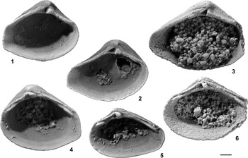
The studied material allows for a complete reconstruction of the hinge of Pseudocorbula (Figs. 3.3–3.6, 4). The anterior lateral tooth AI is generally well developed; it might fade posteriorly (Fig. 3.6) or extend until it abuts on the base of cardinal tooth 3b (Fig. 3.5). Lateral tooth AIII is also well developed and is connected to the prosocline cardinal tooth 3b (Fig. 3.4–3.6). The posterior lateral tooth PI increases in strength toward its posterior end, whereas PIII is formed by projecting shell margin (Fig. 3.6). The anterior (AII) and posterior (PII) lateral teeth of the left valve are also formed by projections of the shell margin (Figs. 3.3, 4.1–4.3). Lateral tooth AII is terminated posteriorly by the stout triangular cardinal tooth 2, which is separated from the slender, prosocline cardinal tooth 4b by a gap (Fig. 3.3).
(1, 4) Pseudocorbula gregaria (Münster in Goldfuss, Reference Goldfuss1837). (1) GPIT-PV-75726, left valve, original specimen of Hohenstein (Reference Hohenstein1913, pl. 2, fig. 16). (4) GPIT-PV-75733, right valve, original specimen of Hohenstein (Reference Hohenstein1913, pl. 3, fig. 1). (2, 3, 6) Pseudocorbula sandbergeri Philippi, Reference Philippi1898: (2) GPIT-PV-75728, left valve, original specimen of Hohenstein (Reference Hohenstein1913, pl. 2, fig. 17); (3) GPIT-PV-75752, left valve, original specimen of Hohenstein (Reference Hohenstein1913, pl. 3, fig. 9); (6) GPIT-PV-75750, right valve, original specimen of Hohenstein (Reference Hohenstein1913, pl. 3, fig. 7). (5) Pseudocorbula nuculiformis (Zenker, Reference Zenker1836), GPIT-PV-75739, right valve, original specimen of Hohenstein (Reference Hohenstein1913, pl. 3, fig. 3). All specimens blacked by graphite emulsion and coated with magnesium oxide. Scale bar = 1 mm.

The main difference from the hinge of Eotrapezium is that the left valve has a single, stout cardinal tooth 2 rather than a slender, prosocline tooth 2b that forms a chevron with AII (Fig. 3.1 versus Fig. 3.3). In this regard, the hinge of Pseudocorbula is comparable to the lucinoid hinge type, but it differs from the standard lucinoid dentition in the absence of tooth 3a. This absence does not only link Pseudocorbula with Eotrapezium; I suggest that it might also have been a stimulant for the formation of a tubercle at the posterior end of AI that functionally substituted the absent tooth 3a and ultimately evolved into the veneroid cardinal tooth 1.
Pseudocorbula has been regarded occasionally as a synonym or subgenus of Myophoriopis von Wöhrmann, Reference von Wöhrmann1889. Notably, Odhner (Reference Odhner1918, fig. 8) suggested that the hinge of Ladinian Myophoriopis lineata Münster in Goldfuss, Reference Goldfuss1837, as figured by Bittner (Reference Bittner1895), could be interpreted as AI, AIII, 1, 3b, PI, PIII / AII, 2a, 2b, PII; thus, this species would have a veneroid hinge type, differing only by the absence of 3a from the general pattern. Unfortunately, the crucial right valve is represented by only one specimen, in which the two anterior lateral teeth AI and AIII unite posteriorly well before the subumbonal region, which makes the interpretation of the cardinal teeth equivocal. In the Treatise, Cox and Chavan (Reference Cox, Chavan and Moore1969) assigned Myophoriopis to the crassatelloid family Myophoricardiidae, implying that the cardinal teeth of the right valve represent 3a and 3b, rather than 1 and 3b. Compared with Pseudocorbula, the similarity seems to be largely confined to the general shape of the valves. The hinge of Myophoriopis differs from that of Pseudocorbula by the presence of two cardinal teeth in the right valve and a striation of the cardinal teeth (Cox and Chavan, Reference Cox, Chavan and Moore1969).
Discussion
The outlined interpretation of the hinge of Pseudocorbula suggests that it is a basal member of the Arcticoidea; however, it differs from more advanced taxa in this superfamily by the absence of an incipient cardinal tooth 1 and of cardinal teeth 2a and 3a. It is closely related to Eotrapezium, which differs from Pseudocorbula in the presence of a chevron-shaped AII–2b teeth complex in the left valve (see the preceding).
The differences between Eotrapezium and Isocyprina are controversial. Arkell (Reference Arkell1934, p. 261, 264) regarded Eotrapezium as a younger synonym of Isocyprina because of the virtually identical hinge structure. Cox (Reference Cox1947, p. 144, figs. 1, 2) confirmed the near-identical hinge morphologies of the two taxa and noted that “[Eotrapezium represents] a Rhaetic and Lower Liassic group, differing from Isocyprina mainly in external characters”; however, he considered the external differences as significant for maintaining both genera. Later, Keen and Casey (Reference Keen, Casey and Moore1969, p. N648) classified Eotrapezium as a subgenus of Isocyprina due to the presence (Isocyprina) or absence (Eotrapezium) of tooth 2a in the left valve. However, this distinction is unseen in their figures, which have been reproduced from Cox (Reference Cox1947, figs. 1, 2), who explicitly stated the absence of 2a in both genera (p. 144). Gardner (Reference Gardner2005, fig. 9) refigured the hinge of the left valve of Isocyprina sharpi Cox, Reference Cox1947 and interpreted an elevation at the posterior part of lateral tooth AII as cardinal tooth 2a; he further suggested that the posteriormost part of AII is the anterior limb of a bifid cardinal tooth 2a. I disagree about this interpretation and regard Gardner’s (Reference Gardner2005) tooth 2a as an integral part of lateral tooth AII because a cardinal tooth is per definition set off from the lateral tooth (Bernard, Reference Bernard1895; Cox, Reference Cox and Moore1969). Regardless of the interpretation of the hinge elements, the hinge morphology of Pseudocorbula distinguishes this genus from its geologically younger descendants, which justifies its classification in a separate subfamily.
The type species of Pseudocorbula is Middle Triassic (Anisian–Ladinian) in age (Diener, Reference Diener and Diener1923). Species assigned to Pseudocorbula have been reported from the Lower Triassic of Siberia (P. kharaulakhensis Kurushin, Reference Kurushin1992) and tentatively from the Lower Triassic of Pakistan (Pseudocorbula? sp.; Wasmer et al., Reference Wasmer, Hautmann, Hermann, Ware, Roohi, Ur-Rehman, Yaseen and Bucher2012), but the hinge details of these species are unknown. Judging from these stratigraphic occurrences, the suggested phylogenetic interpretation of Pseudocorbula suggests an Early or early Middle Triassic origin of Venerida. Accordingly, this order originated soon after the end-Permian mass extinction, as did other bivalve taxa that characterize the modern shelly fauna on the seafloor, such as oysters (Hautmann and Hagdorn, Reference Hautmann and Hagdorn2013; Hautmann et al., Reference Hautmann, Ware and Bucher2017) and scallops (Hautmann, Reference Hautmann2010). However, the evolutionary details differ among these three taxa. The origin of modern scallops (Pectinoidea) was characterized by the evolution of two morphological innovations: the ctenolium and the alivincular–alate ligament (Waller, Reference Waller1984, Reference Waller and Morton1990, Reference Waller2006; Hautmann, Reference Hautmann2004, Reference Hautmann2010), which allowed them to outcompete the ancestral Aviculopectinoidea (s.l.) during the Triassic (Hautmann et al., Reference Hautmann, Friesenbichler, Grădinaru, Jattiot and Bucher2021). The evolution of oysters included a number of morphological innovations, too. In addition to left-pleurothetic cementation, oysters differ from putative ancestral pterioids in the monomyar condition, the absence of a pallial line, and the alivincular–arcuate ligament (Hautmann, Reference Hautmann2001a, Reference Hautmannb, 2004; Hautmann et al., Reference Hautmann, Ware and Bucher2017). In contrast to scallops, however, oysters did not obtain dominance in their guild during the Triassic; rather, they diversified moderately but failed to outcompete the coexisting cementing bivalve families Prospondylidae, Plicatulidae, and Dimyidae (Hautmann, Reference Hautmann2001a, Reference Hautmannb; Fürsich and Hautmann, Reference Fürsich and Hautmann2005). Finally, Venerida failed to radiate at all after their first appearance in the Early or Middle Triassic; the only possible Triassic evolutionary event was the origin of Eotrapezium close to the end of the period. The major evolutionary breakthrough of this order had to await the evolution of siphons in the Jurassic (Cox, Reference Cox1947; Stanley, Reference Stanley1968), which enabled them to colonize deep infaunal habitats.
Acknowledgments
Access to collection material was kindly provided by H. Hagdorn (Muschelkalkmuseum Ingelfingen) and I. Werneburg (University of Tübingen). H. Hagdorn is additionally thanked for his hints on Muschelkalk literature and remarks on the manuscript. K. Hryniewicz and an anonymous reviewer as well as editor S. Schneider provided helpful comments on the manuscript.
Competing interests
The author declares none.



 Open access

