A 24-year-old woman was seen in the emergency department following a second unprovoked nocturnal generalized tonic–clonic seizure that had been witnessed by her husband. After initial moaning, clonic movements were seen for an estimated duration of 2 minutes. The first seizure had occurred during pregnancy a year earlier. No petechial rash had been recorded during the first episode. Neurological examination, EEG, and MRI of the brain were normal.
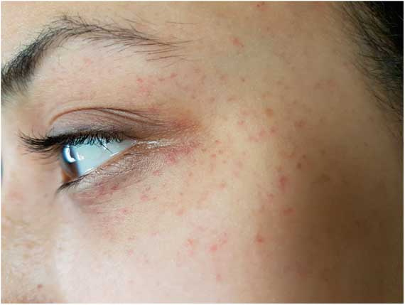
Significant clinical findings were a left lateral tongue bite and bilateral symmetric periorbital petechiae (Figure 1). No other injury was present. Further questioning revealed a history of epilepsy in her sister. Hematological parameters were normal, with normal platelet counts and prothrombin/partial thromboplastin time (PT/PTT) values. Although the clinical history was strongly suggestive of epilepsy in this patient, the presence of periorbital petechiae further supported recognition of the nocturnal spell as a seizure.
Bilateral symmetric periorbital postictal petechiae were observed in a patient after a nocturnal generalized tonic–clonic seizure.

Periorbital petechiae are a rare finding that can be helpful in making a diagnosis of epilepsy when the history is unclear.Reference Reis and Kaplan 1 , Reference Roth and Zumsteg 2 They resolve spontaneously. Concomitant subconjunctival hemorrhages have been previously described and attributed to capillary rupture during the tonic phase of seizures.Reference Rigby and Sadler 3 , Reference Desai and Mehta 4 Similar eruptions can be seen with traumatic asphyxiation, allergic reactions, and coagulopathy, which should be excluded by history and laboratory evaluation. Proposed mechanisms for their occurrence in epilepsy include pressure-induced capillary leakage, as seen in prolonged coughing and vomiting. Other possible mechanisms include cytokines inducing platelet dysfunction, neuronal release of vasoactive substances, and vagal nerve dysfunction.Reference Desai and Mehta 4 Petechial rashes are not specifically recorded in seizure severity scales, and further study would be required to establish the relationship between their occurrence and seizure duration and severity.Reference Aghaei-Lasboo and Fisher 5
Disclosures
Muhammad Ahmer Wali and Matthias Georg Ziller hereby declare that they have nothing to disclose.
Statement of Authorship
Both authors contributed to data collection, writing, and critical revisions.