


Non-technical Summary
Hispaniola, an island in the Caribbean, is known for its Dominican amber deposits dated to the Miocene, approximately 16 million years ago. Dominican amber preserves one of the most morphologically and taxonomically diverse assemblages of insects in the world. Hypoponera, a genus of ant that is globally distributed, was long believed to be present in the Miocene Caribbean fauna but had yet to be confirmed. Here, we formally describe a new species (Hypoponera electrocacica), confirming its presence in the prehistoric Caribbean. Hypoponera electrocacica n. sp. has features of its close living relatives and could be important to understanding the complex evolutionary history of the genus.
Introduction
The ponerine genus Hypoponera Santschi, Reference Santschi1938 is widely distributed and abundant. The genus contains > 150 described species (Bolton, Reference Bolton2025) and, like many ant taxa, its composition is likely a significant underestimate of true species-level diversity. Known species are small, putatively strict predators, found in soil and leaf-litter (Hanisch et al., Reference Hanisch, Drager, Yang, Tubaro and Suarez2020) that comprise colonies as small as just one or two dozen workers or up to 1,500 individuals (Beckers et al., Reference Beckers, Goss, Deneubourg and Pasteels1989; Yamauchi et al., Reference Yamauchi, Kimura, Corbara, Kinomura and Tsuji1996). Hypoponera spp. are frequently sampled in tropical and subtropical environments and appear to constitute a significant component of those ecosystems—specimens were present in 75% of 110 leaf litter samples from five continents (Ward, Reference Ward, Agosti, Majer, Alonso and Schultz2000).
Despite the cosmopolitan nature of Hypoponera and their near ubiquity in standardized sampling, the genus is defined as a kind of morphological vacuum. The group lacks any unique synapomorphies, and diagnoses are determined through a process of triangulation via generalized features (Bolton and Fisher, Reference Bolton and Fisher2011). Such features include triangulate mandibles with small teeth, small and compressed frontal lobes, a rounded subpetiolar process, but also in most cases, an absence of other, more ‘interesting’ features. Such a configuration has led to frustration and exasperation among taxonomists. A more endearing description comes from J. Longino (Reference Longino2005, https://ants.biology.utah.edu/genera/hypoponera/key.html): “If Ponerinae is a Mr. Potato Head game, Hypoponera is the potato.” It is perhaps this lack of distinctiveness that has precluded formal species descriptions of fossil Hypoponera species.
A single fossil Hypoponera has been described to date. First described > 150 years ago in Mayr’s landmark monograph on Baltic amber (Mayr, Reference Mayr1868) as Ponera atavia, Hypoponera atavia (Mayr, Reference Mayr1868) was established by Dlussky in Reference Dlussky2002. Dlussky’s transfer was based on examined lectotype material that Mayr noted; this transfer was justified in part by a simplified subpetiolar process that excluded Ponera Latreille, Reference Latreille1804. Although there has been no formal divergence dating estimation of Hypoponera, recent genus-level estimates suggest that it diverged from its closest relatives—a complex containing Centromyrmex Mayr, Reference Mayr1866, Psalidomyrmex André, Reference André1890, and Plectoctena Smith, Reference Smith1858—from the late Cretaceous to the mid-Paleocene (Doré et al., Reference Doré, Borowiec, Branstetter, Camacho, Fisher, Longino, Ward and Blaimer2025) to as late as the Paleocene to mid-Eocene (Borowiec et al., Reference Borowiec, Zhang, Neves, Ramalho, Fisher, Lucky and Moreau2025). This recent divergence estimate analysis included H. atavia as a node calibration. Although not formally described, putative specimens of Hypoponera have been mentioned as inclusions within Ethiopian (Bouju and Perrichot, Reference Bouju and Perrichot2020; Perrichot et al., Reference Perrichot, Boudinot, Engel, Xu, Bojarski and Szwedo2022), Zhangpu (Wang et al., Reference Wang, Shi, Xu, Spicer and Perrichot2021), and Dominican amber (Wilson, Reference Wilson1985).
Wilson (Reference Wilson1985) mentioned reviewing Hypoponera specimens in the Museum of Comparative Zoology, however, none of these were described. Moreover, many of the specimens referenced by Wilson in the collection have been more recently identified as other small ponerine genera, many of which were considered species of Pachycondyla Smith, Reference Smith1858 until recently (Schmidt and Schattuck, Reference Schmidt and Shattuck2014). Here, we describe a second Hypoponera fossil—the first known from the Western Hemisphere. Although Hypoponera fossils might be of limited phylogenetic utility given the current taxonomic infrastructure, these occurrences offer a significant opportunity for future chronological assessments of this highly prevalent genus.
Materials and methods
We imaged the holotype (MNHNSD FOS 18.126) with a Nikon SMZ25 stereomicroscope and DS-Ri2 camera with the NIS-Elements software (https://www.microscope.healthcare.nikon.com/products/software/nis-elements) at the New Jersey Institute of Technology (New Jersey, USA). All images are digitally stacked photomicrographic composites of several individual focal planes, obtained using the NIS-Elements software. We obtained X-ray based computed tomographic (CT) images of the holotype specimen at the New Jersey Institute of Technology Otto H. York Center for Environmental Engineering and Science, using a Bruker SkyScan 1275 micro-CT scanner. We scanned the specimen at a voltage of 25 kV and a current of 175 μA for 400 ms exposure times averaged over five frames per rotation with a voxel size of 5.96 μm. Z-stacks were generated using NRecon (Micro Photonics, Allentown, Pennsylvania) and reconstructed using 3D Slicer v5.9 (Fedorov et al., Reference Fedorov, Beichel, Kalpathy-Cramer, Finet and Fillion-Robin2012).
We took measurements both directly on the amber specimen using light microscopy and corroborated measurements using the three-dimensional (3D) models produced from CT scanning. Morphological measurements generally followed Sosiak and Barden (Reference Sosiak and Barden2021) for ecological modeling, along with measurements more typical of Hypoponera (Bolton and Fisher, Reference Bolton and Fisher2011). Morphological terminology generally followed Bolton (Reference Bolton1994) for most body structures, as well as Harris (Reference Harris1979) for sculpture, although incorporating Ponerinae-specific terminology following Keller (Reference Keller2011) and Bolton and Fisher (Reference Bolton and Fisher2011).
Repositories and institutional abbreviations
The specimen reported in this study (MNHNSD FOS 18.126) is deposited at the Museo Nacional de Historia Natural ‘Prof. Eugenio de Jesús Marcano,’ Santo Domingo, in the Dominican Republic.
Systematic paleontology
Order Hymenoptera Linnaeus, Reference Linnaeus1758
Family Formicidae Latreille, Reference Latreille1802
Subfamily Ponerinae Lepeletier de Saint-Fargeau, Reference Lepeletier de Saint-Fargeau1835
Genus Hypoponera Santschi, Reference Santschi1938
Type species
Ponera abeillei André, 1881, now Hypoponera abeillei (André, Reference André1881).
Hypoponera electrocacica new species
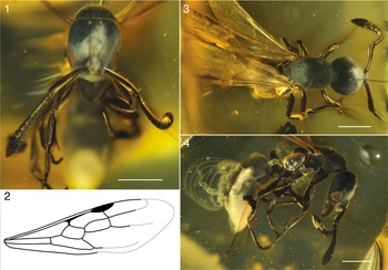
Photomicrographs and illustration of Hypoponera electrocacica n. sp., holotype specimen, MNHNSD FOS 18.126. (1) Head in front face view. (2) Illustration of forewing venation. (3) Body in dorsal view. (4) Body in lateral view. Scale bars = 0.5 mm.

Volume rendering of Hypoponera electrocacica n. sp., holotype specimen, MNHNSD FOS 18.126. (1) Body in lateral view. (2) Body in dorsal view. (3) Lateral enlarged view of petiole; arrows denote the prora and subpetiolar projection; the subpetiolar projection is traced with a dashed line. Scale bars = 0.5 mm (1, 2), 0.2 mm (3).

Holotype
MNHNSD FOS 18.126, alate queen, deposited in the Museo Nacional de Historia Natural, ‘Prof. Eugenio de Jesús Marcano,’ Santo Domingo, Dominican Republic (Figs. 1, 2). Preserved within a 10 × 10 mm section of transparent, yellow amber with internal cracks along the head of the inclusion. The ant is partially coated in a white, finely hairy fungus across part of the head and terminal abdominal segments.
Diagnosis (gyne)
Head trapezoidal; in full face view, anterior clypeal margin blunt and convex; mandibles with seven or eight large teeth, dorsal surface coarsely striate; anapleural sulcus weakly produced, propodeal dorsal margin continuous from mesonotum, weakly convex, approximately half the length of declivitous margin, meeting declivitous face at an obtuse angle, declivitous margin straight to weakly concave; petiolar node subtriangular in profile with the node tall and narrow, subpetiolar process anteriorly projecting, rounded; prora present as a wide lip-shaped, transverse projection.
Occurrence
Early Miocene, Burdigalian (~16 Ma) preserved as an inclusion within fossil resin from the northern mines of the Santiago Providence, Dominican Republic.
Description (gyne)
Head. Head much longer than wide, slightly trapezoidal in front view, wider posterad than anterad; posterior margin straight, lateral margin broadly convex (Fig. 1.1); in full-face view, anterior clypeal margin blunt and convex. Scape just surpassing posterior cephalic margin, funicular segments 7–10 broader than long, width increasing apically, forming an antennal club. Mandible with seven or eight large teeth, no denticles, dorsal surface coarsely striate, outer surface straight, at most slightly concave medially. Eyes large (alate condition), located far forward on the side of the head.
Mesosoma. Pronotal anterior margin in lateral view broadly convex, dorsal margin convex; pronotum rounded in dorsal view, without humeral angle; mesonotal dorsal margin broadly convex, alate condition; anapleural sulcus weekly produced, mesometapleural suture well marked (Fig. 1.4, 2.1); propodeum unarmed, propodeal dorsal margin continuous from mesonotum, weakly convex, approximately half the length of declivitous margin, meeting declivitous face at an obtuse angle, declivitous margin straight to weakly concave; propodeal spiracle opening round, located midway between propodeal declivity and metapleural gland; mesometapleural suture well impressed; metapleural gland opening posteriorly. Mesotibia lacking ‘traction setae’ on external face; metatibial apex with one spur, claws simple, aroleum absent.
Tegula distinct, round, slightly longer than broad. Wings slightly infuscate, entirely and finely setose. Forewing (Fig. 1.3) with costal vein tubular to large and conspicuous pterostigma; costal, basal, and subbasal cells closed; marginal cells 1 and 2 closed, submarginal cells 1 and 2 closed; medial vein consistently strong and tubular from base to lateral wing margin. Cross-vein 1m-cu present, discal cell 1 and subdiscal cell 1 closed; cubital vein consistently tubular, fading out toward lateral half; lateral half of wing not visible.
Hindwing not visible.
Metasoma. Petiolar node subtriangular in profile with the node tall and narrow, the anterior face slightly concave and posterior face vertical, margins converging to rounded short dorsal face. Subpetiolar process anteriorly projecting, rounded (Fig. 2.3). Anterior margin of third abdominal tergite forming a rounded angle at dorsal margin; helcium situated ventrally on anterior face of third abdominal tergite, prora present as a wide lip-shaped, transverse projection. Gaster weakly arched; dorsal margin broadly convex in lateral view, ventral margin relatively straight. Pygidium without stout setae or spines.
Sculpture/pilosity. Most sculpturing smooth with abundant, shallow, and fine punctae; lateroventral surface of head and mesopleuron finely punctured. Body with abundant standing hairs. Occipital margin of head with at least four erect setae. Abundant erect setae on dorsum of mesosoma, small erect setae on dorsum of gaster. Body seemingly dark to black, legs, antenna, and mandible reddish brown.
Etymology
The specific epithet electrocacica (from the Latin electrum, meaning ‘amber,’ and the feminine form of the Taino word cacique, meaning ‘chief’) is a feminine singular adjective in the nominative case. It refers to the alate ant or queen trapped in amber.
Zoobank lsid
urn:lsid:zoobank.org:act:75120710-CDFF-46BB-907E-AE25BEBD5F85
Measurements (in mm)
Holotype alate: Head width 0.593, head length 0.723, Weber’s length 1.012, procoxa length 0.375, eye length 0.216, mesosoma height 0.405, eye height total 0.42, eye height dorsal 0.263, eye height ventral 0.159, eye length total 0.69, eye length anterior 0.233, eye length posterior 0.453, mandible length in profile 0.289, mandible length in frontal view 0.358, metafemur length 0.514, scape length 0.51, and mesosoma width 0.468.
Remarks
Hypoponera electrocacica n. sp. is a strikingly modern-looking representative of the genus. The head shape and mandibles are reminiscent of modern Caribbean Hypoponera species like H. opaciceps (Mayr, Reference Mayr1887) and H. punctatissima (Roger, Reference Roger1859), whereas the body resembles H. opacior (Forel, Reference Forel1893). However, H. electrocacica n. sp. differs from previously described species based on the following combination of characters: scape at least reaching the occipital margin of the head (vs failing to reach apex of head in both H. opaciceps and H. punctatissima), mandible with seven or eight teeth (vs three or four in in both H. opaciceps and H. punctatissima), blunt clypeal margin, finely punctate lateroventral side of the head and mesopleuron, anapleural sulcus weekly impressed (vs strongly impressed in H. punctatissima), and petiole higher than the propodeum and narrow (vs petiole approximately as high as propodeum and broad).
Concluding remarks
Here, we confirm the presence of the genus Hypoponera within a Caribbean lagerstätte, confirming a previously observed trend (Wilson, Reference Wilson1985; De Andrade Reference De Andrade1994, Reference De Andrade2004) of a diverse ponerine community in the early Miocene. Hypoponera is still present in the Caribbean today, although the species on the island are widespread Neotropical species (Lubertazzi, Reference Lubertazzi2019). Lubertazzi (Reference Lubertazzi2019) mentioned that many morphospecies are yet to be reviewed, hinting at a potentially rich extant diversity that could provide more insight into the long-term stasis or turnover of the island.
Data availability statement
All data can be found in Dryad [https://doi.org/10.5061/dryad.sf7m0cgmq] and Zenodo [https://doi.org/10.5281/zenodo.17794736] Digital Repositories.
Acknowledgments
We thank G. de los Santos and A. Sanchez of the Museo Nacional de Historia Natural ‘Prof. Eugenio de Jesús Marcano,’ República Dominicana for their assistance in depositing the specimen in the Dominican Republic. This material is based on work supported by the National Science Foundation under grant no. 2144915 to PB.
Competing interests
The authors declare none.


 Open access