


Introduction
Freshwater parasitic copepods, particularly members of the Ergasilidae and Lernaeidae, are significant pathogens and vectors of fish diseases, impacting fish population dynamics and health (Boxshall and Defaye, Reference Boxshall and Defaye2008; Boxshall and Hayes, Reference Boxshall, Hayes, Smit, Bruce and Hadfield2019). Despite their ecological importance, these parasites remain relatively understudied, and their role in aquatic ecosystems is still not completely clarified. Current knowledge of their diversity varies regionally due to inconsistent research efforts.
Within the Palearctic realm, the Mediterranean and the Middle East are biogeographical regions, each defined by its unique combination of climatic, geological and hydrological conditions that have shaped the evolution and diversity of organisms living in these areas. Both regions are recognized for their high level of biodiversity and endemism, which has long attracted scientific interest (Cuttelod et al., Reference Cuttelod, García, Malak, Temple and Katariya2009; Freyhof et al., Reference Freyhof, Ekmekçi, Ali, Khamees, Özuluğ, Hamidan, Küçük, Smith, Smith, Barrios, Darwall and Numa2014, Reference Freyhof, Kaya, Ali and Jawad2021). However, research on parasitic copepods of the Ergasilidae and Lernaeidae in these regions remains limited and rather uneven. More extensive studies, including the descriptions of new species (Dermoergasilus cichlidus Ali and Adday, Reference Ali and Adday2019; Ergasilus luteusi; Al-Sahlany et al., Reference Al-Sahlany, Adday and Ali2024; Pseudolamproglena boxshalli; Al-Nasiri et al., Reference Al-Nasiri, Ho and Mhaisen2012) in recent years (Al-Nasiri et al., Reference Al-Nasiri, Ho and Mhaisen2012; Ali and Adday, Reference Ali and Adday2019; Al-Sahlany et al., Reference Al-Sahlany, Adday and Ali2024), have been conducted only in a few countries, mainly in Turkey (e.g. Soylu and Soylu, Reference Soylu and Soylu2012; Koyun et al., Reference Koyun, Ulupınar and Gül2015; Öktener, Reference Öktener2021) and Iraq (e.g. Mhaisen and Abdul-Ameer, Reference Mhaisen and Abdul-Ameer2021; Mhaisen and Al-Daraji, Reference Mhaisen and Al-Daraji2023). In the Mediterranean, particularly in its European part, research on parasitic copepods is also limited to specific regions, several studies were conducted in Bosnia and Herzegovina (Nedić et al., Reference Nedić, Skenderović and Riđanović2014; Skenderović et al., Reference Skenderović, Adrović, Hajdarević, Hadžiahmetović Jurida, Čekmić and Bajrić2015, Reference Skenderović, Adrović, Jazić, Zuko and Hadzimustafic2021), Croatia (Tomašec, Reference Tomašec1953; Fijan, Reference Fijan1974, Reference Fijan1982), Greece (Zarfdjian and Economidis, Reference Zarfdjian and Economidis1989; Ragias et al., Reference Ragias, Athanassopoulou and Sinis2005), Spain (e.g. Simon Vicente et al., Reference Simon Vicente, Ramajo and Encinas1973; Almeida et al., Reference Almeida, Almodóvar, Nicola and Elvira2008), Portugal (e.g. Hermida et al., Reference Hermida, Saraiva and Cruz2008; Bao et al., Reference Bao, Costal, Garci, Pascual and Hastie2016) and Italy (Grandori, Reference Grandori1925; Fratello and Sabatini, Reference Fratello and Sabatini1972; Macchioni et al., Reference Macchioni, Chelucci, Torracca, Prati and Magi2015). However, most of these studies do not represent comprehensive research integrating both morphological and molecular approaches, and parasitic copepods have often also remained unidentified (Saraiva and Valente, Reference Saraiva and Valente1988; Vagianou et al., Reference Vagianou, Athanassopoulou, Ragias, Di Cave, Leontides and Golomazou2006; Nedić et al., Reference Nedić, Skenderović and Riđanović2014; Stamou et al., Reference Stamou, Kourkoutmani and Michaloudi2022).
Until now, 30 species belonging to 6 genera of Ergasilidae [Dermoergasilus Ho & Do, 1982 (3), Ergasilus von Nordmann, 1832 (19), Mugilicola Tripathi, 1960 (2), Nipergasilus Yamaguti, 1939 (1), Neoergasilus (Yin, Reference Yin1956) (2) and Paraergasilus Markevich, 1937 (3)) and 10 species belonging to 3 genera of Lernaeidae (Lamproglena von Nordmann, 1832 (5), Lernaea Linnaeus, 1758 (3) and Pseudolamproglena (Boxshall, Reference Boxshall1976) (2)] have been recorded in the Mediterranean and Middle East regions. A detailed checklist of all records of species of the Ergasilidae and Lernaeidae in the Mediterranean and the Middle East is provided in Supplementary Table S1.
Within the Ergasilidae, Ergasilus is currently the most abundant and diverse genus, with up to 19 species reported in these areas. The most widespread species is Ergasilus sieboldi von Nordmann, 1832, which has been reported from various freshwater hosts from the Iberian Peninsula to Iraq. In contrast, other Ergasilus species have shown more restricted distributions (e.g. Ergasilus boleophthalmi Adday & Ali, 2011; Ergasilus iraquensis Amado in Amado, da Rocha, Piasecki, Al-Daraji & Mhaisen, 2001; Ergasilus luteusi Al-Sahlany et al., Reference Al-Sahlany, Adday and Ali2024; Ergasilus pararostralis Amado in Amado, da Rocha, Piasecki, Al-Daraji & Mhaisen, 2001 and Ergasilus synanciensis Amado in Amado, da Rocha, Piasecki, Al-Daraji & Mhaisen, 2001 are all restricted to Iraq) or greater host specialization, such as Ergasilus gibbus von Nordmann, 1832, which predominantly parasitizes fishes from the family Anguillidae. From the genus Neoergasilus have been recorded 2 species, Neoergasilus longispinosus (Yin, Reference Yin1956) on cyprinids in Algeria (Boucenna et al., Reference Boucenna, Khelifi, Boualleg, Allalgua and Bensouilah2018; Berrouk et al., Reference Berrouk, Tolba, Touarfia and Boualleg2020, Reference Berrouk, Sid, Lahoual, Sahtout, Kaouachi and Boualleg2022) and Neoergasilus japonicus, a globally invasive species (Ondračková et al., Reference Ondračková, Tkachenko, Vetešník, Hronek and Janáč2024) of various fish families, has been recorded in several countries in both regions (e.g. Soylu and Soylu, Reference Soylu and Soylu2012; Mirzaei et al., Reference Mirzaei, Khovand and Kheirandish2016; Berrouk et al., Reference Berrouk, Sid, Lahoual, Sahtout, Kaouachi and Boualleg2022). Three species of Paraergasilus (Paraergasilus brevidigitus Yin, 1954; Paraergasilus inflatus Ho et al., Reference Ho, Khamees and Mhaisen1996 and Paraergasilus longidigitus Yin, 1954) were recorded in Iraq, Algeria and Turkey (Ho et al., Reference Ho, Khamees and Mhaisen1996; Koyun et al., Reference Koyun, Altunel and Öktener2007; Berrouk et al., Reference Berrouk, Sid, Lahoual, Sahtout, Kaouachi and Boualleg2022). In addition, an introduced non-native species, Nipergasilus bora (Yamaguti, 1939), has been recorded on fish hosts of the family Mugilidae in several Mediterranean countries (Paperna, Reference Paperna1975; Ben Hassine, Reference Ben Hassine1983; Koyun et al., Reference Koyun, Altunel and Öktener2007). Two species of Mugilicola (Mugilicola bulbosa Tripathi, 1960 and Mugilicola kabatai Piasecki et al., Reference Piasecki, Khamees and Mhaisen1991) parasitizing on fishes of the family Mugilidae and 3 species of Dermoergasilus (Dermoergasilus amplectens (Dogiel & Akhmerov, 1952), D. cichlidus and Dermoergasilus varicoleus Ho et al., Reference Ho, Jayarajan and Radhakrishnan1992) found on a variety of fish species were reported only in Iraq (Piasecki et al., Reference Piasecki, Khamees and Mhaisen1991; Ho et al., Reference Ho, Khamees and Mhaisen1996; Amado Pinto da Motta et al., Reference Amado Pinto da Motta, Falavigna da rocha, Piasecki, Al-Daraji and Mhaisen2001; Ali and Adday, Reference Ali and Adday2019; Al-Mosawi and Adday, Reference Al-Mosawi and Adday2024).
Within the Lernaeidae, 5 species of the genus Lamproglena have been recorded, but only L. pulchella was found in Europe, with its distribution extending as far as Iraq (Mhaisen et al., Reference Mhaisen, Ali and Adday2024). Other Lamproglena species, Lamproglena chinensis Yü, 1937; Lamproglena compacta Markevich, 1936 and Lamproglena jordani (Paperna, Reference Paperna19640 have been recorded on the Cyprinidae and Leuciscidae in Iraq, Iran and Israel, respectively, while Lamproglena monodi Capart, 1944 was recorded on Cichlidae in Egypt (e.g. Reference Paperna1964; Pazooki and Masoumian, Reference Pazooki and Masoumian2012; Hassan et al., Reference Hassan, Mahmoud, Metwally and Mokhtar2013). The genus Lernaea is predominantly represented by the invasive cosmopolitan species L. cyprinacea, which is distributed in almost all Mediterranean and Middle Eastern countries (Ondračková et al., Reference Ondračková, Tkachenko, Vetešník, Hronek and Janáč2024). Additionally, two other species, Lernaea ctenopharyngodontis Yin, 1960 and Lernaea oryzophila Monod, 1932, have been recorded from cyprinid hosts in Iran and Iraq, respectively (Al-Nasiri et al., Reference Al-Nasiri, Mhaisen and Al-Nasiri2001; Pazooki and Masoumian, Reference Pazooki and Masoumian2012). The genus Pseudolamproglena is absent in Europe, but 2 species (Pseudolamproglena annulata Boxshall, Reference Boxshall1976 and Pseudolamproglena boxshalli; Al-Nasiri et al., Reference Al-Nasiri, Ho and Mhaisen2012) were recorded on Cyprinidae, Leucisdidae and Mugilidae in Iraq (Boxshall, Reference Boxshall1976; Al-Nasiri et al., Reference Al-Nasiri, Ho and Mhaisen2012).
In this study, we provide an updated overview of the parasitic copepod fauna in freshwater fish species from the Mediterranean and the Middle Eastern regions. The research is based on extensive sampling conducted from 2014 to 2023. This comprehensive dataset offers new insights into the diversity, distribution and host associations of parasitic copepods in these areas, filling important gaps in the current knowledge.
Materials and methods
Fish collection
During several parasitological surveys between 2014 and 2023, 169 fish species (1484 specimens) were examined for the presence of metazoan parasites. Examined fish included mainly representatives of the Cyprinidae and Leuciscidae (total of 162 species), several fishes of the other families living in sympatry with cyprinoids were also examined (3 species of Gobionidae, 2 species of Nemacheilidae, 1 species of Cobitidae and 1 species of Mugilidae). Fishes were sampled in 155 localities including Spain (13 localities), Portugal (7 localities), Italy (8 localities), Croatia (15 localities), Bosnia and Herzegovina (11 localities), Albania (11 localities), Greece (27 localities), Turkey (52 localities) and Iraq (11 localities) (see Table 1 and Figure 1; for detailed information see Supplementary material Table S2 and Figure S1).
Map of sampling localities with records of parasitic copepods. (P – Portugal, S – Spain, I – Italy, C – Croatia, BIH – Bosnia and Herzegovina, A – Albania, G – Greece, TUR – Turkey, IRQ – Iraq).

List of sampled localities with coordinates (only positive records of parasitic copepods listed, all localities are listed in Supplementary Table S2)

The fish sampling was carried out following local regulations. All applicable institutional, national and international guidelines for the care and use of animals were followed. All fish specimens were transported alive to the field laboratory, sacrificed by severing the spinal cord, and dissected within 48 hours following the classical parasitological dissection procedure (Scholz et al., Reference Scholz, Vanhove, Smit, Jayasundera and Gelnar2018). All fish species used in this study were originally collected and previously used for the studies of monogenean parasites including molecular identification of fish (cytochrome b) (see Šimková et al., Reference Šimková, Benovics, Rahmouni and Vukić2017; Benovics et al., Reference Benovics, Desdevises, Vukić, Šanda and Šimková2018, Reference Benovics, Vukić, Šanda, Rahmouni and Šimková2020, Reference Benovics, Nejat, Abdoli and Šimková2021a, Reference Benovics, Koubková, Civáňová, Rahmouni, Čermáková and Šimková2021b, Reference Benovics, Francová, Volta, Dlapka and Šimková2021c, Reference Benovics, Vukić, Šanda, Nejat, Charmpila, Buj, Shumka, Porcelloti, Tarkan, Aksu, Emiroğlu and Šimková2023, Reference Benovics, Rahmouni, Řehulková, Nejat and Šimková2024; Nejat et al., Reference Nejat, Benovics, Řehulková, Vukić, Šanda, Kaya, Tarkan, Abdoli, Aksu and Šimková2023, Reference Nejat, Benovics, Šanda, Vukić, Kaya, Tarkan and Šimková2025; Rahmouni et al., Reference Rahmouni, Seifertová, Benovics and Šimková2023). All fish sampling and morphological identification in the field was performed by the members of Czech team (Radek Šanda and Jasna Vukić) with contribution of local coworkers in all countries, their names are included in acknowledgements. The present study was part of a larger project concerning host-parasite relationships between monogeneans and their cyprinoid hosts.
Parasite collection and identification
Live copepods were collected from the gills using fine needles and processed for morphological and molecular purposes, as described in Míč et al. (Reference Míč, Řehulková and Seifertová2023). The mounted specimens in GAP (mixture of glycerine and ammonium picrate) or pure glycerine were studied using an Olympus BX61 microscope equipped with phase contrast optics. Drawings of the copepods were made using an Olympus drawing attachment and edited with a graphic tablet (Wacom Intuos5 Touch) compatible with Adobe Illustrator and Adobe Photoshop (Adobe Systems Inc., San Jose, CA, USA). All measurements (in micrometres) were taken using digital image analysis software (Olympus Stream Motion v. 1.9.3) and are presented as the mean followed by the range and the number (n) of specimens measured in parentheses.
For scanning electron microscope analysis, two specimens fixed in 70% ethanol were dehydrated in an increasing ethanol grades, dried in a CPD 030 critical point drying apparatus (Bal-tec, Balzers, Liechtenstein) using liquid CO2, mounted on aluminium stubs with double sided adhesive discs, coated with gold in a SCD 040 sputter coating unit (OC Oerlikon Balzers Coating, Balzers, Liechtenstein) and examined in a VEGA scanning electron microscope (TESCAN) operating at 5 kV.
The type specimens of the copepods collected in the present study were deposited in the Institute of Parasitology, Czech Academy of Sciences, České Budějovice, Czech Republic. Prevalence (percentage of infected fish) and mean intensity of infection (mean number of parasites per infected host) were calculated following Bush et al. (Reference Bush, Lafferty, Lotz and Shostak1997). Morphological terminology follows Huys and Boxshall (Reference Huys and Boxshall1991), and host nomenclature was checked against the World Register of Marine Species (WoRMS, www.marinespecies.org).
Molecular and phylogenetic analyses
Genomic DNA was extracted from each individual parasite specimen (or egg sacs only, when applicable) using DNeasy®Blood & Tissue Kit (Qiagen, Hilden, Germany) following the manufacturer’s protocol. Three genetic markers were used for molecular identification of copepod species: two partial fragments of nuclear ribosomal DNA (rDNA) regions (28S and 18S rDNA) and one fragment of the mitochondrial cytochrome c oxidase subunit I (COI) gene. The primers used for amplification are listed in Table 2. PCR amplification and sequencing were conducted according to the protocols and conditions outlined in Míč et al. (Reference Míč, Řehulková and Seifertová2023), (Reference Míč, Řehulková, Šimková, Razanabolana and Seifertová2024)). Obtained sequences were edited using Sequencher® v. 4.10.1 (Gene Codes Corporation, Ann Arbor, MI, USA), and the newly generated sequences for parasite species were checked by the nBLAST Search Tool (https://blast.ncbi.nlm.nih.gov/Blast.cgi) to assess any similarity to available congeners and deposited in GenBank (for accession numbers, see Table 3).
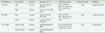
List of primers and PCR conditions used for DNA amplification of partial fragments of ribosomal genes (18S and 28S rDNA) and partial mitochondrial cytochrome oxidase gene (COI) of parasitic copepods

List of Ergasilidae and Lernaeidae species molecularly analysed in this study, including their host species, locality, total number of isolates, GenBank accession numbers for 18S 28S, COI sequences and values of intraspecific genetic distances. For locality ID abbreviations see Table S2

Molecular vouchers (hologenophores, paragenophores; Pleijel et al., Reference Pleijel, Jondelius, Norlinder, Nygren, Oxelman, Schander, Sundberg and Thollesson2008) were deposited in the Institute of Parasitology, Czech Academy of Sciences, České Budějovice, Czech Republic.
To investigate the phylogenetic position of collected parasitic copepods to the representatives of parasitic Cyclopoida, 59 sequences of 28S rDNA of the species belonging to 8 genera of Ergasilidae and 2 genera of Lernaeidae were retrieved from GenBank (for details, see Supplementary Table S3). The sequences were aligned with the G‐INS‐i method in MAFFT online service version 7 (Katoh et al., Reference Katoh, Rozewicki and Yamada2019) and ambiguous positions in the alignment were manually edited in BioEdit (Hall, Reference Hall1999). ModelFinder (Kalyaanamoorthy et al., Reference Kalyaanamoorthy, Minh, Wong, Von Haeseler and Jermiin2017) was employed to select the most appropriate model of DNA evolution. According to the Bayesian Information Criterion (BIC), GTR + F + I + G4 was selected as the best‐fit model. Both maximum likelihood (ML) analysis and Bayesian inference (BI) were used to reconstruct the phylogenetic tree. The ML tree was constructed using an ultrafast bootstrap method (Hoang et al., Reference Hoang, Chernomor, Von Haeseler, Minh and Vinh2018) with 1000 replicates in the IQ‐TREE web server (Trifinopoulos et al., Reference Trifinopoulos, Nguyen, von Haeseler and Minh2016). BI analysis was carried out in MrBayes 3.2.6 (Huelsenbeck and Ronquist, Reference Huelsenbeck and Ronquist2001), the analysis included 2 simultaneous runs of Markov chain Monte Carlo for 106 generations, sampling every 100 generations, with a ‘burn-in’ of 25%. The trees were visualized and edited in FigTree v. 1.4.3 (Rambaut, Reference Rambaut2016) and Adobe Photoshop (Adobe Systems Inc., San Jose, CA, USA). Genetic distances (uncorrected p-distance) were calculated in MEGA v. 11 (Tamura et al., Reference Tamura, Stecher and Kumar2021).
Results
A total of 59 (39 Leuciscidae, 18 Cyprinidae and 2 Gobionidae; 35%) of the 169 fish species sampled in the Mediterranean and the Middle East watersheds were found to be positive for parasitic copepods of the Ergasilidae and Lernaeidae (1004 parasitic copepod adult females). The collected parasites were identified as 6 previously described species of Ergasilidae (E. barbi, E. briani, E. lizae, E. rostralis, N. japonicus and P. longidigitus) and 2 previously described species of Lernaeidae (L. pulchella and L. cyprinacea) based on their morphological and molecular characteristics (Figures 2 and 3). Additionally, two new species, Ergasilus italicus n. sp. found on Protochondrostoma genei (Bonaparte, 1839) in Italy and Pseudolamproglena zahrziensis n. sp. parasitizing Carasobarbus luteus (Heckel, 1843) in Iraq were described. Their morphological characterization and detailed description are provided below. All species are listed in Table 4, including their host(s), locality of collection, localization on fish and values of abundance, prevalence and intensity of infection. The full list of all fish examined (including non-infected fish) is given in Supplementary Tables S4 and S5.
Photomicrographs of representative species from the Mediterranean and the Middle East: (A) E. barbi; (B) antennae of E. barbi; (C) legs of E. barbi, spine on Exp-2 of L1 (white arrow); (D) E. briani; (E) antennae of E. briani; (F) urosome of E. briani, long caudal rami (white arrow); (G) E. lizae; (H) urosome of E. lizae; (I) antenna of E. lizae; (J) E. rostralis; (K) urosome of E. rostralis; (L) rostrum of E. rostralis (white arrow).

Photomicrographs of representative species from the Mediterranean and the Middle East: (A) N. japonicus; (B) antenna of N. japonicus; (C) urosome of N. japonicus; (D) P. longidigitus; (E) L. pulchella; (F) copepodid stage of L. pulchella; (G) copepodid stages of L. cyprinacea; (H) L. cyprinacea.

List of collected parasitic copepods from respective hosts, including localities of their collection and their epidemiological statistics in the Mediterranean and the Middle East

Notes: N – number of fish hosts; NP – number of fish hosts positive for parasitic copepods; S – stage; L – localization on the host; A – abundance; IN – intensity of infection (min – max); P – prevalence;
* – new host records for the species
Ergasilus briani was documented as the most abundant species, its occurrence was confirmed in Turkey, where it was previously recorded (Supplementary Table S1) and now we documented the presence of this species for the first time in Bosnia and Herzegovina, Croatia, Albania and Greece. Ergasilus lizae was found on two localities in Greece, confirming its previous presence. The occurrence of E. barbi in Turkey, P. longidigitus in Albania and N. japonicus in Croatia and Iraq were revealed for the first time. Neoergasilus japonicus was also found in Italy and Turkey, where its presence had been previously documented (Supplementary Table S1). Within Ergasilidae, the highest host range was observed for N. japonicus (14) and E. briani (10).
The occurrence of L. pulchella was confirmed in Italy and Turkey, and for the first time in Bosnia and Herzegovina and Greece. Moreover, this species exhibited the highest host range encompassing 19 host species (Table 4). Lernea cyprinacea was recorded on nine host species in Portugal, Spain, Croatia, Bosnia and Herzegovina and Turkey. It was the only copepod parasite recorded on the Iberian Peninsula in this study. All species of Lernaeidae were found in both copepodid and adult stages.
All Ergasilus, Lamproglena and Pseudolamproglena specimens were found on the gills, adult Lernaea specimens usually burrowed in the skin, N. japonicus was attached to the fins and P. longidigitus was found in the nasal cavity. Juvenile stages of Lamproglena, Pseudolamproglena and Lernaea were found on the gills. Each fish specimen typically harboured only a single species of parasitic copepod. Several cases of mixed infections, such as the co-infestation of gills by adult females of Ergasilidae and copepodid stages of Lernaeidae, were reported. The co-occurrence of N. japonicus on the fins and an ergasilid species on the gills of a single fish was also found.
Family Ergasilidae Burmeister, 1835
Genus Ergasilus Nordmann, 1832
Ergasilus italicus (Míč et al., Reference Míč, Řehulková, Šimková, Razanabolana and Seifertová2024) n. sp.
Type-host: Protochondrostoma genei (Bonaparte, 1839) (Leuciscidae)
Type-locality: Torrente Cerfone, Tiber River drainage, Le Ville, Italy; 43°28′42″N 12°04′25″E
Type and voucher material: Holotype (adult female): IPCAS Cr-40 (1 specimen). Paratype (adult female): IPCAS Cr-40 (1 specimen). Hologenophores (adult females): IPCAS Cr-40 (2 specimens).
Site on host: Gill filaments.
Prevalence and intensity of infection: 44% (4 fish infected/9 fish examined); 1 specimen per infected host.
ZooBank registration: urn:lsid:zoobank.org:act:48903023-9FEB-4CD6-8F8B-2C0E10BF1EF7
Representative DNA sequences: A 1355 bp long 18S rDNA sequence and 640 bp long 28S rDNA sequence obtained from 2 specimens are deposited in the NCBI GenBank database under the accession numbers PX000631 and PX000658, respectively.
Etymology: The species was named after the country Italy where it was first discovered.
Description
Adult female. [Based on 4 specimens; Figures 4–7; measurements in Table 5]. Body length (measured from anterior margin of prosome to posterior margin of caudal rami) 1363 (1360-1365; n = 4). Body elongated and comprises prosome, urosome and caudal rami (Fig. 4A; 7A). Prosome 5-segmented, composed of cephalothorax and 3 free pedigerous somites; cephalosome and first pedigerous somite (PS-1) fused together, without distinct separation. Cephalothorax guitar-shaped, much longer than wide, bearing deep indentation between anterior cephalosome and posterior first pedigerous somite. Cephalic ornamentation comprising anterior circular eyespot and inverted T-shaped marking of thickened chitin situated medially on dorsal side. Paired sensory pores and papillae observed on the rostrum, anterior to eyespot and T-shaped marking, as well as on lateral margins of cephalosome (Figure 4E). Rostrum (Figure 4F) well-developed, with triangular posterior margin. Second to fourth pedigerous somites (PS-2 to PS-4) all wider than long and each markedly narrowing posteriorly.
Ergasilus italicus Míč & Seifertová, 2025 n. sp., paratype female. (A) habitus, dorsal; (B) antenna with spine (sp) and sensillum (se), ventral; (C) mouthparts, ventral; (D) antennule, distal segment 2 aesthetasc (ae), ventral; (E) cephalosome, dorsal; (F) rostrum, ventral.

Measurements (in micrometres) of specimens (n = 4) of Ergasilus italicus n. sp. parasitizing Protochondrostoma genei in Italy

Urosome (Fig. 5A; 7E) comprising fifth pedigerous somite (PS-5), genital double somite and 3 free abdominal somites (AS-1 to AS-3). PS-5 reduced but clearly visible, carrying rudimentary leg 5. Genital double-somite relatively small, wider than long, with transverse row of spinules and pair of hook-shaped ornamentation on ventral side, bearing pair of multiseriate egg sacs dorsally. Free abdominal somites decreasing in width posteriorly. AS-1 wider than long (2.8-3.1 times), slightly larger than AS-2 (1.2 times), bearing transverse row of spinules ventrally at widest part. AS-2 only slightly larger than AS-3 (1.07 times) with transverse row of spinules at midlength. AS-3 (= anal somite) deeply incised posteromedially, with spinules on posterior margin. Caudal rami slightly longer than AS-3 (1.07-1.1 times), slightly longer than wide (1.14-1.16 times) and ornamented with row of spinules towards distal margin. Each caudal rami ornamented ventrally with row of spinules on posterior margin and each bearing 3 terminal setae – innermost longest and thickest, ornamented with transversal rings of inconspicuous scales at posterior 3/4. Egg sacs (Figure 5B) long and multiseriate, much longer than wide (4.2 times), each composed of 2–3 rows of eggs.
Ergasilus italicus Míč & Seifertová, 2025 n. sp., paratype female. (A) abdomen and caudal rami; (B) egg sac, dorsal; (C) leg 5, ventral; (D) interpodal plates, ventral.

Antennule (Figure 4D) 6-segmented, tapering, armed with long and short setae. The margin between the first and second segments inconspicuous (fused dorsally). Setal formula from proximal to distal segments: 3–13–5–3–2 + ae–6 + ae. Antenna (Fig. 4B; 7B) 4-segmented, comprising coxobasis, 3-segmented endopod (Enp-1 to Enp-3) and curved terminal claw. Enp-1 (proximal) longest, nearly 1.55 times longer than coxobasis, tapering distally, bearing one sensillum distally on the concave margin; Enp-2 (medial) elongated, slightly curved, about 0.7 length of Enp-1, with prominent spine distally on anterior margin and with conspicuous groove in cuticle on inner side (Figure 7C, F). Enp-3 inconspicuous, unornamented. Terminal claw long and curved, about 0.7 size of Enp-2, unornamented.
Mouthparts (Fig. 4C; 7H) comprising mandible, maxillule and maxilla; maxilliped absent. Mandible consisting of 3 blades (anterior, middle and posterior); anterior blade with sharp teeth on anterior margin; middle blade with sharp teeth on both margins; and posterior blade with sharp teeth on anterior margin. Maxillule bearing 2 unequally long smooth outer setae and 1 minute inner seta. Maxilla 2-segmented, comprising unarmed syncoxa and basis, distally with numerous sharp teeth on anterior margin.
Swimming legs (L1 to L4) biramous; each comprising coxa, basis, endopod (inner ramus) and exopod (outer ramus) (Figure 6). Intercoxal sclerites (Figure 5D) slender; each with tapering ends directed posterolaterally, unornamented. Interpodal plates slender (Figure 5D), each different in shape and decreasing in size, each with 2 inconspicuous bilateral pores and row of spinules. Armature formula of L1–L4 (spines – Roman numerals; setae – Arabic numerals) shown in Table 6.
Ergasilus italicus Míč & Seifertová, 2025 n. sp., paratype female. (A) leg 1, ventral; (B) leg 2, ventral; (C) leg 3, ventral; (D) leg 4, ventral.

Spine (Roman numerals) and setal (Arabic numerals) formula of swimming legs of Ergasilus italicus n. sp

Coxa of all legs unarmed; coxa of L1-L4 with a row of spinules extending along its outer posterior margin. Basis of all legs armed with proximal outer spine, unornamented. L1–L4 with outer margin of both rami ornamented with rows of spinules; outer and inner margin of first endopodal and exopodal segment, respectively, of all legs partly or completely covered with bristles.
Leg 1 (Fig. 6A; 7E): exopod 3-segmented; first segment with small naked spine arising from outer posterior margin; second segment with small naked spine arising from outer posterior margin and 1 inner plumose seta; third segment with 2 blade-like serrated spines (shorter more proximal) and 5 plumose setae. Endopod 3-segmented; first and second segments each with 1 plumose seta; third segment with 4 plumose setae and 2 blade-like serrated spines.
Light microscope photographs of Ergasilus italicus Míč & Seifertová, 2025 n. sp., paratype female. (A) habitus, dorsal; (B) antenna, ventral; (C) spine on the antenna (arrow); (D) leg 2 with only 1 seta on the second segment of endopod (arrow); (E) leg 1 with spine on the second segment of exopod (arrow), dorsal; (F) groove on the antenna (arrow); (G) leg 5; (H) maxillule with 3 setae (arrow)

Leg 2 (Fig. 6B; 7D): exopod 3-segmented; first segment with small naked spine arising from outer posterior margin; second segment with 1 plumose seta; third segment with 6 plumose setae. Endopod 3-segmented; first and second segments each with 1 plumose seta; third segment with 1 blade-like serrated spine and 4 plumose setae.
Leg 3 (Figure 6C) with same ornamentation and armament described for L2.
Leg 4 (Figure 6D): exopod 2-segmented; first segment elongated, with small naked spine arising from outer posterior margin; second segment with 5 plumose setae. Endopod 3-segmented; first segment with 1 plumose seta; second segment with 2 plumose setae; third segment with 1 slender blade-like serrated spine and 3 plumose setae.
Leg 5 (Fig. 5C; 7G): reduced, but clearly visible, 2-segmented. Basal segment very small, bearing outer seta. Distal segment rectangular with 1 small seta on lateral margin and 1 long apical seta.
Specimens preserved in ethanol faint brown in colour, with dark brown spots in the cephalothorax.
Male: unknown
Remarks
Ergasilus italicus n. sp. has a combination of unique morphological characteristics distinguishing it from other species of Ergasilus. The most prominent distinguishing features are the morphology of the cephalothorax, and the leg armature formula. With an overall mean body length exceeding 1350 μm, E.italicus n. sp. also belongs to one of the largest species currently known.
The leg armature formula of E. italicus n. sp. closely resembles that of E. sieboldi, particularly in the presence of a spine on the outer margin of the second exopod segment of leg 1. However, a key difference lies in the armature of the second endopod segment of legs 2 and 3. While most species of Ergasilus carry 2 inner setae on this segment, E. italicus n. sp. bears only 1 seta. This feature is shared among European species only with E. gibbus and Ergasilus tumidus Markevich, 1940, but E. italicus n. sp. differs from E. gibbus by having: (i) two-segmented leg 5 (vs small one-segmented papilla); (ii) 2 setae on the second endopod segment of leg 4 (vs 1 seta); (iii) 2 unequal setae and 1 minute seta on maxillule (vs only 2 setae on maxillule); and from E. tumidus by having: (i) spine on Enp-2 of the antenna (vs absence); (ii) two-segmented leg 5 (vs small one-segmented papilla); (iii) outer spine on Enp-3 of legs 2 and 3 (vs absence). All other species of Ergasilus recorded in Europe are characterized by the presence of 2 setae on the second endopod segment of legs 2 and 3. The descriptions of Ergasilus boettgeri Reichenbach-Klinke, 1958, Ergasilus osmeri Beneden, 1870 and Ergasilus suboculatus (Hesse, 1871) do not include the leg armature formula. However, based on the available drawings, the shape of the cephalothorax and antenna do not match E. italicus n. sp.
The guitar-shaped cephalothorax has been noted in 12 currently known species worldwide: Ergasilus arthrosis (Roberts, Reference Roberts1970) from the USA; Ergasilus atafonensis Amado & Rocha, 1996 from Brazil; Ergasilus bahiensis Amado & Rocha, 1996 from Brazil; E. barbi from Iraq; E. briani from many countries in Europe and China; Ergasilus curticrus Muriel-Hoyos, Santana-Pineros, Cruz-Quintana & Suarez-Morales, 2015 from Colombia; Ergasilus cyanopictus Carvalho, 1962 from Brazil; E. iraquensis from Iraq; Ergasilus mirabilis Oldewadge & Van As, 1987 from South Africa; E. mosulensis from Iraq and Ergasilus parabahiensis El-Rashidy & Boxshall, Reference Hall1999 from Guyana.
Only 3 species (E. barbi, E. luteusi and E. mosulensis) share the combination of the guitar-shaped cephalothorax and in the same time have only 1 seta on the armature of the second endopod segment of legs 2 and 3. The new species differs from E. barbi by having: (i) only 1 spine on the antenna (vs 3 spines on the antenna); (ii) only 2 setae on leg 5 (vs 3 setae on leg 5); (iii) mean body length over 1350 µm (vs mean body length 813–1138 µm). It is clearly distinguished from E. luteusi by having: (i) cephalosome completely fused with the first pedigerous somite (vs well-developed depression between the cephalosome and the first pedigerous somite; (ii) only 1 spine on the antenna (vs 3 spines on the antenna); (iii) 2 unequal setae and 1 minute seta on maxillule (vs only 2 unequal setae on maxillule). It also differs from E. mosulensis by having: (i) an outer spine on the second segment of the exopod of leg 1 (vs absence); (ii) only 1 spine on the antenna (vs 3 spines on the antenna); (iii) only 2 setae on leg 5 (vs 3 setae on leg 5).
E. italicus n. sp. represents the second ergasilid copepod described solely from Italy (after Ergasilus lagunaris Grandori, Reference Grandori1925).
Family Lernaeidae Cobbold, 1879
Genus Pseudolamproglena (Boxshall, Reference Boxshall1976)
Pseudolamproglena zahrziensis (Míč et al., Reference Míč, Řehulková, Šimková, Razanabolana and Seifertová2024) n. sp.
Type-host: Carasobarbus luteus (Heckel, 1843) (Cyprinidae)
Type-locality: Zahrzi, Tabin River, Tigris River drainage, Iraq; 35°48′32″N 45°01′20″E
Additional localities: Grdi Go, Zalm Stream, Tigris River drainage, Iraq; 35°18′26″N 45°58′18″E and Du Choman, Aw-e Shiler River, Tigris River drainage, Iraq; 35°45′49″N 45°27′12″E
Type and voucher material: Holotype (adult female): IPCAS Cr-41 (1 specimen). Paratypes (adult females): IPCAS Cr-41 (1 specimen). Hologenophores (adult females): IPCAS Cr-41 (3 specimens).
Site on host: Gill filaments.
Prevalence and intensity of infection: 69% (9 fish infected/13 fish examined); 1–4 specimens per infected host.
ZooBank registration: urn:lsid:zoobank.org:act:00366F43-B1C2-4F48-9040-5B8B3FE43BA7
Representative DNA sequences: A 1395 bp long 18S rDNA sequence, 733 bp long 28S rDNA sequence and 3 haplotypes of 620 bp long COI sequences obtained from 4 specimens are deposited in the NCBI GenBank database under the accession numbers PX000651, PX000680 and PV988346–PV988349, respectively.
Etymology: The species was named after the city of Zahrzi in Iraq, near which it was first discovered.
Description
Adult female. [Based on 10 specimens; Figures 8–10].
Pseudolamproglena zahrziensis Míč & Seifertová, 2025 n. sp., paratype female. (A) habitus, ventral; (B) habitus, dorsal; (C) cephalothorax with antennule (a1), antenna (a2), transversal ridge (tr), labrum (lab), maxilla (mx), maxilliped (mxp), ventral; (D) caudal rami; (E) antennule, distal segment 2 aesthetasc (ae); (F) antenna; (G) maxilla; (H) maxilliped.

Body length (measured from anterior margin of head to posterior margin of caudal rami) 2172 µm (1816–2445 µm; n = 10). Body cylindrical and indistinctly segmented (Fig. 8A, B; 10A). Cephalothorax (Fig. 8C; 10B) broad, dorsal surface concave, comprising 16-20% of total body length. First pedigerous somite incorporated into cephalothorax, narrowing posteriorly to form ‘neck’ between cephalothorax and trunk. Second to fourth pedigerous somites separated by intersegmental sutures, subdivided into anterior and posterior portions by a transverse groove, equal in width. Thoracic legs located anterior to groove. Pedigerous somites increasing in size posteriorly.
Genital complex small, narrower than fourth pedigerous somite and with conspicuous dorsal swellings marking the genital apertures situated dorso-laterally. Abdomen elongate (Figure 10D), consisting of two indistinctly divided somites, narrower than fourth pedigerous somite and genital complex. Posterior margin of anal somite bilobate, bearing medially directed caudal rami. Egg string (Figure 9F) uniseriate, containing up to 21 eggs.
Pseudolamproglena zahrziensis Míč & Seifertová, 2025 n. sp., paratype female. (A) leg 1; (B) leg 2; (C) leg 3; (D) leg 4; (E) leg 5; (F) egg sac.

Antennule (Figure 8E) situated on ventral surface near anterior margin of cephalothorax, directed posterolaterally, only apical segment clearly delimited. Armature comprising 8 setae on anterior margin of proximal segment, 3 setae on distal segment and 2 aesthetasc-like structures.
Antenna (Figure 8F) situated lateral to transverse ridge on ventral surface of cephalothorax, curved posteriorly, indistinctly two-segmented with 3 longer and 1 shorter setae.
Oral region (Fig. 8C; 10B) occupied by large trilobed labrum. Transverse ridge present on ventral surface of cephalothorax anterior to trilobed labrum. Maxillule absent. Maxilla (Fig. 8G; 10C) large, indistinctly two-segmented; proximal segment broad, distal segment marked by transverse constriction and armed with robust dorsally curved claw on medial surface. Maxilliped (Figure 8H) indistinctly three-segmented, proximal segment connected by transverse ridge of tissue on ventral surface, middle segment elongated and widening distally, terminal segment round and armed with two setiform spines on medial surface.
Scanning electron micrographs of Pseudolamproglena zahrziensis Míč & Seifertová, 2025 n. sp., paratype female. (A) habitus, ventral; (B) cephalothorax, ventral; (C) maxilla; (D) abdomen and caudal rami, ventral; (E) leg 1 and leg 2; (F) leg 5.

Thoracic legs 1-4 (Figure 9) similar, biramous. Sympod projecting from body surface, bearing single seta lateral to exopod base. Endopod indisctinctly two-segmented, exopod three-segmented. Leg 1 (Fig. 9A; 10E) sympod with serrated distal margin; endopod with one terminal seta; exopod with 3 terminal setae and 1 lateral spine on both middle and proximal segments. Leg 2 (Fig. 9B; 10E) endopod unarmed; exopod with 3 short terminal setae on distal segment. Leg 3 (Figure 9C) endopod unarmed; exopod with 3 short terminal setae on distal segment and 1 lateral spine on proximal segment. Leg 4 (Figure 9D) endopod unarmed; exopod with 3 terminal setae on distal segment and 1 lateral spine on both middle and proximal segments. Leg 5 (Fig. 9E; 10F) simple process with 2 apical setae, positioned anteriorly on ventral surface of genital complex.
Caudal ramus (Figure 8D) armed with 3 setae; 1 on lateral margin and 2 at each posterolateral corner. Two small papillae present on distal margin.
Remarks
Currently, there are 4 species of Pseudolamproglena described, with similar general body shape, but differences in other morphological traits. Pseudolamproglena sinilabis (Kuang, Reference Kuang1980) is the longest of all the species of the genus, with the total length of 2930–3670 µm (Kuang, Reference Kuang1980). In comparison, P. boxshalli is less than 2600 µm, Pseudolamproglena simplex (Boxshall, Reference Boxshall1976) less than 2450 µm (Boxshall, Reference Boxshall1976) and P. annulata less than 2200 µm (Boxshall, Reference Boxshall1976), while the newly described species less than 2450 µm. P. zahrziensis n. sp. is also distinguishable from P. sinilabis in its armature on the antenna with 4 setae (vs 8), bearing three-segmented exopods on all four pairs of legs (vs two-segmented exopods), caudal rami with 3 setae (vs 6) and posession of maxillipeds (vs maxillipeds absent).
P. zahrziensis n. sp. shares the same armature of the maxillipeds (2 setae on the distal segment) with P. simplex, but differs from it by having more distinctly segmented body, the absence of maxillules (vs presence), a presence of trilobed labrum in the oral region (vs hemispheric labrum), caudal rami with 3 setae (vs 6) and differences in the armature of the legs 1-4.
P. zahrziensis n. sp. is similar to P. annulata and P. boxshalli in its distinctly segmented body, the absence of the maxillule and the presence of the large trilobate labrum in the oral region. It also shares the same caudal rami armature (3 setae) and serrated distal margin of sympod with P. boxshalli, and similar antennule armature (8 setae on the proximal segment) and presence of single seta lateral to exopod of legs 1-4 with P. annulata. However, it differs from P. annulata in the armature of the maxillipeds with 2 setae on the distal segment (vs 1), caudal rami with 3 setae (vs 4), serrated distal margin of sympod (vs smooth) and differences in the armature of the legs 1-4. From P. boxshalli it differs in the armature of the maxillipeds with 2 setae on the distal segment (vs 9), the armature of the antennules with 8 setae on the proximal segment (vs 14), presence of single seta lateral to exopod of legs 1-4 (vs absence) and in differences in the armature of the legs 1-4.
Molecular characterisation and phylogenetic relationships of Mediterranean and Middle East Ergasilidae and Lernaeidae
In this study, the first molecular data were obtained for parasite copepod species E. barbi, E. lizae, E. rostralis and L. pulchella. Despite many attempts, no molecular data were obtained for specimens of P. longidigitus, probably due to inappropriate fixation or drying up of the specimens during long term storage. No intraspecific genetic variability was observed for rDNA sequences of E. barbi, E. briani, E. lizae, E. italicus n. sp., P. zahrziensis n. sp. and L. cyprinacea. Two genetic variants of 18S and 28S rDNA were observed for N. japonicus (variant 1: PX000635, PX000637 (18S), PX000662, PX000664 (28S); variant 2: PX000636, PX000638 (18S), PX000663, PX000665 (28S)), which were identical with 2 type of genetic variants previously observed in the Czech Republic (Ondračková et al., Reference Ondračková, Fojtů, Seifertová, Kvach and Jurajda2019). For L. pulchella, 4 closely related genetic variants of 28S rDNA (variant 1: PX000666, PX000667, PX000668, PX000671, PX000674; variant 2: PX000669, PX000670; variant 3: PX000672; variant 4: PX000673), but only one variant of 18S rDNA were found. Intraspecific mean p-distance values are listed in Table 3. From 2 to 4 unique COI haplotypes were obtained for species studied, except E. italicus n. sp. and E. barbi (even if several PCR modifications and primers combinations were tested, PCR failed). The intraspecific COI variation ranged from 0.21 % in E. rostralis to 13.82 % in L. pulchella (Table 3).
The tree topologies obtained by ML and BI methods were almost identical, and the resulting phylogram based on the ML analysis of 28S rDNA sequences with posterior probabilities (BI) and bootstrap values (ML) along nodes is presented in Figure 11. The phylogenetic analyses confirmed the presence of several well-supported clades congruent with previous studies (e.g. Míč et al., Reference Míč, Řehulková and Seifertová2023, Reference Míč, Řehulková, Šimková, Razanabolana and Seifertová2024; Jansen et al., Reference Jansen, Vanhove, Makasa, Vorel, Kmentová and Cruz-Laufer2024; Narciso et al., Reference Narciso, Smit, Perbiche-Neves and da Silva2024). The species of the Ergasilidae formed 6 well-supported clades (A-F, Figure 11). Species of E. barbi, E. lizae, E. rostralis and E. italicus n. sp. were placed in clade A (BI = 0.98; ML = 97), which includes species parasitizing African cichlids [Ergasilus macrodactylus (Sars, 1909), Ergasilus parvus (Míč et al., Reference Míč, Řehulková and Seifertová2023), Ergasilus kandti van Douwe, 1912, Ergasilus megacheir (Sars, 1909), Ergasilus parasarsi (Míč et al., Reference Míč, Řehulková and Seifertová2023) and Ergasilus caparti (Míč et al., Reference Míč, Řehulková and Seifertová2023)] and catfishes (E. mirabilis), the Chinese species Ergasilus yaluzangbus Kuang & Qian, 1985, and unidentified species of Ergasilus from the endemic Floridichthys polyommus Hubbs, 1936 in the Yucatán Peninsula. Ergasilus lizae was placed in a basal position within the African subclade A.1., E, barbi and E. rostralis formed a well-supported subclade A.2 (BI = 1; ML = 100) with the Chinese species E. yaluzangbus, positions of the new described species E. italicus n. sp. and an unidentified Mexican species were unresolved within clade A. The species of Ergasilus briani and N. japonicus were included in clade B, E. briani together with 3 other Chinese species (E. tumidus, Ergasilus scalaris Markevich, 1940 and Ergasilus parasiluri (Yamaguti, 1936)) formed subclade B.1 (BI = 0.99; ML = 99), and two genetic variants of N. japonicus from Mediterranean and Middle East were included in subclade B.2 (BI = 1; ML = 100).
Phylogenetic tree of Ergasilidae and Lernaeidae reconstructed by Maximum Likelihood. The tree is based on the partial 28S rDNA sequences. Values along the branches indicate posterior probabilities from Bayesian Inference and bootstrap values from Maximum Likelihood (dashes indicate values below 0.7 and 50, respectively). New sequences are in bold and newly described species are underlined. Detailed information about localities and accession numbers are given in Table 3.

Within Lernaeidae, species were formed by 2 big clades, corresponding to subfamilies Lamprogleninae and Lernaeinae (BI = 1; ML = 100). All observed genetic variants of L. pulchella and the new described species P. zahrziensis n. sp. clustered together with African species Lamproglena hoi Dippenaar, Luus-Powell & Roux, 2001 and Lamproglena clariae Fryer, 1956 (BI = 0.98; ML = 95). The newly obtained sequence of L. cyprinacea was placed with others L. cyprinacea sequences isolated from diverse fish hosts from different parts of world and Chinese species Lernaea cruciata (Lesueur, 1824), L. ctenopharyngodontis and Lernaea polymorpha (Yü, Reference Yü1938) (BI = 1; ML = 100).
Discussion
In the present study, parasitic copepods were recorded on 60 fish host species across 9 countries. These findings, which include the identification of 10 species from six genera, contribute to the growing knowledge of parasitic copepod diversity in the Mediterranean and Middle East regions. Comparisons with previous records of 30 species belonging to 6 genera of Ergasilidae and 10 species of 3 genera of Lernaeidae in the Mediterranean and Middle Eastern regions reveal both consistencies and novel findings.
Within the Ergasilidae, 7 species representing 3 genera were found, with the highest species richness observed for the genus Ergasilus. No species were recorded from the genera Dermoergasilus, Mugillicola and Nipergasilus that had previously been reported in Mediterranean areas and Iraq. From the 19 previously described Ergasilus species in the Mediterranean and Middle Eastern regions, only E. barbi, E. briani, E. lizae and E. rostralis were found in this study.
Ergasilus briani was found to be the most prevalent and abundant species, exhibiting the widest distribution range. In this study, its presence has been recorded for the first time in Bosnia and Herzegovina, Croatia, Albania and Greece. However, the Mediterranean regions have historically been under-sampled for fish parasites. Recent records of this species in North Macedonia, Algeria or Turkey (Alas et al., Reference Alas, Öktener and Türker2015; Berrouk et al., Reference Berrouk, Tolba, Touarfia and Boualleg2020, Reference Berrouk, Sid, Lahoual, Sahtout, Kaouachi and Boualleg2022; Blazhekovikj-Dimovska and Stojanovski, Reference Blazhekovikj-Dimovska and Stojanovski2022), combined with the present findings, suggest a broader distribution range than previously assumed. No specimens have yet been detected in the Iberian or Apennine peninsulas. Ergasilus briani was originally described by Markevich (Reference Markevich1933) from Russia (then the USSR) and has since been documented across much of the Palearctic region. Parasitic females of this species predominantly infest various cyprinid fishes, typically residing on the inner side of the gill filaments (Alston et al., Reference Alston, Boxshall and Lewis1993). Our morphological identification of specimens of E. briani matched the most recent redescription by Alston et al. (Reference Alston, Boxshall and Lewis1996). Additionally, molecular comparison with Chinese specimens deposited in the GenBank database [DQ107572 (18S), DQ107532 (28S); Song et al., Reference Song, Wang, Yao, Gao and Nie2008] showed high similarity (99.71% for 18S, 98.51% for 28S).
Ergasilus barbi, previously confirmed only in Iraq, was now recorded at 5 localities in Turkey, with the highest abundance found on Barbus escherichii (Steindachner, 1897) in Kütahya (a total of 165 specimens recorded on 5 host specimens). It was originally described by Rahemo (Reference Rahemo1982) from Arabibarbus grypus (Heckel, 1843) in the Tigris River near Mosul, Iraq. In the same study, morphologically very similar E. mosulensis was also described. Ho et al. (Reference Ho, Khamees and Mhaisen1996) later provided a redescription due to discrepancies between their collected specimens and the original paratypes. The only difference between the two species is a presence of an outer spine on the second exopod segment of leg 1 in E. barbi (vs absence in E. mosulensis). Until now, E. barbi had not been recorded outside of Iraq. However, given that E. mosulensis has previously been reported in Atatürk Dam Lake, Turkey (Jawad and Öktener, Reference Jawad and Öktener2007; Öktener et al., Reference Öktener, Trilles and Leonardos2007, Reference Öktener, Ali and Alas2008; Öktener and Alaş, Reference Öktener and Alaş2009; Öktener, Reference Öktener2021), it is plausible that both species may have expanded from Iraq. Nevertheless, comprehensive morphological and molecular analyses are recommended to reliably differentiate E. barbi from E. mosulensis and to avoid possible misidentification, especially given that no molecular data are currently available for E. mosulensis.
The findings of Ergasilus rostralis in low abundances at 2 localities in Iraq are in accordance with its previous geographical records. It was first discovered on coastal water fishes from Kerala, India on three species of grey mullet in Veli Lake (estuarine), Trivandrum and in Neendakara (estuarine), Quilon (Ho et al., Reference Ho, Jayarajan and Radhakrishnan1992). Later, it was recorded from the Shatt Al-Arab River, Basrah Province, Iraq, where it is currently known to infect 20 species of both fresh and marine fish (Ho et al., Reference Ho, Khamees and Mhaisen1996; Al-Daraji and Mhaisen, Reference Al-Daraji and Mhaisen2023; Mhaisen and Al-Daraji, Reference Mhaisen and Al-Daraji2023). Phylogenetic analysis revealed a close relationship with E. barbi, suggesting that both species may have originated in Iraq or close area. The occurrence of E. rostralis in India could be a secondary introduction or expansion, but molecular data from India are still lacking.
In this study, E. lizae was recorded only in Greece, which represents the second record of this species in the country (Ragias et al., Reference Ragias, Athanassopoulou and Sinis2005). In the Mediterranean and Middle East areas, it was previously reported from several countries. Generally, E. lizae is considered an almost cosmopolitan species, primarily restricted to fish hosts from the family Mugilidae, but it is not highly host-specific and may also infect other fish species such as cichlids, eels or cyprinids, especially when they occur in the same area as mullets (Paperna, Reference Paperna1975). Ergasilus lizae was first reported by Krøyer (Reference Krøyer1863) from the gills of the mullet in USA (New Orleans), although no drawings were provided. Later, Ben Hassine and Raibaut (Reference Ben Hassine and Raibaut1980) synonymised Ergasilus nanus Beneden, 1870 with E. lizae even though Kabata (Reference Kabata1979) concluded, based on a comparison of his specimens with Roberts (Reference Roberts1970) description, that they are distinct species. Ergasilus lizae was redescribed by Kabata (Reference Kabata1992) from specimens from Australia and most recently from Mexico (Morales-Serna and Camacho-Zepeda, Reference Morales-Serna and Camacho-Zepeda2024). In our sampling, the morphology of specimens of E. lizae was more consistent with the former descriptions of Kabata (Reference Kabata1992) instead of the redescription from Mexico. Moreover, some authors claim E. lizae to be a marine species (Morales-Serna and Camacho-Zepeda, Reference Morales-Serna and Camacho-Zepeda2024), while others report it from brackish waters (Paperna, Reference Paperna1975; Yalım et al., Reference Yalım, Emre, Emre and Kaymak2023) or even freshwater (Kabata, Reference Kabata1992). We identified E. lizae in the Sperchios River (near the village of Ypati), which has over 60 tributaries and forms a large delta before emptying into the Maliakos Gulf, ultimately reaching the Aegean Sea (Piria et al., Reference Piria, Simonović, Kalogianni, Vardakas, Koutsikos, Zanella, Ristovska, Apostolou, Adrović, Mrdak, Tarkan, Miloševič, Zanella, Bakiu, Ekmekçi, Povž, Korro, Nikolić, Škrijelj, Kostov, Gregori and Joy2018). In this river, which is influenced by seawater, salinity levels in the estuarine area can fluctuate, creating brackish conditions that may be suitable for E. lizae. In addition, El-Rashidy (Reference El-Rashidy1999) suggested that E. lizae might also represent a complex of cryptic species with similar morphology. The discrepancies among authors regarding whether it is marine or freshwater species, as well as synonymization of E. nanus, might actually support this hypothesis of cryptic species complex. The phylogenetic analyses revealed a close relationship between E. lizae and African Ergasilus species, including E. kandti, a species recorded from the upper parts of Egypt, which belongs to the Mediterranean and Middle East region. This finding could suggest a possible evolutionary or biogeographical link between African and Mediterranean species.
The new described species, E. italicus n. sp., is the first description of a new species of Ergasilidae in Europe in this century. Only 4 specimens were found on the gills of the endemic fish Protochondrostoma genei, distributed in Italy and Slovenia. Previously, only 3 known species of the genus Ergasilus (E. lagunaris, E. lizae and E. sieboldi) were recorded in Italy (Grandori, Reference Grandori1925; Aisa et al., Reference Aisa, Desideri, Guerrieri and Bonelli1983; Lui et al., Reference Lui, Manera, Giari, Mulero and Dezfuli2013). Additionally, an unidentified Ergasilus sp. was used in the study examining mast cell responses (Dezfuli et al., Reference Dezfuli, Giari, Lui, Lorenzoni and Noga2011). However, neither morphological nor molecular data were provided to support its identification. Based on the single photo it is difficult to presume which species it might be, but it does not appear to match E. italicus n. sp. Ergasilus lagunaris was described from the Venetian Bay in 1925 (Grandori, Reference Grandori1925), but it was documented on a single occasion and has not been referenced since. The description of E. lagunaris is outdated and only based on a male specimen, which should not be a standard for describing new species of parasitic copepods. Both E. lizae and E. sieboldi from Scardovari Lagoon and Lake Trasimeno have also been recorded on a single occasion only. There is a considerable lack of scientific papers on ergasilids or lernaeids from Italy, and our discovery may indicate the potential for further new records from this area. Phylogenetic analyses confirmed the inclusion of E. italicus n. sp. in clade A, which includes the African Ergasilus species, E. barbi, E. lizae, E. rostralis, E. yaluzangbus and unidentified Ergasilus sp. from Mexico, but its closer relationship with these species was not supported.
From 2 Neoergasilus species previously recorded in the Mediterranean and Middle East, N. japonicus was recorded in this study from 14 host species of the Leuciscidae and Cyprinidae in Italy, Croatia, Turkey and Iraq. Neoergasilus japonicus was originally described from Taiwan in Lake Jitsugetsutan (Harada, Reference Harada1930) as Ergasilus japonicus (Harada, Reference Harada1930) from cyprinid fishes, but later transferred by Yin (Reference Yin1956) to the genus Neoergasilus. This species is native to eastern Asia, including Taiwan, China, Japan, Korea and the Russian Far East (Nagasawa and Uyeno, Reference Nagasawa and Uyeno2012). Since its discovery it has spread throughout the world, often introduced along with live fishes and exhibits extremely low host specificity (Suárez-Morales et al., Reference Suárez-Morales, Paredes-Trujillo and González-Solís2010) and is currently classified as an invasive parasite (Ondračková et al., Reference Ondračková, Tkachenko, Vetešník, Hronek and Janáč2024, Reference Ondračková, Kvach, Tkachenko, Pravdová, Seifertová, Bartáková and Jurajda2025). Our new geographical records in Croatia and Iraq further support the hypothesis that N. japonicus is readily dispersed, likely through natural water flows, fish translocations, or human-mediated activities. Present results suggest that its current distribution is likely underestimated, with a potential for even wider dissemination than currently documented. Phylogenetic reconstruction confirmed its placement in clade B with the closest relationships with ergasilids from China (Song et al., Reference Song, Wang, Yao, Gao and Nie2008). For both rDNA sequences, 2 types of genetic variants were found, which is consistent with the findings of Ondračková et al. (Reference Ondračková, Fojtů, Seifertová, Kvach and Jurajda2019). However, no morphological differences were observed, and it might only be a case of intraspecific variability due to introduction from various places on multiple occasions. Genetic data are currently available for only two species of this genus, namely N. japonicus, which forms a monophyletic well-supported group within clade B, and the newly described Neoergasilus africanus (Fikiye et al., Reference Fikiye, Van As, Truter, Smit and Hadfield2024) parasitizing Clarias gariepinus (Burchell, 1822) in South Africa (Fikiye et al., Reference Fikiye, Van As, Truter, Smit and Hadfield2024), which has been placed at the base of an entire clade of Ergasilidae, raising doubts about the monophyly of the genus.
Only 1 species of the genus Paraergasilus, namely P. longidigitus has been found for the first time in Albania with very low abundances (3 specimens overall). In the studied areas, it was previously documented only from Alburnus alburnus (Linnaeus, 1758) in Turkey (Koyun et al., Reference Koyun, Altunel and Öktener2007). In the Palearctic region, it is a widely distributed species, typically inhabiting the nasal cavity of fish. This specific localization may lead to its underreporting if dissections are not conducted thoroughly. In general, the members of the genus Paraergasilus are known for their low host specificity, with over 20 cyprinid species identified as hosts, while some species are also capable of parasitising bivalve molluscs (Chernysheva and Purasjoki, Reference Chernysheva and Purasjoki1991). Unfortunately, we were unable to obtain any molecular data for P. longidigitus due to drying off of fixed specimens during long-term storage, but the morphological examination and comparison matched the most recent study of Kvach et al. (Reference Kvach, Tkachenko, Seifertová and Ondračková2021).
Within Lernaeidae, only 3 species belonging to 3 genera were recorded in this study. Lernaea cyprinacea was found in both adult and copepodid forms in various stages of the life cycle in Portugal, Spain, Italy, Croatia, Bosnia and Herzegovina and Turkey, thereby confirming its extensive distribution in these regions and very low host specificity. It is one of the most recognized parasitic copepods and is currently regarded as a cosmopolitan species with a broad geographic distribution, spanning North and South America, Europe, Asia, Southern Africa and Australia (Avenant-Oldewage, Reference Avenant-Oldewage, Woo and Buchmann2012). No intraspecific variability was observed in our dataset, and all available genetic sequences of Lernaea species are strikingly similar. The phylogenetic analysis suggests that L. cruciata, L. ctenopharyngodontis and L. polymorpha are not genetically distinct from L. cyprinacea. This observation raises doubts about the distinctiveness of these Lernaea species, suggesting that they may not be separate species, but rather variations of a single, widely distributed species, L. cyprinacea, with greater intraspecific genetic variability. Another possible explanation is that the rDNA genetic markers, commonly used for phylogenetic analysis and delimitation of species of Lernaeidae, may not provide enough information to accurately distinguish species within Lernaea. Moreover, several Lernaea species have previously been synonymized with L. cyprinacea (e.g., Lernaea carassii Tidd, 1933 and Lernaea elegans Leigh-Sharpe, 1925) (Harding, Reference Harding1950) and multiple subspecies have also been described in the past (Yü, Reference Yü1938; Hu, Reference Hu1948; Gnanamuthu, Reference Gnanamuthu1951).
In the Mediterranean and Middle Eastern regions, L. pulchella was previously recorded only from Italy, Iraq and Turkey and now it was found as adults on 20 different host species across Italy, Bosnia and Herzegovina, Greece and Turkey. It is presumably the only Lamproglena species occurring in Europe, with a distribution extending across most of the Palearctic region, reaching as far as Iraq (Rahemo and Ami, Reference Rahemo and Ami2013), and with its most recent discovery in North Macedonia (Blazhekovikj-Dimovska and Stojanovski, Reference Blazhekovikj-Dimovska and Stojanovski2024). Even though this species appears to be quite uniform in its morphological traits, several discrepancies between the original description and later records have been observed. The molecular analysis of L. pulchella revealed the presence of 4 distinct but closely related 28S rDNA genetic variants, suggesting that this taxon may represent a complex of cryptic species rather than a single species. Despite genetic variation, individuals exhibited highly similar morphological traits, with only minor differences observed in their overall size. Specimens were collected from multiple locations and fish hosts, with main differences between sequences from Turkey and European countries. This may highlight the necessity for further studies to clarify the taxonomic status of L. pulchella and potentially new species might have to be described in the future. Since no distinct morphological traits were observed for the description of new species, we identify all of them as L. pulchella.
The newly described species, P. zahrziensis n. sp., is the third species of genus Pseudolamproglena recorded from Iraq, following P. annulata and P. boxshalli, and the fifth within the genus, distinguished by several unique morphological features. The genus Pseudolamproglena appears to be geographically restricted to the Middle and Far East, but it has rarely been reported outside of Iraq since its initial description and does not seem to exhibit high prevalence or dispersal tendencies. The genus Pseudolamproglena is distinguished from Lamproglena mainly by distinctive somatic segmentation and the structure and armature of the maxillae and maxillipeds (Boxshall, Reference Boxshall1976). The molecular data obtained in this study represent the first genetic records for this genus. Molecular analysis placed P. zahrziensis n. sp. firmly within the family Lernaeidae, clustering closely with sequences of L. pulchella from the Middle East and Mediterranean, as well as L. hoi from South Africa. The close phylogenetic relationship between P. zahrziensis n. sp. and species of Lamproglena suggests that the members of Pseudolamproglena may not represent a distinct genus, but instead could be highly modified copepods within the Lamproglena clade. However, further molecular data, particularly from additional Pseudolamproglena species and other members of the Lamproglena clade, are necessary to resolve this issue definitively. Until more comprehensive genetic analyses are available, the status of Pseudolamproglena as a separate genus remains uncertain. It is possible that future studies will reveal that Pseudolamproglena should be synonymized with Lamproglena.
Conclusion
This study provides new insights into the distribution and taxonomy of parasitic copepods in the Mediterranean and the Middle East, expanding known host ranges and identifying new localities for several species. The findings underscore the importance of continued research on these parasites, as their diversity and biogeographical patterns remain underexplored. Molecular analyses revealed significant phylogenetic challenges within the families Ergasilidae and Lernaeidae, particularly regarding the polyphyletic nature of Ergasilus and the uncertain species boundaries in the genera Lernaea and Lamproglena. The study suggests that taxonomic revisions are necessary, potentially requiring the reclassification of some genera. Comprehensive morphological and molecular studies are needed to resolve taxonomic uncertainties and assess the true diversity and distribution of these parasitic copepods.
Supplementary material
The supplementary material for this article can be found at https://doi.org/10.1017/S0031182025100814.
Data availability statement
Type and voucher specimens were deposited in the Institute of Parasitology, Czech Academy of Sciences, České Budějovice, Czech Republic (accession codes IPCAS Cr-40 and IPCAS Cr-41). The sequences produced in this study were deposited in GenBank of NCBI at https://www.ncbi.nlm.nih.gov/ (accession codes PX000625-PX000680; PV988324-PV988349).
Acknowledgements
The authors are grateful to Kateřina Čermáková, Jaroslav Červenka, Milan Gelnar, Maria Lujza Červenka Kičinja, Farshad Nejat Pashaki, Tomáš Pakosta, Chahrazed Rahmouni, Eva Řehulková, Jiří Vorel, Kristýna Voříšková, Petra Zahradníčková (Masaryk University, Czech Republic) and Zeynep Zehra İpek (Recep Tayyip Erdogan University, Rize, Turkey) for their help with fish dissection and parasite collection. We are thankful to Stefano Porcellotti (Associazione Ichthyos Italia, Italy), Paolo Sala, Pietro Volta (CNR IRSA Water Research Institute Verbania, Italy), Ignacio Doadrio (Museo Nacional de Ciencias Naturales, Spain), Carla Sousa-Santos (Marine and Environmental Sciences Centre, Portugal), Stamatis Zogaris (Hellenic Centre for Marine Research, Greece), Spase Shumka (Agricultural University of Tirana, Albania), Denik Ulqini (Universiteti i Shkodres ‘Luigj Gurakuqi’, Albania), Dario Marić (Dobrič b.b., Bosnia and Herzegovina), Ivan Bogut (Josip Jujar Strossmayer University of Osijek, Croatia), Ivana Buj and Zoran Marčić (University of Zagreb, Croatia), Cüneyt Kaya (Recep Tayyip Erdoğan University, Turkey), Serhan A. Tarkan (Muğla Sıtkı Koçman University, Turkey), Özgür Emiroğlu and Sadi Aksu (Eskisehir Osmangazi University, Turkey) for arranging permissions and help with the field work. Further thanks to Asghar Abdoli, Hussein Valikhani and Amir Shahinpour (Shahid Beheshti University, Iran), Mohammed Azeez Saeed and Salahaddin University in Erbil for providing sampling permits in Iraq and opportunity to conduct field collection in the region.
Author contributions
RM and MS conceived and designed the study. RM, AŠ, MB and MS collected parasitological material, RM performed morphological characterization and described the species, RŠ and JV collected fishes. RM and MS performed molecular and phylogenetic analyses. IH performed SEM. RM and MS wrote the first draft of the manuscript. All authors substantially contributed to the final draft and approved the final version of the manuscript.
Financial support
This study was financially supported by the Czech Science Foundation (project numbers GA15-19382S and GA20-13539S). RM was supported by the Masaryk University, Czech Republic, project no. MUNI/A/1762/2024.
Competing interests
The authors declare there are no conflicts of interest.
Ethical standards
All applicable institutional, national and international guidelines for the care and use of animals were followed. This study was approved by the Animal Care and Use Committee of the Faculty of Science, Masaryk University in Brno (Czech Republic).


 Open access