





Background: learning the ropes
A degree in Biological Sciences at Edinburgh University led me to a PhD in the Genetics department, supervised by Henrik Kacser, who is remembered for his influential theories on the control of metabolic flux and for being a pioneer of Systems Biology(Reference Kacser and Burns1,Reference Martinoga2) . My project, using the fungus Neurospora, explored flux control in amino acid pathways(Reference Flint, Tateson and Barthelmess3). My first jobs were lectureships at Nottingham University and the University of the West Indies (Barbados). I then secured an MRC grant that took me back to Edinburgh as I suddenly realised how quickly things were changing in my field – by this time (the early ‘80s) the recombinant DNA revolution was in full swing. Taking advantage of expertise in my old department, I was able to clone a Neurospora gene (ornithine transcarbamylase) and study the phenomenon of general amino acid control at the level of transcription and protein expression.
Contrasting systems for microbial carbohydrate utilisation by gut bacteria
In 1985, I became a full-time research scientist at the Rowett Institute in Aberdeen, with the brief of applying recombinant DNA techniques to improve productivity in ruminant animals (I was regarded as a ‘genetic engineer’). At the start I knew nothing of the remarkable diversity of rumen micro-organisms or their extreme sensitivity to oxygen, but I soon became fascinated by the ability of native rumen organisms to degrade the plant material on which the animals depend for nearly all their dietary energy. Gene libraries were constructed from the gram-positive cellulose-degrader Ruminococus flavefaciens and screened for activity against cellulose, xylan and beta-glucan. This yielded some of the first polysaccharidase gene sequences from rumen bacteria, revealing that polypeptides could carry more than one catalytic domain(Reference Flint, Martin and McPherson4). Also present were short, non-catalytic, regions (dockerins) that we recognised as indicative of an enzyme complex. We went on to establish that R. flavefaciens produced the most sophisticated cellulosome complex known (and the first in a gut bacterium) incorporating some 220 gene products(Reference Rincon, Ding and McCrae5,Reference Rincon, Dassa and Flint6) . This work owed much to the dedication of Marco Rincon in my lab, also to our collaboration with Ed Bayer’s lab in Israel, and a genome sequence made available by Bryan White in Illinois.
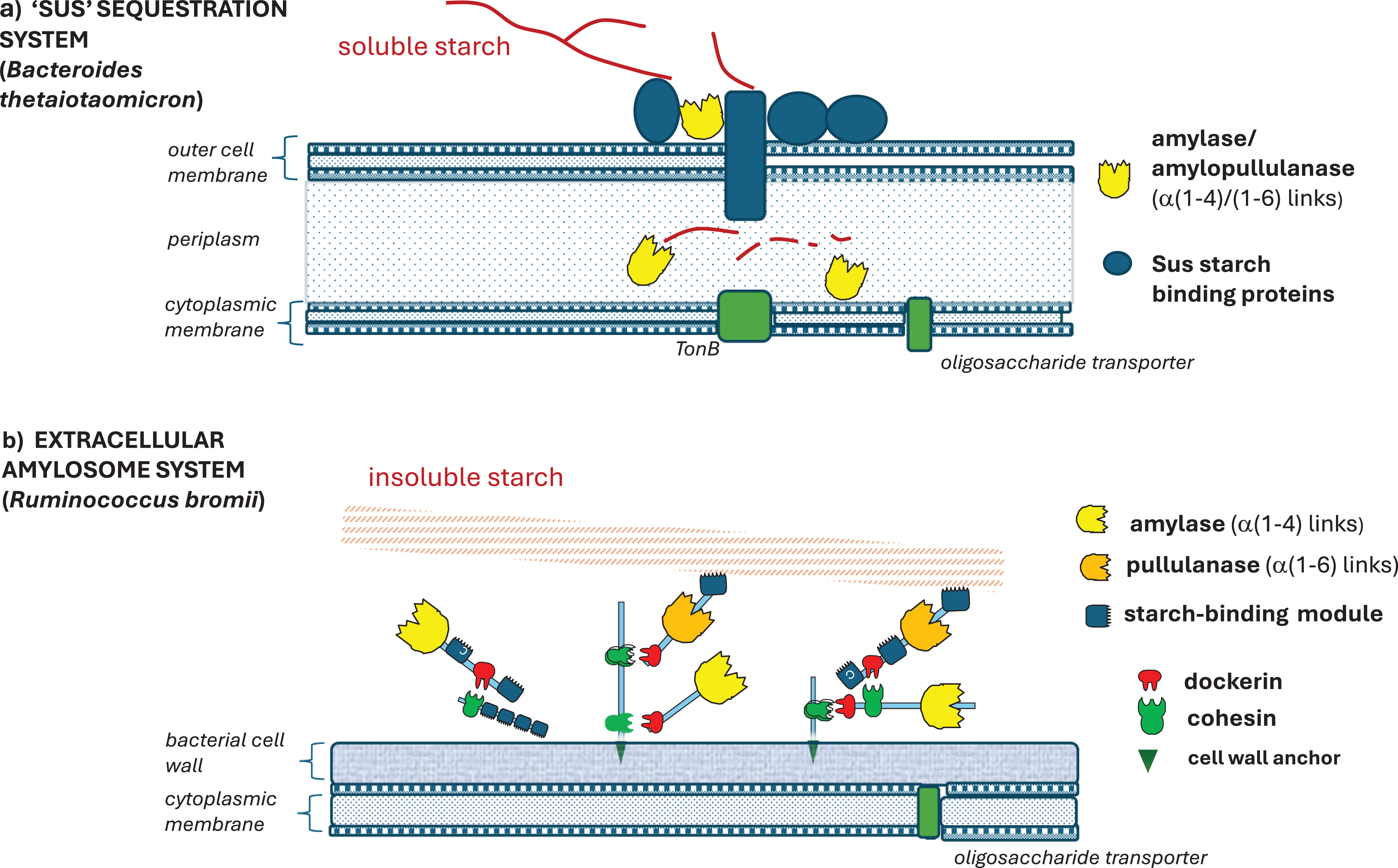
In parallel, my lab cloned DNA fragments coding for xylanases from the gram-negative anaerobe Prevotella bryantii, identifying a gene cluster that also encoded an unusual two-component regulator(Reference Miyamoto, Hirase and Mercer7). Genome sequencing in the USA completed the picture, showing that this cluster was in fact a Polysaccharide Utilization Locus (or PUL)(Reference Dodd, Moon and Swaminathan8), resembling those in human colonic Bacteroides. Bacteroides PUL encode surface proteins that trap soluble carbohydrates which are then hydrolysed within the gram-negative periplasmic space(Reference Reeves, Wang and Salyers9,Reference Martens, Koropatkin and Smith10) (Fig. 1). The contrast between this organisation (referred to as a ‘sequestration’ system by its discoverer, Abigail Salyers) and the extracellular enzyme complex of R. flavefaciens was highlighted in an invited review(Reference Flint, Bayer and Rincon11). The ‘Sus (starch utilization system)-type’ systems of gram-negative Bacteroidetes appear geared to the utilisation of soluble polysaccharides; whereas the elaborate cell-attached systems found in certain specialised Firmicutes evolved to degrade insoluble fibre (Fig. 1).
Contrasting enzyme systems involved in the utilisation of dietary starches by gut bacteria. (a) The Starch Utilization System (Sus) first described by Abigail Salyers in the human colonic bacterium Bacteroides thetaiotaomicron (Reference Reeves, Wang and Salyers9,Reference Martens, Koropatkin and Smith10) . This system allows binding of soluble starch fragments that are largely broken down by enzymes in the periplasmic space; there is no evidence that it can act on insoluble resistant starches. (b) The extracellular ‘amylosome’ system of the gram-positive anaerobe, Ruminococcus bromii (Reference Ze, Ben David and Laverde-Gomez60,Reference Mukhopadhya, Morais and Laverde-Gomez61) . The enzymes, which carry their own starch-binding modules, are organised into complexes via dockerin:cohesin interactions and held onto the bacterial cell surface via sortase-mediated attachment. R. bromii is a highly effective degrader of insoluble resistant starches.

Figure 1. Long description
Panel A: A diagram of the Starch Utilization System (Sus) in Bacteroides thetaiotaomicron. The system includes outer cell membrane, periplasm, and cytoplasmic membrane. Soluble starch is broken down by amylase and amylopullulanase enzymes in the periplasmic space. Sus starch binding proteins facilitate this process. TonB and oligosaccharide transporter are also involved. Panel B: A diagram of the extracellular amylosome system in Ruminococcus bromii. This system degrades insoluble starch using enzymes with starch-binding modules. The enzymes are organized into complexes via dockerin and cohesin interactions and attached to the bacterial cell surface via cell wall anchor. The system includes bacterial cell wall and cytoplasmic membrane, with oligosaccharide transporter present.
Understanding the microbial community of the human large intestine
Identifying resident human colonic bacteria that help to maintain health
In the late 1990’s my research switched to the anaerobic microbial community of the human large intestine. Our initial target was to define the bacteria responsible for producing butyrate which, surprisingly, were little known despite the well-documented role of butyrate in maintaining colonic health(Reference Hamer, Jonkers and Venema12). Our approach was to isolate bacteria from faecal samples using strictly anaerobic techniques developed for the rumen, accompanied by 16S rRNA gene sequencing, which revealed that many isolates were not closely related to known species(Reference Barcenilla, Pryde and Martin13). We also selectively isolated human colonic bacteria that could utilise lactate for growth, recovering new species that convert lactate and acetate to butyrate(Reference Duncan, Louis and Flint14,Reference Duncan and Flint15) . Interest in the human gut microbiota was growing rapidly at this time, but with frequent claims that most gut bacteria were ‘unculturable’. For this reason, we examined bacterial diversity by direct PCR amplification and sequencing of 16S rRNA genes in DNA recovered from colon samples, proving that the butyrate-producing bacteria we had cultured were highly abundant within the colonic microbiota(Reference Hold, Pryde and Russell16). This was the first time that sequencing of directly amplified 16S rRNA genes had been used to analyse the microbiota of human colon samples (as opposed to faecal samples(Reference Suau, Bonnet and Sutren17)) although a subsequent paper from David Relman’s lab in California, using the same primers, performed vastly more sequencing(Reference Eckberg, Bik and Bernstein18). The use of anaerobic rumen fluid-based media appears to have been the key to our success in isolating ‘novel’ species(Reference Aminov, Walker and Duncan19), but for convenience we designed a rumen fluid-free medium containing a mixture of SCFA (YCFA medium) that supports growth of most of our isolates(Reference Lopez-Siles, Khan and Duncan20). Faecalibacterium prausnitzii requires acetate(Reference Barcenilla21), but, along with related species of Ruminococcaceae, also has complex vitamin requirements(Reference Soto-Martin, Warnke and Farquharson22,Reference Laverde Gomez, Mukhopadhya and Duncan23) . Oxygen sensitivity is also a major factor; whereas the better-studied colonic Bacteroides strains can survive in air for several hours, many of our new Firmicutes isolates could not survive longer than a few minutes(Reference Flint, Duncan and Scott24). Surprisingly, F. prausntzii, although an obligate anaerobe, proved able to exploit very low oxygen concentrations, a capability that may help it survive in proximity to the gut wall(Reference Khan, Duncan and Stams25).
DNA sequencing methods were being improved at a bewildering pace during this period. Soon they allowed draft bacterial genomes to be completed within days, and metagenomic analyses to be performed through mass sequencing of DNA from mixed samples, followed by sophisticated bio-informatic analysis, as shown by a landmark paper from the EU MetaHit study(Reference Qin, Li and Raes26). Reference genomes from cultured organisms proved critical in assigning these metagenome sequences, however, and we can note that 14 out of the 56 most abundant cultured species detected in the MetaHit analysis came from our isolates. Many of these butyrate-producing species have since been reported to show reduced abundance in a wide range of disease states(Reference Hamer, Jonkers and Venema12,Reference Sokol, Pigneur and Watterlot27–Reference Forslund, Hildebrand and Nielsen29) . indicating that they represent important components of a healthy gut microbiota.
New insights into butyrate and propionate formation and lactate utilisation
Many colonic butyrate-producing bacteria, including those related to Roseburia and Faecalibacterium, consume acetate when grown in pure culture(Reference Barcenilla, Pryde and Martin13). These bacteria were shown to depend on butyryl-CoA:acetate CoA-transferase rather than butyrate kinase for the final step in butyrate formation. The butCoAT gene from a R. hominis strain was overexpressed to determine the enzyme’s specificity(Reference Charrier, Duncan and Reid30) and the diversity of human gut bacteria carrying this gene explored using degenerate PCR primers(Reference Louis, Young and Holtrop31). Net acetate uptake varied with growth conditions, especially the initial culture pH. We were able to explain this by proposing that variable amounts of external acetate could lead to the formation of acetyl-CoA and butyrate via the CoA-transferase reaction(Reference Louis and Flint32). Based on recent work in Germany(Reference Li, Hinderberger and Seedorf33,Reference Buckel and Thauer34) , the reduction of crotonate by butyryl-CoA dehydrogenase (BCD) together with electron-transferring flavoprotein (ETF) produces reduced ferrodoxin that in turn drives additional energy gain via ion gradients(Reference Louis and Flint35). We predicted that greater net acetate uptake per mol of glucose fermented should be linked to an increase in energy yield and butyrate formation(Reference Louis and Flint32). Evidence consistent with this shift in stoichiometry also came from incubations of mixed faecal bacteria with different carbohydrate energy sources in which % butyrate among the SCFA products was higher with an initial pH of 5.5 than one of 6.5 for any given % of butyrate producers within the mix(Reference Reichardt, Vollmer and Holtrop36).
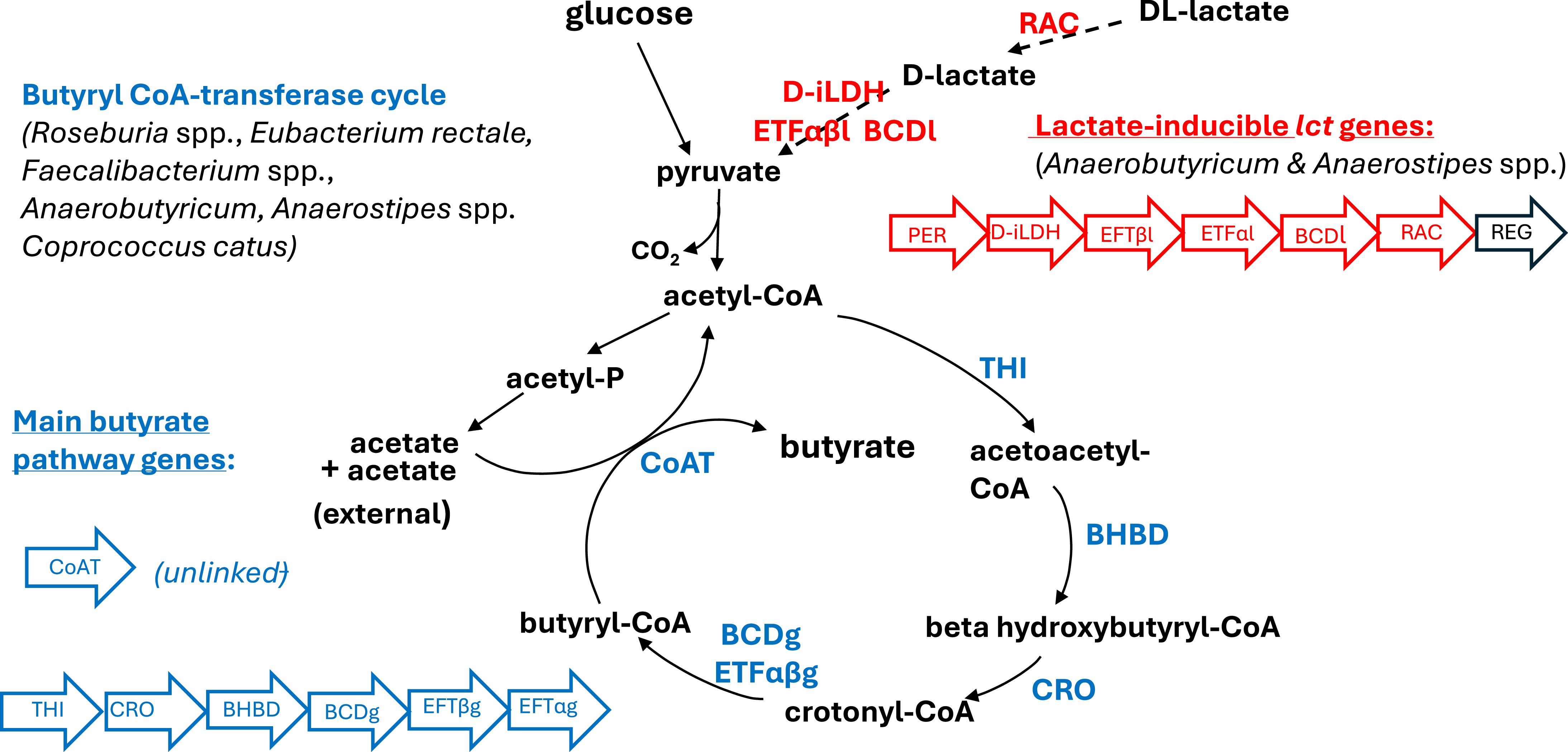
The formation of lactate from butyrate was assumed to require lactate oxidation to pyruvate, an energetically unfavourable reaction under anaerobic conditions(Reference Weghoff, Bertsch and Muller37). With the advent of RNAseq technology, we were able to study gene expression during growth on lactate compared with glucose. This revealed a massive (log2 > 14) up-regulation of six clustered genes (lct) concerned with lactate utilisation in our isolate (L2-7) of Anaerobutyricum soehngenii (formerly Eubacterium hallii)(Reference Shetty, Zuffy and Bui38,Reference Shetty, Boeren and Bui39) during growth on lactate(Reference Sheridan, Louis and Tsompanidou40). Unexpectedly, these include an acyl-CoA dehydrogenase that is an alternative to the BCD enzyme normally expressed during growth on glucose (Fig. 2). The down-regulation of the main butyrate pathway BCD and ETF genes strongly suggests that on lactate, it is the lct-encoded BCD and ETF that function together with the D-iLDH to ensure close linkage of lactate oxidation with the reduction of crotonate. In the rumen, lactate is mainly converted via the acrylate or succinate pathways to propionate. We also discovered the acrylate pathway in the human gut anaerobe Coprococcus catus, although this organism produces butyrate when growing on carbohydrates(Reference Sheridan, Louis and Tsompanidou40).
Butyrate formation from carbohydrates and from lactate in dominant human colonic anaerobes. Many Firmicutes bacteria employ butyryl-CoA:acetate CoA-transferase to produce butyrate. Genes and enzymes involved in the main pathway from acetyl-CoA are shown here in blue, with five clustered genes and the unlinked gene that encodes the CoA-transferase indicated. The ability of Anaerobutyricum and Anaerostipes spp. to produce butyrate from lactate depends on a second, highly inducible, gene cluster (lct), indicated in red. The cluster encodes a second butyryl-CoA dehydrogenase (BCDl) enzyme that appears to replace the function of the ‘normal’ enzyme (BCDg) during growth on lactate(Reference Sheridan, Louis and Tsompanidou40). The alternative butyrate kinase pathway (not shown here) is used to produce butyrate in some Coprococcus and Clostridium species. Key: THI thiolase; BHBD beta-hydroxybutyrate dehyrogenase; CRO crotonase; BCD butyryl-CoA dehydrogenase; ETFβg, ETFαg electron-transferring flavoprotein (growth on carbohydrate); CoAT butyryl-CoA:acetate CoA-transferase; PER lactate permease; D-iLDH D-lactate dehydrogenase; ETFβl, ETFαl electron-transferring flavoprotein (lactate inducible); RAC lactate racemase; REG lct regulatory protein. Gene orders shown are for A. soehngenii L2-7(Reference Louis and Flint35,Reference Sheridan, Louis and Tsompanidou40) . Anaerostipes hadrus lacks the racemase gene and can only utilise D-lactate.

After surveying the distribution of fermentation pathways in human gut anaerobes, based on available metagenome sequences and amplification of diagnostic genes, we concluded that propionate and butyrate are generally produced from carbohydrates by different bacterial species in the human colon. At least one butyrate-producing Firmicutes possesses the propanediol pathway that converts fucose or rhamnose to propionate(Reference Scott, Martin and Campbell41), and, as noted above, C. catus can produce propionate via the acrylate pathway, but the bulk of propionate production comes from the Bacteroidetes, via succinate(Reference Reichardt, Duncan and Young42).
Changes in gut microbiota and metabolites in obese volunteers on weight loss diets
We collaborated in several studies run by Alex Johnstone, through the Human Nutrition Unit at the Rowett Institute, looking at the impact of weight loss diets in overweight subjects. These studies benefitted from complete control over food intake, with all meals prepared by the HNU kitchen. At first our microbiota analysis was limited to enumeration by FISH (fluorescent in situ hybridisation) microscopy using a panel of probes targeting 16S rRNA genes. FISH analysis had some advantages, being free of uncertainties around sample storage and DNA extraction that dogged some early sequence-based studies. This method delivers absolute cell counts, rather than relative abundance, and revealed that the numbers of Roseburia/E.rectale cells(Reference Aminov, Walker and Duncan19) correlated very well with butyrate concentrations in faecal samples(Reference Duncan, Belenguer and Holtrop43). A second study reported more extensive data on faecal metabolite concentrations, revealing a major impact of weight loss diets low in carbohydrate and fibre on phytochemicals; this, together with the decrease in butyrate, suggested such diets may pose risks for colonic health if maintained in the long term(Reference Russell, Gratz and Duncan44).
We found no evidence in these studies for a deficiency in Bacteroidetes in obese individuals(Reference Duncan, Lobley and Holtrop45). This contrasted with results from an earlier sequence-based study(Reference Ley, Turnbaugh and Klein46) which reported a major phylum-level change in the ratio of Firmicutes to Bacteroidetes in obese compared with lean individuals. Our findings agreed with another FISH-based study(Reference Schwiertz, Tars and Schafer47), and with subsequent reports from sequence-based or metagenomic analysis(Reference Le Chatelier, Nielsen and Qin48). Nevertheless, there is some evidence that a few species within the microbiota may influence fat deposition, either promoting it(Reference Woting, Pfeiffer and Loh49) or decreasing it(Reference Goodrich, Waters and Poole50).
It was also suggested that the gut microbiota should promote weight gain because they supply additional calories to the host via the SCFA produced by the fermentation of fibre(Reference Backhed, Ding and Wang51,Reference Turnbaugh, Ley and Mahowald52) . The net contribution of gut micro-organisms to ‘energy harvest’ is not quite so clearcut, however, based on further analysis and experimental findings(Reference Fleissner, Huebel and El-Bary53). In particular, we know that the same amount of sugar consumed as fibre yields substantially fewer calories via the absorption of fermentation products by comparison with sugar absorbed directly. Thus, replacing digestible starch with the same amount of resistant starch (RS), for example, will increase microbial fermentation in the colon, but it delivers fewer net calories and may also contribute to satiety(Reference Flint54).
Evidence for diet-responsive species within the human gut microbiota
It soon became obvious that FISH microscopy was too slow and laborious compared to newer sequence-based methods. Our next human study, that compared weight maintenance diets incorporating different types of fibre (RS or wheat bran), employed 16S rRNA targeted qPCR techniques developed by my colleague Petra Louis, 16S rRNA gene sequencing with Alan Walker (at the Sanger Institute) and microarray analysis through a collaboration with Willem De Vos in the Netherlands(Reference Walker, Ince and Duncan55,Reference Salonen, Lahti and Salojärvi56) . We were still concerned to avoid artefacts, however, and all samples were processed rapidly before freezing. Only later did we settle on a glycerol storage procedure to allow freezing without the risk of losing DNA from certain bacterial groups. This proved to be our most significant human study for understanding of the gut microbiota. Up to this point, there was a view that a healthy adult individual’s microbiota composition was stable through time. Our study however revealed significant microbiota changes with the different dietary regimens, providing evidence of ‘diet-responsive’ species, especially in the case of diets high in RS(Reference Walker, Ince and Duncan55). The two greatest responders (in relative abundance) were the Firmicutes Ruminococcus bromii and Eubacterium rectale – except that two of our 14 individuals harboured no R. bromii on any diet. Extraordinarily, these were the only two individuals who showed significant residual starch in their faecal samples. The clear implication was that R. bromii was uniquely well equipped to degrade this substrate, making it a ‘keystone species’ for the release of energy from this substrate(Reference Ze, Duncan and Louis57,Reference Ze, Le Mougen and Duncan58) . R. bromii produces acetate, ethanol and formate as products in pure culture but co-culture with an acetogen consumes the formate(Reference Laverde Gomez, Mukhopadhya and Duncan23), suggesting that acetate will be its main product in the colon. A subsequent human study of similar design that contrasted extreme plant-based and animal-based diets demonstrated still more wide-ranging shifts in species composition(Reference David, Maurice and Carmody59).
Understanding how Ruminococcus bromii degrades resistant starch
Work on cultured strains established the superiority of R. bromii over Bacteroides thetaiotaomicron (in which the classic ‘Sus’ starch utilisation system was first described(Reference Reeves, Wang and Salyers9)) in degrading RS in pure culture, with the latter only able to use soluble starches efficiently. Cross-feeding of breakdown products between R. bromii and other species was also demonstrated(Reference Ze, Duncan and Louis57). A genome sequence for our R. bromii strain enabled us to establish its amylase complement. A real surprise was that five large extracellular amylases carried dockerin sequences, suggesting that they must be part of an enzyme complex, and protein:protein interactions were quickly established with the help of our Israeli cellulosome collaborators. We decided to call these new complexes (the first involving starch-degrading enzymes) ‘amylosomes(Reference Ze, Ben David and Laverde-Gomez60)’ (Fig. 1). But how widespread was this system? We compared the genome sequences of the very few extant strains of R. bromii (one from the USA, two from Scotland, one from South America and a rumen strain from Australia), establishing that the major amylosome components were very highly conserved(Reference Mukhopadhya, Morais and Laverde-Gomez61). Around this time, we also uncovered a cellulosome system in a human colonic bacterium (Ruminococcus champanellensis) that can degrade crystalline cellulose(Reference Ben David, Dassa and Borovok62,Reference Chassard, Delmas and Robert63) . This closely resembles the system previously documented in its rumen relative, R. flavefaciens. These cases show that the nutritional niche, rather than the mammalian host species, is the key determinant of the enzyme system. The R. bromii genomes also revealed a full set of sporulation genes, and sporulation was confirmed experimentally(Reference Mukhopadhya, Morais and Laverde-Gomez61). Sporulation is likely to be more common than previously assumed among ‘non-sporing’ anaerobes(Reference Browne, Forster and Anonye64), helping to explain how such highly oxygen-sensitive species can colonise new hosts.
Microbial fermentation of soluble v. insoluble fibres
When genome sequences first became available for human gut anaerobes, by far the largest number and diversity of genes for carbohydrate active enzymes (notably glycosyl hydrolases) was evident among Bacteroides species(Reference El Kaouturi, Armougam and Gordon65). It was widely assumed that this group must therefore be largely responsible for fibre degradation in the large intestine. As we have seen, however, Bacteroides are poorly equipped to utilise insoluble fibre and were not prevalent among the ‘diet-responsive’ species detected in our human studies, most of which were Firmicutes(Reference Salonen, Lahti and Salojärvi56). A small subset of Firmicutes were the primary colonisers of wheat bran detected using an in vitro model system(Reference Duncan, Russell and Quartieri66). The remarkable GH gene diversity in Bacteroides is however likely to confer great versality in the utilisation of soluble carbohydrates of dietary and host origin(Reference Martens, Koropatkin and Smith10). In a subsequent human dietary study(Reference Chung, Walker and Bosscher67), we found that another group of Bacteroidetes (Prevotella spp. especially P. copri), responded strongly to soluble xylan derivatives (AXOS). 8 of the 21 volunteers harboured Prevotella before the intervention, with bifidobacteria showing the greatest response in the 13 individuals who lacked Prevotella. The reasons for this striking variation in the occurrence of Prevotella, indicating apparent mutual exclusion between Prevotella and Bacteroides, are still unknown, although dietary fibre intake may be a contributing factor(Reference Wu, Chen and Hoffmann68,Reference De Filippo, Cavalieri and Di Paolo69) .
Studying the impact of soluble fibres and pH change in chemostat communities
Continuous cultures can be run in vitro under anaerobic conditions with a constant inflow of nutrient medium (and matching outflow) at a controlled pH (chemostats)(Reference Macfarlane, Hay and Gibson70). This provides an excellent opportunity to study the behaviour of microbial communities established from a faecal inoculum and supplied with soluble polysaccharide fibres. We used a single stage system to examine the impact of alternative substrates (apple pectin or inulin) over a stepped pH range from 5.5 to 6.9 (and in reverse) on the composition of separate microbial communities derived from three healthy donors(Reference Chung, Walker and Louis71). A striking conclusion was the extreme specificity of responses to the substrate, with different species of Bacteroides being promoted by inulin and by pectin. Another chemostat study explored the impact of substrate diversity upon microbiota diversity. The community established with a simple homopolymer (inulin) gave rise to a less diverse community than a highly complex substrate such as apple pectin, or mixtures of up to six different soluble fibre sources(Reference Chung, Walker and Vermeiren72). A more diverse gut microbiota is suggested to offer health benefits(Reference Le Chatelier, Nielsen and Qin48).
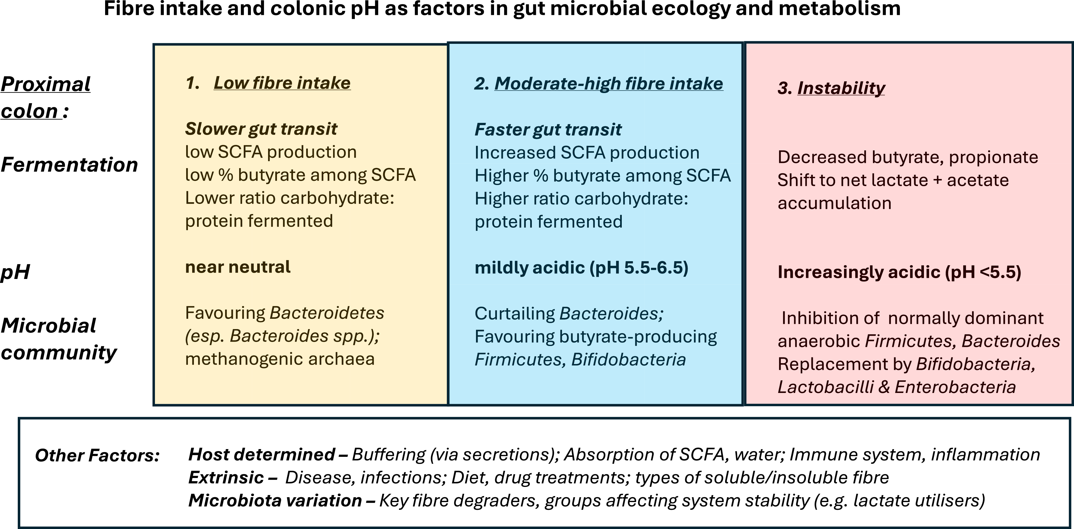
It was notable that Bacteroides became less dominant at lower pH values(Reference Chung, Walker and Louis71). We had observed this phenomenon previously in chemostats supplied with a mixture of polysaccharides (mainly soluble starch), with pH 5.5 promoting butyrate production(Reference Walker, Duncan and MacWilliam Leitch73). Bacteroides are evidently more sensitive to slightly acidic pH than are the butyrate-producing Firmicutes, which become able to compete for the substrates at the lower pH. This hypothesis was supported by measuring the growth rates of pure cultures at different initial pH values(Reference Duncan, Louis and Thomson74). Significantly, the inhibition of B. thetaiotaomicron growth at pH 5.5 was increased by concentrations of SCFA normally found in the colon (Fig. 3). These findings suggest that Bacteroides will compete more successfully in the less acidic distal regions of the colon in vivo.
Fibre intake and pH as factors determining microbial ecology and metabolism in the human colon: a simplified picture. Typically, pH is mildly acidic in the proximal colon, then increases along the transverse and distal colon. Three conditions for the proximal colon are distinguished here. 1. At low fibre intakes (highlighted in beige), concentrations of SCFA resulting from fermentation are low and colonic pH close to neutrality. Gut transit may be slow and protein fermentation (signalled by production of BCFA) may be significant compared to fermentation of carbohydrate fibre. 2. At moderate or high (including recommended) intakes of fermentable fibre (highlighted in blue), colonic pH becomes mildly acidic due to the SCFA formed by fermentation. % butyrate among SCFA increases, and % BCFA decreases (reflecting increased fermentation of carbohydrate relative to protein). 3. (highlighted in mauve), a variety of factors (disease, infection, pH too low) can cause highly unbalanced patterns of fermentation, with accumulation of lactate and acetate (acidosis). In each case, the likely shifts in the microbial community are indicated, based on evidence from chemostat communities(Reference Chung, Walker and Louis71,Reference Walker, Duncan and MacWilliam Leitch73,Reference Wang, Rubio and Duncan75) , human dietary studies(Reference Duncan, Belenguer and Holtrop43,Reference Russell, Gratz and Duncan44,Reference LaBouyer, Holtrop and Horgan83) and theoretical modelling(Reference Kettle, Louis and Holtrop79,Reference Kettle, Louis and Flint81) . The second condition (blue) is considered optimal for gut and systemic health. It should be stressed that a great many factors combine to influence gut metabolism and microbial populations, contributing to inter-individual and temporal variation. These are listed in the bottom panel. Also, faecal SCFA concentrations measured in human volunteer studies reflect fermentation along the colon, especially in the distal colon, where pH is higher and propionate-producing Bacteroides are likely to proliferate. Our modelling used available information to predict the situation in the proximal colon in vivo.

In another set of chemostat experiments, we asked how constant infusion of lactate (coming either from the intestinal microbiota, or from host tissues) might affect a colonic microbial community. Infusion of 20mM DL lactate had no discernible effect on microbiota composition or metabolite concentrations if the pH was maintained at 6.5, indicating efficient consumption by lactate-utilising bacteria (LUB)(Reference Wang, Rubio and Duncan75). At pH 5.5, however, the community readily tipped into a state in which lactate and acetate were the major products and the normally dominant anaerobes (Bacteroides and Firmicutes) were replaced by Actinobacteria, Lactobacilli and Enterobacteria(Reference Wang, Rubio and Duncan75,Reference Louis, Duncan and Sheridan76) (Fig. 3). As the pH was held constant, this points to differential inhibition of growth of the various bacterial groups by lactate. The potential for using LUB such as A. soehngenii as therapeutic probiotics(Reference Duncan and Flint15) is now being actively explored(Reference Gilijamse, Hartstra and Levin77). The ability of pathogenic enterobacteria such as Salmonella to benefit from lactate availability(Reference Gillis, Hughes and Spiga78) is a significant health concern.
Theoretical modelling of the human colonic microbiota
Steady state systems are amenable to mathematical modelling. With expert colleagues from Biomathematics and Statistics Scotland (BioSS), we developed a model based on 10 Microbial Functional Groups, chosen to align approximately with phylogenetic groups of bacteria and archaea (methanogens) and to represent the range of metabolic activities considered above. Detailed assumptions on pathways, substrate preferences, metabolic outputs, growth rates and pH responses were built into the model, based on our experimental observations on representative isolates. The resulting simulations came remarkably close to our chemostat data for butyrate concentration(Reference Walker, Duncan and MacWilliam Leitch73) with a good approximation to the pH-driven switch from Firmicutes to Bacteroidetes(Reference Kettle, Louis and Holtrop79,Reference Widder, Allen and Pfeiffer80) . Simulation of the lactate infusion experiments required modifying our 10 microbial functional groups (MFG) model by assuming different Ki’s for different MFG(Reference Wang, Rubio and Duncan75). This reproduced the phenomena of perturbation and recovery observed with 10mM lactate infusions at pH 5.5. Furthermore, the outcome was predicted to depend critically on the assumed populations within the microbial inoculum of the two MFG groups representing LUB.
Helen Kettle took on the still more challenging task of translating our 10 MFG model into the context of the human intestine, with the additional complexities of periodic intake, gut compartments, variations in gut transit and the absorption of SCFA and water(Reference Kettle, Louis and Flint81). This was the first attempt to consider detailed microbial community dynamics in the context of the human colon in an exact, quantitative manner (an earlier treatment had considered only two microbial groups(Reference Cremer, Arnoldini and Hwa82)). While some of the assumptions may require subsequent modification in the light of new information, the model allows such adjustments to be quickly tested in silico.
Explanations for, and significance of, the ‘butyrate shift’ seen in human studies
Analysis of available data from 10 human studies conducted at the Rowett Institute over a 10-year period confirmed some highly significant relationships. Looking at baseline samples from 163 volunteers, there was a positive correlation between total SCFA concentration in faecal samples and the % butyrate among total SCFA(Reference LaBouyer, Holtrop and Horgan83). Faecal pH measurements, available from half of these studies, confirmed that lower pH correlated with higher total SCFA concentration. The question then is, why should mildly acidic pH favour butyrate production? One explanation is the stoichiometric shift associated with the CoA-transferase pathway, that can result in more butyrate per mol of glucose fermented at slightly acidic pH(Reference Louis and Flint32). A second is our evidence that some butyrate-producers become more able to compete for soluble carbohydrate substrates as Bacteroides growth is suppressed by slightly acidic pH(Reference Chung, Walker and Louis71,Reference Walker, Duncan and MacWilliam Leitch73) . A third is that bifidobacteria and other lactate-producers are also stimulated at acidic pH, with much of the lactate being routed to butyrate by LUB. While this butyrate shift might perhaps be preventable by better buffering of the pH in the proximal colon, evidence that an enhanced butyrate supply is beneficial for the colonic mucosa(Reference Roediger84) suggests that the pH drop (due to the rapid fermentation of fibre) is likely to be host-controlled(Reference Flint, Louis and Duncan85). Thus, a pH environment in the proximal colon that favours butyrate production can be seen as a mechanism for ensuring an adequate supply of butyrate as the major energy source for the colonic epithelium, with consequent health benefits. Earlier indications of this phenomenon, based on much smaller numbers of volunteers, have suggested that reduced butyrate production occurs in individuals exhibiting methanogenesis and/or slow gut transit(Reference Lewis and Heaton86,Reference Abell, Conlon and McOrist87) .
The other significant change we observed with increasing total [SCFA] was a decrease in % of branched-chain fatty acids (BCFA)(Reference LaBouyer, Holtrop and Horgan83). Since BCFA derive exclusively from branched-chain amino acids, this can be explained simply if the increase in total [SCFA] reflects increased fermentation of carbohydrate fibre relative to protein (Fig. 3).
Conclusions – where next for research on the gut microbiota in relation to nutrition?
Can DNA sequencing tell us everything about the microbiota?
The past 25 years have seen an explosion of interest in the human gut microbiota and its role in human health, driven primarily by the vastly improved speed and throughput of nucleic acid sequencing technologies that bypass the need for microbial culturing. This has transformed our knowledge of diversity within the human gut microbiota across the globe, and for different age groups, dietary habits and disease states(Reference Dominguez-Bello, Costello and Conteras88–Reference Yatsunenko, Rey and Manary92). Most information on gut microbiota profiles has come from single ‘snapshot’ faecal samples however and there is scope for more controlled intervention studies that address defined questions.
Sequence information, by itself, has limitations. While metagenomes can yield functionally relevant information, this depends critically on accurate gene annotation. Work involving cultured micro-organisms, including isolation of new taxa, plays an essential role in advancing fundamental scientific knowledge and informing gene annotation. Importantly, culturing can also give rise to novel therapeutic probiotics and prebiotic strategies. Another promising area is the investigation of secondary metabolites produced by gut anaerobes, some of which act as antimicrobial agents or signal molecules(Reference Donia, Cimermancic and Schulze93,Reference Hatziioanou, Gherghisan-Filip and Saalbach94) .
The relative proportions of microbial taxa, or genes, estimated from metagenomes are hard to relate to physiology and human metabolism in any truly quantitative manner. For this we need to deal in absolute cell counts, rates of substrate degradation and metabolite flows, while also considering mucosal absorption and gut transit. Only then is it possible to develop deterministic theoretical models that simulate microbial fermentation in the gut(Reference Cremer, Arnoldini and Hwa82). Our own mathematical modelling, based on 10 MFG, relied on cultured isolates for assumptions about growth rates, pH sensitivity, substrate preferences and biochemical pathways(Reference Wang, Rubio and Duncan75,Reference Kettle, Louis and Holtrop79,Reference Kettle, Louis and Flint81) . While our model was designed to predict the major products of carbohydrate fermentation, similar approaches could be used for other important metabolites (e.g. vitamins, phytochemicals and bile acids) given enough information to define the relevant functional groups.
Inter-individual variation in the gut microbiota
Gut microbiota composition changes throughout life from infancy to adulthood to old age, and with dietary change, disease and medication(Reference Forslund, Hildebrand and Nielsen29,Reference Walker, Ince and Duncan55,Reference David, Maurice and Carmody59,Reference Dominguez-Bello, Costello and Conteras88,Reference Claesson, Jeffery and Conde89) . Nevertheless, microbiota composition in adults shows individuality and stability over time. An early proposal to subdivide adult human microbiota profiles into three discrete ‘enterotypes’ proved controversial(Reference Arumugam, Raes and Pelletier95), although the difference between individuals for whom Prevotella or Bacteroides is the predominant genus of gram-negative anaerobe has been found repeatedly(Reference Wu, Chen and Hoffmann68,Reference De Filippo, Cavalieri and Di Paolo69) . It is still unclear whether dietary differences, some form of mutual exclusion, or a combination of these factors, are responsible. Furthermore, evidence that the human genotype influences gut microbiota composition is growing rapidly(Reference Turpin, Espin-Garcia and Xu96). For example, genetic variation in the production of digestive enzymes and in genes specifying components of the immune system(Reference Sidiq, Yoshihama and Downs97) can influence the microbiota.
There is a strong argument that we should be aiming at individual, rather than universal, dietary recommendations. Some people suffering from irritable bowel syndrome (IBS) are intolerant of fibre and their health can improve with diets that lack fermentable material(Reference Simren, Barbara and Flint98). Exclusions also apply to those suffering from coeliac disease or food allergies, while adult lactose-intolerance affects much of the world’s population(Reference Udigos-Rodriguez, Matallana-Gonzalez and Sanchez-Mata99).
Prebiotics v. complex fibre sources from food?
In vitro chemostat studies reveal that microbial community responses to single types of soluble fibre can be highly species-specific(Reference Chung, Walker and Louis71). Nevertheless, different species can be promoted depending on the initial inoculum, and in vivo dietary supplementation studies with prebiotics show that different species may be promoted in different individuals(Reference Chung, Walker and Bosscher67). This means that impacts on health will not be uniform. On the other hand, any increase in fermentable fibre tends to decrease the pH of the proximal colon, altering the competition between different groups of gut bacteria (Fig. 3). This probably explains why low pH-tolerant bifidobacteria are promoted by a such wide range of ‘prebiotic’ fibres. An important benefit from ‘natural’ fibre sources is that potentially beneficial phytochemicals intrinsic to plant cell walls are released and further transformed during microbial breakdown(Reference Russell, Gratz and Duncan44,Reference Duncan, Russell and Quartieri66) . Furthermore, they contain mainly insoluble fibre that is likely to be more slowly fermented within the colon. Also, a varied intake of natural fibre sources is more likely to increase microbiota diversity by comparison with intake of a single prebiotic fibre.
Microbiota diversity
There is evidence that individuals with higher microbiota diversity show better biomarkers for metabolic health than those with lower diversity, while in low diversity groups both microbiota diversity and health scores can be improved with dietary change(Reference Le Chatelier, Nielsen and Qin48,Reference Cotillard, Kennedy and Kong100) . It has also been shown that gut microbiota diversity is greater in ‘hunter-gatherer’ communities than in urban populations(Reference Schnorr, Candela and Rampelli101). ‘Western’ style diets, early use of antibiotics and caesarian birth, are all considered factors that tend to decrease microbiota diversity(Reference Blaser102–Reference Sonnenberg, Smits and Tikhonov104). Increasing and diversifying dietary fibre intake should help to increase microbiota diversity. On the other hand, it has been argued that in many individuals from industrialised countries, key microbial species (or functional groups) have been irreversibly lost, or never acquired at birth(Reference Blaser102). We know surprisingly little about the ability of gut anaerobes to recolonise the adult gut, either naturally or by deliberate introduction, and this seems a topic worthy of more research. It would be particularly relevant to know whether new species can be acquired in humans following a diet shift, or a change in drug (including antibiotic) treatment, that creates new niches. More generally, a broader evolutionary perspective would be valuable(Reference Tannock105).
The success of Faecal Microbiota Transfer (FMT) in restoring a healthy microbiota in patients suffering from Clostridium difficile infection(Reference Konig, Siebenhaar and Hogenauer106) has encouraged interest in using ‘health-promoting’ bacteria as therapeutics, and this is an increasingly active area. Candidates can now include obligate anaerobes, some of which are known to produce spores, while others can be delivered in ways that protect them against oxygen. In addition to single organisms(Reference Gilijamse, Hartstra and Levin77), the use of ‘cocktails’ of isolated gut bacteria has also been examined(Reference Petrof, Gloor and Vanner107). Genome sequencing can help to ensure safety by confirming the absence of genes connected with pathogenicity or antibiotic resistance.
Bottom line
Can we define a healthy microbiota, other than to say it is that found in a healthy person? The extent of redundancy and diversity within the microbial community means this cannot be done in detail at the species level, but the presence of key functional groups and keystone species offers a better approach. The supply of health-protective nutrients, roles in fibre degradation, anti-inflammatory action and activities such as lactate utilisation that help to stabilise the community, can all be considered. The most obvious feature of a ‘healthy’ gut microbiota, however, must be the near absence of infectious pathogens, together with the protective barrier provided against them by the resident microbial community(Reference Lawley, Clare and Walker108). We know that the prevalence of different pathogens varies widely with geography. So far, most detailed work on gut microbial ecology has been pursued in Europe, North America and Japan, but there is a clear need for more intensive study of the dominant representatives of the gut microbiota worldwide, including their interactions, their relationship to diet and their role in preserving health(Reference De Filippo, Cavalieri and Di Paolo69,Reference O’Keefe, Li and Lahti109) .
Acknowledgements
I would like to thank my colleagues at the Rowett Institute who contributed over the past 40 years to the work described here including Sylvia Duncan, Jennifer Martin, Petra Louis, Alan Walker, Karen Scott, Wendy Russell, Alex Johnstone, Susan Pryde, Georgina Hold, Carol Leitch, Nicole Reichardt, Jenny Laverde, Indrani Mukhopadhya, Alvaro Belenguer, Rustam Aminov, Colin Stewart, Gerald Lobley, Macro Rincon, Adela Barcenilla, Xiaolei Ze, Faith Chung and Paul Sheridan. Indeed, I am grateful to all the talented PhD students, postdoctoral researchers, visiting scientists and co-workers who contributed so much to our research over the years. Thanks also to Helen Kettle, Grietje Holtrop and Graham Horgan of BioSS (Biomathematics and Statistics Scotland) for their amazing contributions on mathematical modelling and statistical analysis. Finally, I must acknowledge the immense value of our many collaborations with laboratories across Europe (UK, Spain, France, Netherlands, Germany, Finland and Italy) and in Israel, USA, China and Japan.
Financial support
No financial support was involved in the preparation of this manuscript. Sources of support for the research described here are given in the individual papers cited.
Author contributions
The author had sole responsibility for all aspects of the preparation of this manuscript.
Competing interests
None.

 Open access

