Non-technical Summary
The origin and evolution of mammals have been the subject of paleobiological work for decades. Bone histology, the study of bone tissue composition, is one of the best lines of evidence for assessing growth and development in the fossil record. However, little developmental information is available for nonmammalian cynodonts, the lineage of extinct mammal relatives that gave rise to mammals. Here, I report the bone histology of two Triassic cynodonts from Tanzania. By comparing femur size and bone tissue composition, I test whether size is correlated with age in two coeval species, Scalenodon angustifrons and Luangwa drysdalli. My sample includes 15 individuals that span a size range from ~30% to 100% maximum femur size for each species. My results show that Scalenodon and Luangwa have disparate life histories despite being similarly sized contemporaries. Luangwa is characterized by an initial phase of slow growth that transitions to rapidly deposited bone tissue early in ontogeny, interpreted as a growth spurt. This increase in growth rate is seen in small- and middle-sized individuals but is resorbed and remodeled in the largest, skeletally mature individual. By contrast, Scalenodon is characterized by fast growth in early ontogeny. However, femur size is not correlated with changes to slower growth rates, as multiple individuals show slower-growing tissue regardless of size. This indicates that growth rates are highly flexible in Scalenodon and that size is not an accurate representation of maturity for this species. Together, these results demonstrate that contemporary nonmammalian cynodont species show different life histories. The underlying mechanisms to explain different life histories in these taxa are likely due to (1) intrinsic differences in growth rates and (2) varying degrees of developmentally flexible growth. The implication of this work is that intraspecific variation in growth dynamics may be more widespread than currently understood in cynodonts and that size is not a good indicator of maturity for some species.
Introduction
Cynodonts represent the last major radiation of the synapsid lineage before the evolution of mammals and are therefore crucial for understanding mammal origins (e.g., Kemp Reference Kemp1982; Kielan-Jaworowska et al. Reference Kielan-Jaworowska, Cifelli and Luo2004; Ruta et al. Reference Ruta, Botha-Brink, Mitchell and Benton2013; Abdala Reference Abdala, Alderton and Elias2021). The earliest nonmammalian cynodonts (hereafter referred to as cynodonts) appear in the late Permian (Botha et al. Reference Botha, Abdala and Smith2007; Pusch et al. Reference Pusch, Kammerer and Fröbisch2024). During the Triassic, cynodonts diversified into two subgroups—the Cynognathia and Probainognathia (Hopson and Kitching Reference Hopson and Kitching1972, Reference Hopson and Kitching2001; Abdala Reference Abdala2007; Liu and Olson Reference Liu and Olsen2010). The probainognathian lineage gave rise to Mammaliaformes, a group that continues to the present day, whereas the cynognathian lineage became the more diverse and speciose subgroup during the Middle Triassic but eventually became extinct in the Late Triassic (Angielczyk and Kammerer Reference Angielczyk, Kammerer, Zachos and Asher2018). The clade that Cynognathia owes its success to is the immensely speciose Traversodontidae, characterized by their specialized herbivorous postcanine dentition of transversely expanded teeth (Abdala and Ribiero Reference Abdala and Ribeiro2010; Liu and Abdala Reference Liu, Abdala, Kammerer, Angielczyk and Fröbisch2014; Abdala and Gaetano Reference Abdala, Gaetano and Tanner2018).
Traversodontids, along with other nonmammalian cynodonts more broadly, share many mammalian traits, including a bony secondary palate that separates oral and nasal passages, an attachment point to the vertebral column of double occipital condyles, multi-cusped teeth that precisely occlude, a more upright and parasagittal stance, and a regionalized vertebral column with a separate thoracic region with a lumbar region that lacks ribs (Crompton Reference Crompton1963; Hopson Reference Hopson1969; Crompton et al. Reference Crompton, Joysey and Kemp1972; Crompton and Jenkins Reference Crompton and Jenkins1973; Jenkins Reference Jenkins1973; Kemp Reference Kemp1980a; Sidor and Hopson Reference Sidor and Hopson1998; Jones et al. Reference Jones, Angielczyk, Polly, Head, Fernandez, Lungmus, Tulga and Pierce2018). The evolution of these mammalian traits has long been hypothesized to be linked to the evolution of mammalian endothermy (e.g., Hopson Reference Hopson1973; de Ricqles Reference de Ricqlès1974; McNab Reference McNab1978; Farmer Reference Farmer2000; Kemp Reference Kemp2006) but assessing the physiological capabilities of extinct taxa is notoriously difficult. However, the cynodont fossil record can inform our understanding of the evolution of mammalian life histories, developmental patterns, and growth rates—the data that some physiological inference models are drawn from (e.g., Faure-Brac and Cubo Reference Faure-Brac and Cubo2020).
Bone histology provides detailed and direct evidence of the rate of skeletal growth in extinct animals by comparison with living animals with known growth rates (Amprino Reference Amprino1947; de Margerie et al. Reference Margerie, Cubo and Castanet2002, Reference Margerie, Robin, Verrier, Cubo, Groscolas and Castanet2004; Montes et al. Reference Montes, Castanet and Cubo2010). For example, living mammals have high postnatal growth rates until skeletal maturity is reached, at which point growth stops (e.g., Marín-Moratalla et al. Reference Marín-Moratalla, Cubo, Jordana, Moncunill-Solé and Köhler2014. These rapid growth rates are reflected in the disorganized bone tissue of mammalian long bone cortices (see a review in de Buffrénil et al. Reference Buffrénil, de Muizon, Dumont, Laurin, Lambert, de Buffrénil, de Ricqlès, Padian and Zylberberg2021). The specific duration that different mammalian species take to reach skeletal maturity depends on a number of life-history trade-offs (e.g., Weaver et al. Reference Weaver, Fulghum, Grossnickle, Brightly, Kulik, Mantilla and Whitney2022), but generally, mammals have consistently faster growth rates when compared with non-avian reptiles.
Previous studies that reconstruct cynodont growth patterns using bone histology show highly variable growth rates and life histories (reviewed in Kulik Reference Kulik2023). However, ontogenetic data are not available for many of the nonmammalian cynodont species that have been histologically studied thus far (e.g., de Ricqles Reference de Ricqlès1969; Ray et al. Reference Ray, Botha and Chinsamy2004; Botha-Brink et al. Reference Botha-Brink, Abdala, Chinsamy and Chinsamy-Turan2012, Reference Botha-Brink, Soares and Martinelli2018; Botha and Huttenlocker Reference Botha, Huttenlocker, de Buffrénil, de Ricqlès, Zylberberg and Padian2021). Yet many individuals show peripheral slower-growing tissue (Kulik Reference Kulik2023). Understanding the timing of the onset of peripheral slow-growing tissue, indicating the attainment of sexual/skeletal maturity, is critical for interpreting cynodont life history. Furthermore, few studies can compare contemporaneous taxa collected from the same localities. Here, using a sample derived from individuals collected from a stratigraphically and temporally constrained interval, I explore the impacts that (1) shared ancestry, (2) similar body size, and (3) similar dietary niche have on bone tissue development, growth, and life history.
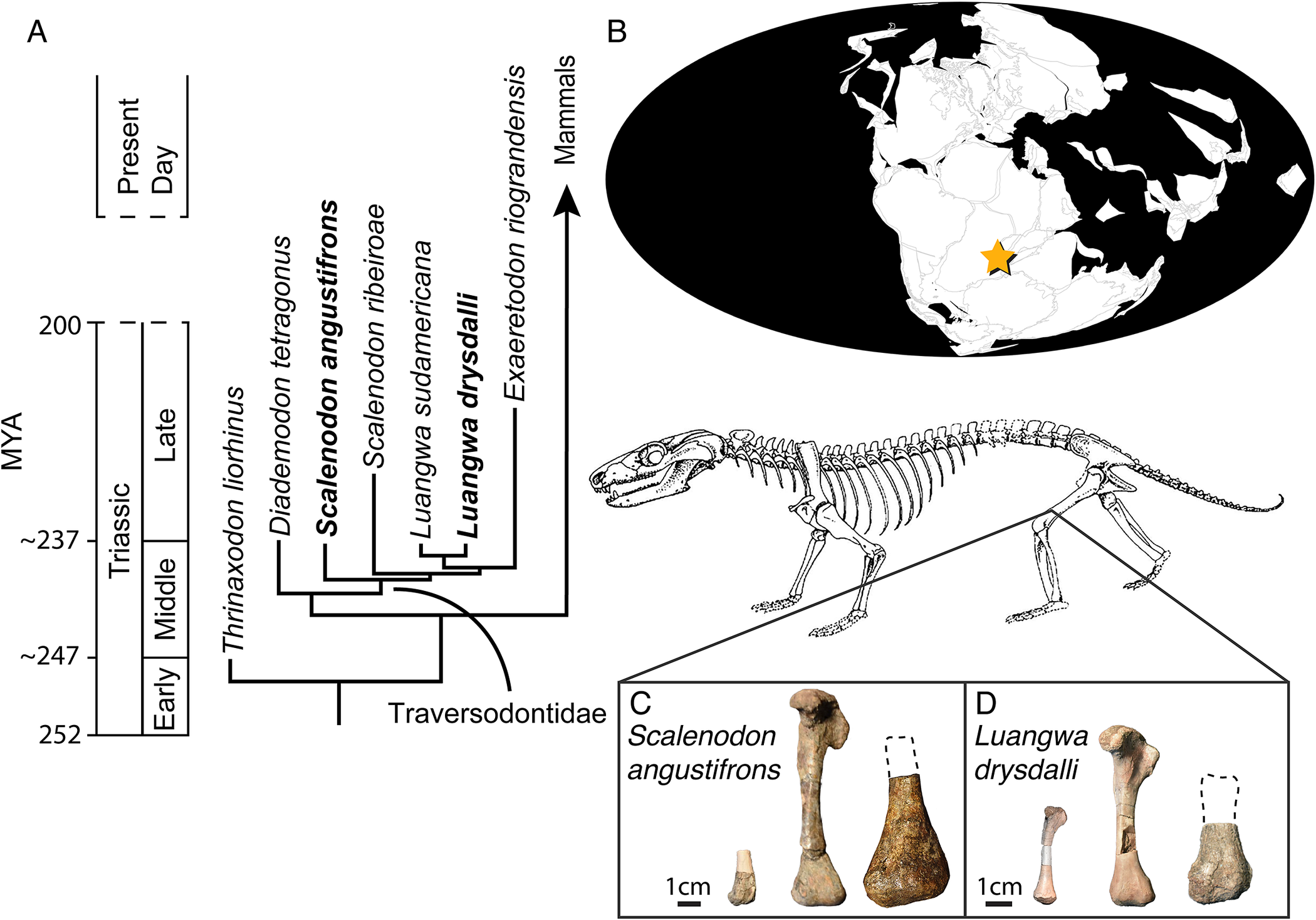
Two early-diverging traversodontid species, Scalenodon angustifrons and Luangwa drysdalli, are ideal taxa to examine the effects that shared ancestry, similar body size, and a herbivorous ecology have on growth and life history, as both species co-occur in the Manda Beds of Tanzania. In addressing these questions, I present the largest interspecific and intraspecific investigation of bone tissue composition for nonmammalian cynodonts from a spatially and temporally constrained locality. Scalenodon angustifrons and L. drysdalli are known from hundreds of long bones recovered from the middle–upper Lifua Member of the Manda Formation, Tanzania (Fig. 1). By leveraging a large dataset derived from one geographic area and geologic interval, I control for relative paleoenvironmental conditions. This is also the most complete and intensively sampled investigation of histological variation in cynodonts and provides a much-needed case study to understand intrinsic and extrinsic effects on growth and development in cynodonts. I also investigate histological evidence for developmental plasticity and its bearing on life-history evolution in the forerunners of mammals.
A, Scalenodon angustifrons and Luangwa drysdalli are two early-diverging Traversodontids recovered from B, the Ruhuhu Basin of Tanzania (star). The histological sample includes a range of femora, a selection of which are figured here; note that the midshafts have been removed in the largest femora (dotted outlines). C, Scalenodon femora from left to right include size class 1 (NMT RB860), size class II (NMT RB564.A, NMT RB866). D, Luangwa femora from left to right include size class I (NMT RB1078), size class II (NMT RB1491), and size class III (NMT RB882). Scale bar, 1 cm. Map modified from the Paleobiology Database and representative traversodont skeletal reconstruction of Massetognathus was modified from Jenkins (1970).

Institutional Abbreviation
NMT: National Museum of Tanzania.
Materials and Methods
Geologic Context
The fossils described here were collected from the middle–upper Lifua Member in the Ruhuhu basin of Tanzania. Unfortunately, no absolute dates are available for the Lifua Member. Biostratigraphic correlations to dated sequences in South America suggest an Anisian to Carnian age (e.g., Peecook et al. Reference Peecook, Steyer, Tabor and Smith2017; Abdala Reference Abdala, Alderton and Elias2021; Pretto et al. Reference Pretto, Müller, Moro, Garcia, Neto and Da Rosa2022). Namely, the middle–upper Lifua Member shares several taxa with the late Middle Triassic (Ladinian) Dinodontosaurus Assemblage Zone, including Aleodon, Luangwa, and Scalenodon (Peecook et al. Reference Peecook, Steyer, Tabor and Smith2017; Pretto et al. Reference Pretto, Müller, Moro, Garcia, Neto and Da Rosa2022).
In the Lifua Member, vertebrate remains are hosted in a series of floodplain mudrock deposits where fossils are usually recovered from shallow exposures (Smith et al. Reference Smith, Sidor, Angielczyk, Nesbitt and Tabor2017). Most middle–upper Lifua fossils show similar taphonomic signatures of disarticulation and weathering, and elements are entirely encased in calcareous or micritic matrix. Smith et al. (Reference Smith, Sidor, Angielczyk, Nesbitt and Tabor2017) interpreted the depositional environment of these fossil-bearing localities as having accumulated around distal floodplain and pond deposits between meander belts. Compared with the underlying Kingori Sandstone Formation, the Lifua Member lithology documents a shift toward increasing rainfall and ephemeral ponds that likely hosted the diversity of cynodonts and other Triassic vertebrates found here (Smith et al. Reference Smith, Sidor, Angielczyk, Nesbitt and Tabor2017).
The histological sample was collected from seven localities (Table 1). It is difficult to precisely correlate the fossil-bearing localities because of the densely vegetated and patchy outcrops, but the localities appear to be within tens of meters of each other stratigraphically, near the middle of the member. In addition to hundreds of mostly disarticulated cynodont fossils collected from these outcrops, numerous other Middle Triassic tetrapods have been described, including temnospondyls (Steyer et al. Reference Steyer, Peecook, Arbez, Nesbitt, Tolan, Stocker, Smith, Angielczyk and Sidor2021), small reptiles (Werning and Nesbitt Reference Werning and Nesbitt2016; Tsuji Reference Tsuji2017), and archosaurs, including the early-diverging dinosauriform Asilisaurus kongwe (Griffin and Nesbitt Reference Griffin and Nesbitt2016b; Nesbitt et al. Reference Nesbitt, Butler, Ezcurra, Barrett, Stocker, Angielczyk and Smith2017, Reference Nesbitt, Langer and Ezcurra2020), a large indeterminate silesaurid (Barrett et al. Reference Barrett, Nesbitt and Peecook2015), and the pseudosuchian archosaurs Mambawakale ruhuhu (Butler et al. Reference Butler, Fernandez, Nesbitt, Leite and Gower2022) and Mandasuchus tanyauchen (Butler et al. Reference Butler, Nesbitt, Charig, Gower and Barrett2017).
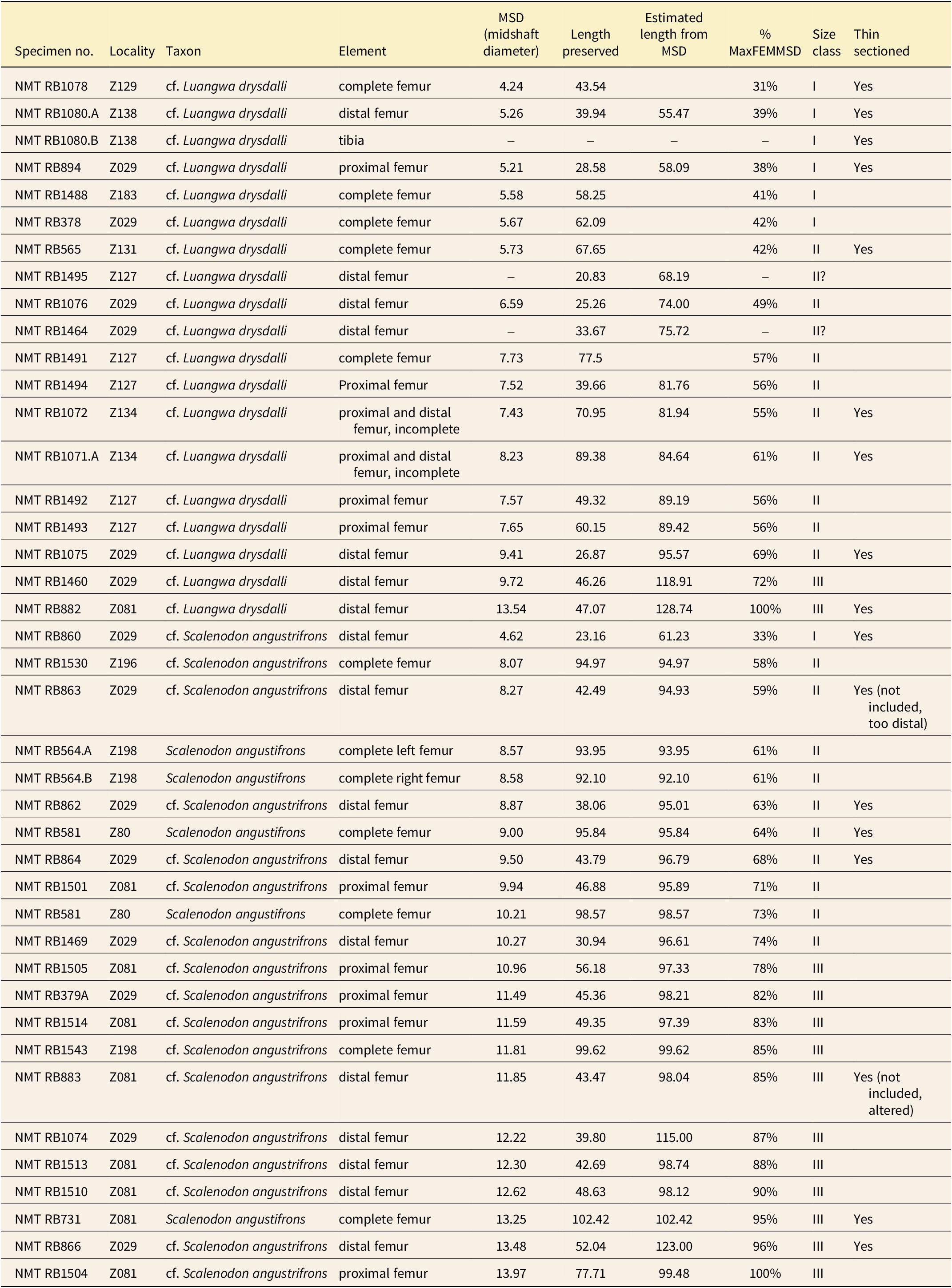
Specimen locality information and femoral measurements of referable Scalenodon angustifrons and Luangwa drysdalli fossils. All measurements in millimeters (mm). Midshaft diameter (MSD), preserved length, estimated length, and proportional midshaft diameter are reported. Histologically sampled individuals, size classes based on estimated midshaft diameter, and preservation details are also noted.
The specimens and paleohistological slides generated from this work are permanently reposited at the National Museum of Tanzania but are temporarily housed at the Burke Museum of Natural History and Culture, University of Washington, Seattle, Washington, U.S.A. (for a full list, see Table 1).
Size Class Estimation
To contextualize potential size/age correlations in the histological sample, measurements from the referable femora to S. angustifrons and L. drysdalli that were not histologically sampled were collected (Table 1). Many of these femora are incomplete, particularly for Scalenodon, the more abundant taxon. The lack of complete femora prompted length estimates and size classes based on midshaft diameter (MSD), femoral head depth, and lateral condyle width. Length estimates were calculated for each taxon to provide a range of femoral lengths between 43 and 128 mm in L. drysdalli and 61 and 123 mm in S. angustifrons. However, due to poor correlation between midshaft diameter and length in complete specimens, midshaft diameter was used to bin femora into size classes rather than estimated length. Because of the arbitrary nature of size class cut offs, additional ontogenetic information from the degree of articular surface ossification and histological changes in bone tissue composition were used to confirm the relative maturity stage of individuals in each size class.
Size class I includes specimens that are less than 50% maximum size, size class II spans elements 50–75% maximum size, and size class III corresponds to specimens that are larger than 75% maximum size. Estimated maximum femoral lengths overlap for S. angustifrons and L. drysdalli and are between 120 and 130 mm. However, Luangwa midshafts are more gracile when compared with Scalenodon femora at the same size (Fig. 2). Cross-sectional profiles also serve to differentiate femora, as Luangwa have triangular profiles due to the expansion of the lesser trochanter and distal ridge that extends distally to the mid-diaphysis. Scalenodon cross sections are equidimensional, appearing square closer to the proximal portion of the midshaft or rounded toward the distal diaphysis.
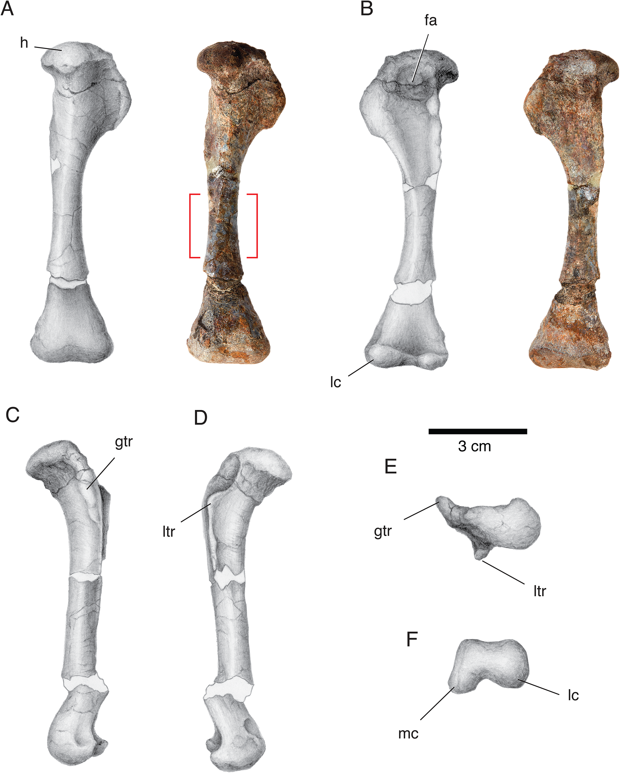
The femur of cf. Scalenodon angustifrons (NMT RB564.A). A left femur in dorsal (A), ventral (B), lateral (C), medial (D), proximal (E), and distal (F) views; red bracket indicates location where specimens were sectioned. Abbreviations: fa, adductor fossa; gtr, greater trochanter; h, head of femur; lc, lateral condyle; ltr, lesser trochanter; mc, medial condyle.

Elements in size class I have poorly ossified joint surfaces and show very spongy and pitted surfaces indicative of epiphyseal cartilage attachment in immature individuals, at least among saurian reptiles (Griffin et al. Reference Griffin, Stefanic, Lessner, Riegler, Formoso, Koeller and Nesbitt2020). By comparison, the joint surfaces of elements in size classes II and III are more robustly built and more completely ossified, although slight pitting remains on the distal surface of elements from size class II. The relative degree of epiphyseal ossification combined with histological features is well correlated for L. drysdalli, whereas the smallest individuals of S. angustifrons show a more advanced degree of ossification compared with Luangwa femora at the same size.
Thin Section Preparation
Thin sections were made following standard osteohistological techniques outlined in Lamm (Reference Lamm, Padian and Lamm2013). Each thin section was made in the transverse plane nearest to or at the midshaft, depending on specimen preservation. Only femora with intact midshafts were included. Transverse cross sections were stabilized with Paleobond and cut on a high-precision diamond-edged saw (Isomet 1000). These pieces were then molded and cast using Moldstar silicone rubber and Smooth-On plastic resin to create research-quality replicas. The casts were then glued to the remaining un-imbedded proximal and/or distal ends of the specimen to preserve its three-dimensional morphology for future research.
Sectioned midshafts were embedded in Epothin Epoxy/Resin 2, sectioned to a thickness of approximately 2 mm on a high-precision diamond-edged saw (Isomet 1000), and glued to glass slides using Devcon 2-Ton epoxy. Before 2021, slides were ground on a Metaserv 3000 lapidary plate until the specimen was 80 μm thick or until optical clarity was reached. From 2021 onward, slides were ground to optical clarity using an automated lapidary plate (Exakt 400CS). High-magnification and composite images were taken using a Nikon Eclipse LV100POL microscope under plain and cross-polarized light with a ¼ plate lambda filter. Composite images were processed using Nikon NIS-Elements AR (v. 5.20.02) and Nikon NIS-Elements BR (v. 5.24.03) imaging software. The Extended Depth Focus function was used to autofocus and z-stack photomicrographs to improve the image quality of microstructural details. Ontogenetic maturity was assessed through bone tissue composition, articular surface texture and morphology, and relative size compared with other cynodont specimens from the middle–upper Lifua Member.
Referred Material of Scalenodon angustifrons
The paleontological sample includes associations of skeletal remains and isolated femora referable to Scalenodon angustifrons: NMT RB581, a mostly articulated skeleton of S. angustifrons including the skull with lower jaws, at least 26 articulated vertebrae, articulated partial scapula, coracoid, complete humerus, and proximal ulna, proximal partial humerus, complete left femur (thin sectioned), distal right femur, parts of both tibiae and fibulae, several ribs, and unprepared pieces. NMT RB731, a partial skeleton including a skull and occluded mandible, a complete humerus, distal humerus articulated with a proximal radius and ulna, complete right femur (thin sectioned), partial proximal tibia, numerous caudal vertebrae, and other unprepared elements. Ten distal femora (five thin sectioned), and five proximal femora. For a complete list of femora referable to S. angustifrons, see Table 1.
Identification. The partial skeleton (NMT RB581) is identified as S. angustifrons based on craniodental features outlined in Crompton (Reference Crompton1955) and described in Kulik (Reference Kulik2023). Morphological similarities to associations of cranial and postcranial remains support the referral of the isolated distal and proximal femora that were histologically sectioned to S. angustifrons. Tibiae that were histologically sampled were found in association with referable femora or craniodental remains.
Description
There are no genus-level autapomorphies present in the femora of any of the Triassic-aged cynodonts known from southern Pangea (Jenkins Reference Jenkins1971; Kemp Reference Kemp1980a,Reference Kempb; Martinelli et al. Reference Martinelli, Kammerer, Melo, Neto, Ribeiro, Da-Rosa, Schultz and Soares2017; Sidor and Hopson Reference Sidor and Hopson2017), but morphological similarities support the referral of isolated and incomplete femora to S. angustifrons based on the following descriptions.
In S. angustifrons, the proximal view of the head of the femur has a crescent-shaped outline that tapers laterally toward the greater trochanter (Fig. 2). The anteromedial border of the head makes up the largest and most rounded portion of the head, whereas the articulating surface flattens between the head and the greater trochanter (Fig. 2E). The greater trochanter is long and squared off ventrally, with a thin, dorsally curved profile when viewed laterally (Fig. 2C). The lesser trochanter is developed into a short, flat shelf that makes up the medial border of the adductor fossa, which has a gently curved and shallow surface for muscle attachment. This is in contrast to Luangwa (Fig. 3) and Aleodon (Fig. 4), which have sharp adductor fossae with large basins and very prominent lesser trochanters.
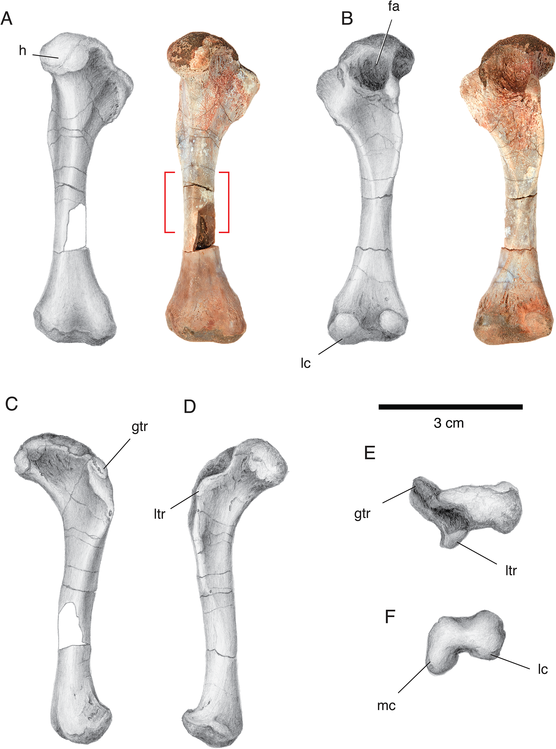
The femur of cf. Luangwa drysdalli (NMT RB1491). A left femur in dorsal (A), ventral (B), lateral (C), medial (D), proximal (E), and distal (F) views; red bracket indicates location where specimens were sectioned. Abbreviations: fa, adductor fossa; gtr, greater trochanter; h, head of femur; lc, lateral condyle; ltr, lesser trochanter; mc, medial condyle.

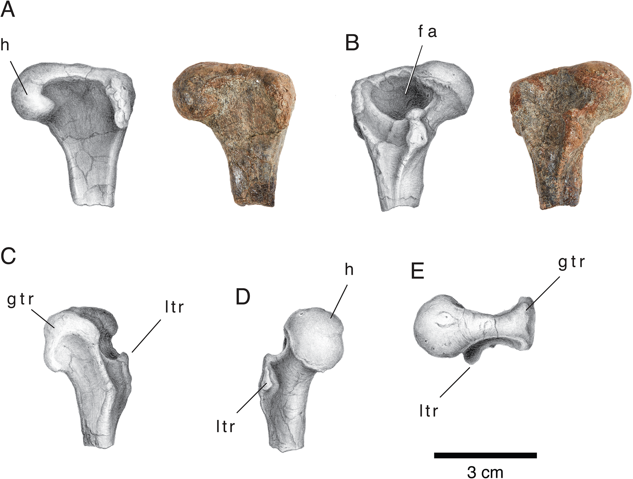
The proximal femur of cf. Aleodon brachyrhamphus. Proximal right femur, reflected for consistency in dorsal (A), ventral (B), lateral (C), medial (D), and proximal (E) views. Abbreviations: fa, adductor fossa; gtr, greater trochanter; h, head of femur; ltr, lesser trochanter.

The distal femoral profile of S. angustifrons lacks a well-developed patellar groove, leaving a smooth dorsal surface of the femur (Fig. 2F). This is unlike the condition seen in Cricodon metabolus, where the distal profile is dorsoventrally flattened and a gentle constriction separates the medial and lateral condyles (Crompton Reference Crompton1955; Sidor and Hopson Reference Sidor and Hopson2017). In S. angustifrons, the medial condyle extends ventrally, in line with the medial side of the shaft and does not hook laterally, like the condition seen in Luangwa drysdalli (Fig. 3F).
Femora referable to Aleodon brachyrhamphus are distinguished from other Lifua Member cynodonts based on their distinct proximal profiles characterized by an anterior expansion and thickening of the greater trochanter, giving a T-shaped outline (Fig. 4). Moreover, Aleodon midshafts are larger, more robust, and rectangular compared with Scalenodon and Luangwa femora at similar sizes. In Aleodon, the head of the femur is very pronounced and rounded (Fig. 4D,E). In ventral view, the lesser trochanter bounds a deep adductor fossa (Fig. 4B) and has a rugose expanse for muscle attachment that wraps medially on its proximal surface (Fig. 4D). The combined morphological features of Aleodon femora are distinct from Scalenodon and Luangwa, which bear more similarities to each other.
Referred Material of Luangwa drysdalli
The paleontological sample includes isolated skulls of L. drysdalli and isolated referable postcranial material: NMT RB93, a partial right side of a skull preserving the upper postcanine toothrow. NMT RB58, snout and occluded lower jaws, postcanine teeth are not exposed. NMT RB59, snout and occluded mandible with exposed postcanine teeth in labial view. Five complete femora are referable to L. drysdalli (NMT RB378, NMT RB565, NMT RB1078, NMT RB1488, NMT RB1491), along with seven distal fragments, three of which were sectioned, and six proximal fragments, two of which were sectioned. For a complete list of referred femora, see Table 1.
Identification. There are no definitive associations of cranial and postcranial remains of L. drysdalli in collections housed at the Burke Museum nor are there figured or described skeletons that include a description of the hindlimb (e.g., Abdala and Smith Reference Abdala and Smith2009). However, the identification of cranial remains, as outlined by Brink (Reference Brink1963) and Abdala and Sa-Teixeira (Reference Abdala and Sa-Teixeira2004), and the referral of isolated femora based on Kemp (Reference Kemp1980a) permit the assignment of isolated femora to L. drysdalli. Craniodental features outlined in Abdala and Sa-Teixeira (Reference Abdala and Sa-Teixeira2004) include: reduced temporal region, oval upper postcanines with an anterolabial cingulum, a posterior cingulum with small cuspules behind the transverse crest of the upper postcanines, serrated canines (neogomphodontia), and an external (labial) cingulum on upper postcanines with an anterolateral accessory cusp (absent in Scalenodon).
Description
The femoral anatomy of L. drysdalli shares similarities with S. angustifrons, but notable differences in the development of the lesser trochanter and medial condyle permit their distinction and referral to L. drysdalli. However, no associations of cranial and postcranial remains have been recovered from the Manda Beds, and the only figured specimen of a femur referable to L. drysdalli is a composite reconstruction in Kemp (Reference Kemp1980a). Therefore, I refer isolated femora to L. drysdalli using the following morphological features described below. Additionally, histological details such as the prevalence of parallel-fibered bone in small individuals and the triangular aspect of the femoral midshaft in thin section provide additional features to identify L. drysdalli femora from S. angustifrons femora.
In proximal view, the head of the femur bears a broad articular surface that is expanded on the medial side to make up a well-defined head, like in S. angustifrons (Fig. 3A). The proximal profile of the head is constricted slightly at the point of attachment with the lesser trochanter. The articular surface continues to taper laterally toward the greater trochanter, at which point a sharp, medially directed angle separates the surface of the greater trochanter from the rest of the head as the trochanter curves mediodorsally (Fig. 3B,E). This sharp deflected angle of the greater trochanter is unlike the condition seen in S. angustifrons, which bears a more gradual curve. Furthermore, the greater trochanter is more robust than in S. angustifrons and bears a sharper, squared-off ventral contact with the rest of the metaphysis (Fig. 3B). In lateral view, the greater trochanter is wider than in S. angustifrons and similarly contributes to a broad, flat ventral surface.
On the ventral side, the lesser trochanter is very well developed and contributes to a deep adductor fossa that is bordered by a shelf that extends from the lateral contact of the lesser trochanter to the rest of the proximal shaft. The lesser trochanter has a teardrop-shaped surface for muscle attachment that is well ossified, even in small individuals. It continues as a long crest down the ventral surface of the shaft and gently tapers at the midshaft. In cross-sectional profile, L. drysdalli femora are triangular, with the medial edge of the midshaft making up the apex of the triangle.
In distal profile, the medial and lateral condyles are separated by a broad and flat patellar groove that is slightly more developed than in S. angustifrons. Distal femora referable to L. drysdalli can be identified based on the morphology of the medial condyle that bears a well-rounded “head” that hooks laterally (Fig. 3F). The lateral condyle is similarly well developed as a rounded articular surface but is considerably larger than the medial head and is equidimensional in distal profile. The distal epiphyseal surface of cf. L. drysdalli femora is separated from the rest of the metaphysis by a constriction that encircles the distalmost shaft; this is unlike the condition in S. angustifrons, where the metaphysis merges seamlessly with the epiphyseal surface.
Results
Bone Tissue Composition of Luangwa and Scalenodon
Bone tissues seen in skeletally mature Luangwa drysdalli (Figs. 5, 6) and Scalenodon angustifrons (Figs. 7, 8) conform to the three-stratum structure described in a review of extant mammal histology by de Buffrénil et al. (Reference Buffrénil, de Muizon, Dumont, Laurin, Lambert, de Buffrénil, de Ricqlès, Padian and Zylberberg2021). Three strata are formed by a central disorganized layer bounded by two organized layers of endosteally and periosteally directed growth, respectively. The central layer comprises primary periosteal tissue that varies in composition depending on the species. In L. drysdalli, parallel-fibered bone transitions to woven bone in the central stratum, whereas a woven-parallel complex with disorganized vascular canals is seen in S. angustifrons. However, parallel- and woven-fibered bone can occur simultaneously and often in different proportions within the same section, forming a spectrum of tissue types that can be difficult to define. Here, I follow the terminology outlined in Francillion-Veilliot (Reference Francillon-Vieillot, de Buffrénil, Castanet, Géraudie, Meunier, Sire, Zylberberg, de Ricqlès and Carter1990) with updates to bone matrix terminology described in de Buffrénil and Quilhac (Reference Buffrénil, Quilhac, de Buffrénil, de Ricqlès, Padian and Zylberberg2021). In some individuals, the central stratum also includes compacted coarse cancellous bone (CCCB; sensu Enlow Reference Enlow1962) that formed at the metaphyseal level earlier in ontogeny and was maintained and compacted during growth, whereby it reached the diaphyseal region (e.g., Figs. 6E,H, 7A).
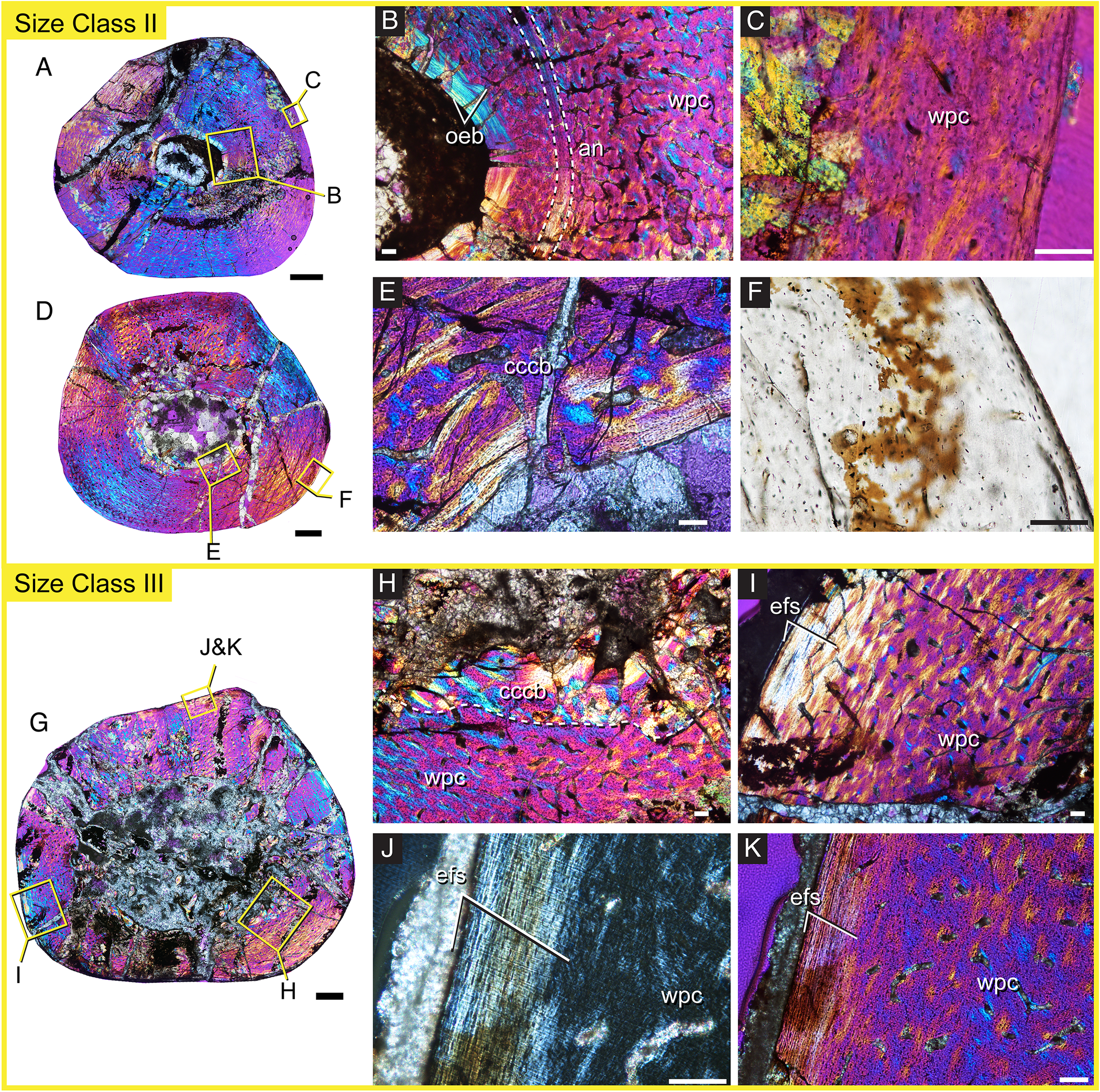
Femoral histology of cf. Luangwa drysdalli from size class I. The femur NMT RB1078 (A) represents the earliest record of growth and is composed of parallel-fibered bone with a midcortical annulus (white arrowhead) that interrupts a mosaic woven-parallel complex (B, plain-light on left side, cross-polarized light with lambda plate on right side, 20× magnification in C). The associated femur NMT RB1080.A (D) and tibia NMT RB1080.B (G) show organized endosteal bone (F) and a growth spurt of woven-parallel bone in early ontogeny (E). In the tibia (G), the growth mark is bounded by an annulus (dotted line in I). Rapid bone deposition continues at the subperiosteal surface (H). Scale bar, 1 mm (A, D, G); 100 μm in all photomicrographs (B, C, E, F, H, and I). Abbreviations: oeb, organized endosteal bone; pfb, parallel-fibered bone; mc, medullary cavity; wpc, woven-parallel complex. High-resolution images can be found under MorphoSource project ID: 636652.
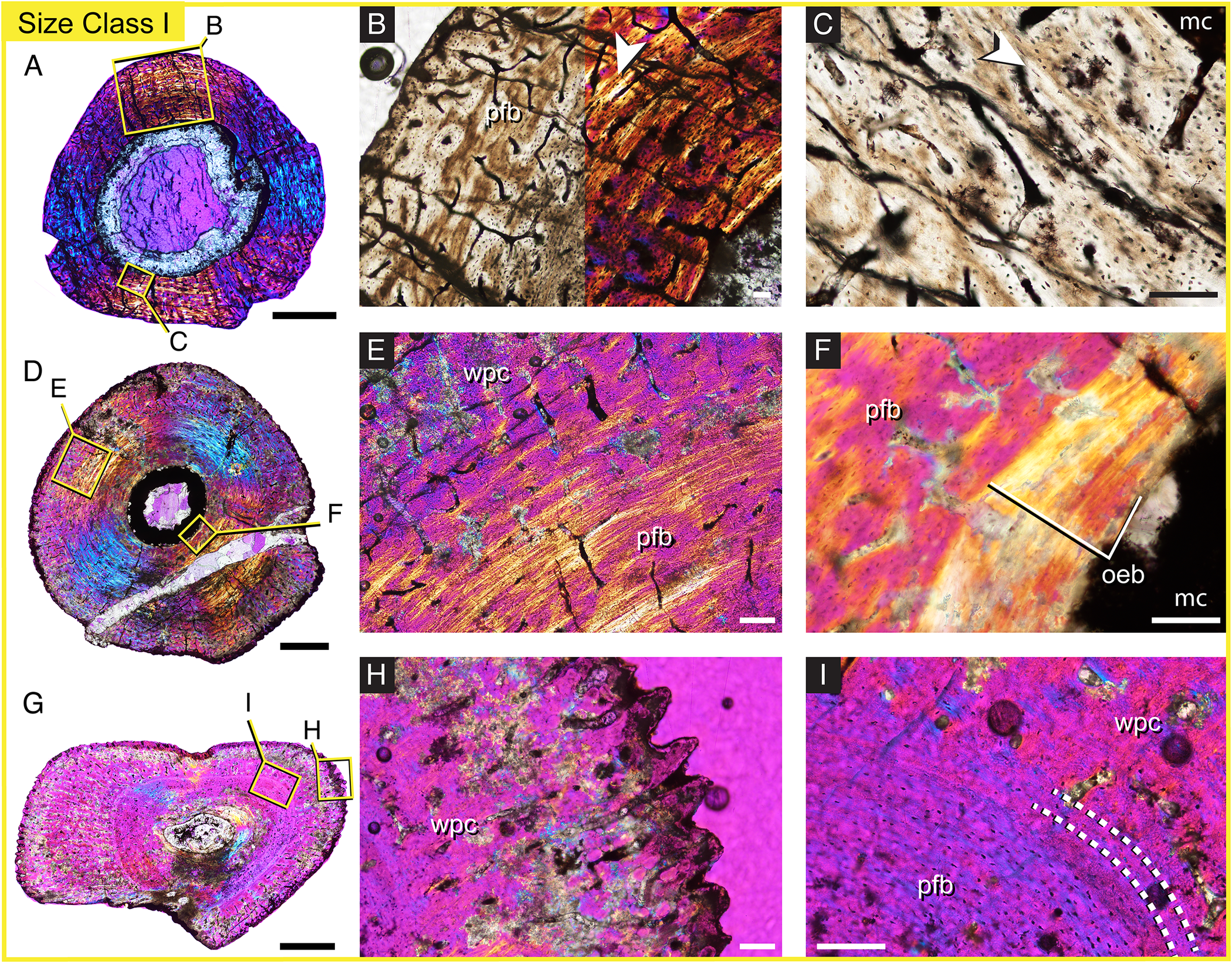
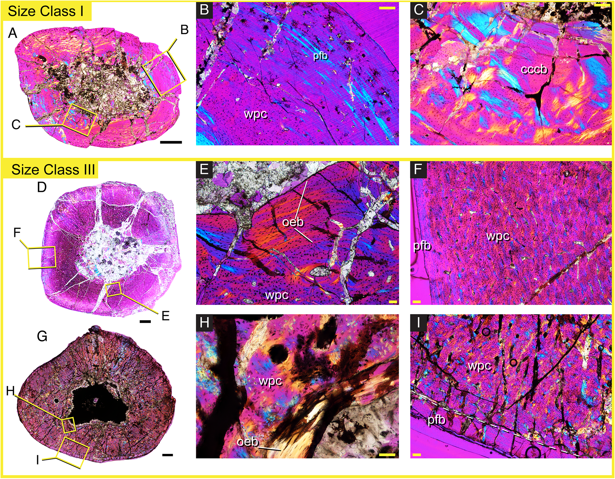
Femoral histology of cf. Luangwa drysdalli from size classes II and III. The thick cortex in NMT RB1071 (A) is composed of remnants of the incipient slower-growing bone bounded by an annulus (white dotted lines) after which, woven-parallel bone is deposited (B, C). NMT RB1072 (D) shows deep cortical remodeling and compacted coarse cancellous bone (CCCB) (E) and a primary cortex composed of a woven-parallel complex (F). NMT RB866 from size class III (G) retains none of the deep parallel-fibered bone and instead has CCCB (H) and an external fundamental system (I). J, 20× magnification of the external fundamental system (EFS) in cross-polarized light without the lambda filter used in K. Scale bar, 1 mm (A, D, G); 100 μm in all photomicrographs (B, C, E, F, H, I, J, and K). Abbreviations: an, annulus; cccb, compacted coarse cancellous bone; efs, external fundamental system; oeb, organized endosteal bone; wpc, woven-parallel complex. High-resolution images can be found under MorphoSource project ID: 636652.

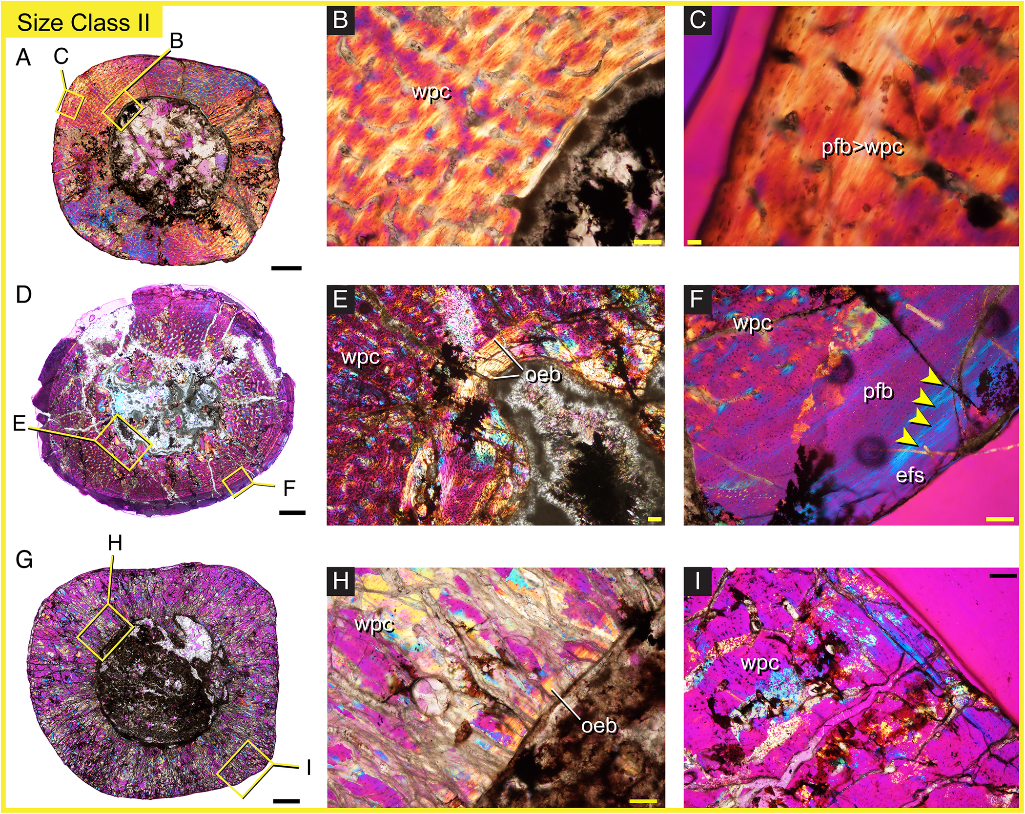
Femoral histology of cf. Scalenodon angustifrons from size classes I and III. The cortices of femora from size classes I (NMT BR860, A) and III (NMT RB731, D; NMT RB866, G) are composed of woven-parallel bone that transitions peripheral parallel-fibered bone, highlighting that this taxon does not follow a stereotyped pattern in which bone tissue composition changes predictably with increasing size. Woven-parallel bone (B) is bounded between compacted coarse cancellous bone (CCCB; C), deep parallel-fibered bone and peripheral parallel-fibered bone (B) in NMT RB860 (A). Organized endosteal bone (E, H) bounds thick cortices of a woven-parallel complex that transitions to a thin region of parallel-fibered bone (F, I) in NMT RB731 and NMT RB866. Scale bar, 1 mm (A, D, G); 100 μm in all photomicrographs (B, C, E, F, H, and I). Abbreviations: cccb, compacted coarse cancellous bone; oeb, organized endosteal bone; pfb, parallel-fibered bone; wpc, woven-parallel complex. High-resolution images can be found under MorphoSource project ID: 636652.
Femoral histology of cf. Scalenodon angustifrons size class II. Thick cortices are composed of rapidly growing tissue in a woven-parallel complex in NMT RB862 (A–C), with variable amounts of endosteal organized bone in NMT RB864 (D, E) and NMT RB581 (G, H). NMT RB864 (D) shows peripheral parallel-fibered bone and an external fundamental system (EFS) composed of avascular lamellar tissue and four lines of arrested growth (LAGs; yellow arrows in F), whereas NMT RB581 (G) shows a woven-parallel complex (I). Scale bar, 1 mm (A, D, G);100 μm in all photomicrographs (B, C, E, F, H, and I). Abbreviations: efs, external fundamental system; oeb, organized endosteal bone; pfb, parallel-fibered bone; wpc, woven-parallel complex. High-resolution images can be found under MorphoSource project ID: 636652.

The deepest cortical tissue that composes the inner stratum includes organized endosteally directed lamellar bone (e.g., Figs. 5C,F, 6B, 7E,H). When CCCB is not present in the deep cortex, a reversal line separates the organized endosteal bone from the primary periosteal growth, illustrated in Figure 7E. From this, it is evident that the local morphogenesis of the femoral shaft in both Scalenodon and Luangwa experienced multiple episodes of medullary expansion and resorption throughout ontogeny.
The outer stratum consists of slow-growing parallel-fibered to lamellar tissue that is poorly vascularized or avascular, termed the outer circumferential layer or external fundamental system (EFS). This peripheral tissue can also include closely spaced lines of arrested growth (LAGs). Together, the EFS and any co-occurring LAGs are indicative of skeletal maturity and represent the final smoothing of the subperiosteal surface whereby no additional appositional growth occurs. Skeletally mature individuals were recovered in both taxa examined here (Figs. 6I, 8F). However, the skeletally mature femur of Scalenodon is not representative of maximum size and has a midshaft width that is 68% maximum known size. Larger individuals of Scalenodon show sustained rapid growth or only a minimal amount of peripheral parallel-fibered bone, indicating that the ability to sustain rapid bone deposition varied intraspecifically. By contrast, the skeletally mature femur of Luangwa represents an individual that is 100% maximum size. Differences in the bone tissue compositions and the proportions of bone tissue types of each taxon indicate divergent life histories as described in detail in the following sections.
Bone Histology of Luangwa drysdalli
Eight individuals referable to L. drysdalli were sampled from a size range that spans 43.5mm -128.7mm estimated femoral length. Size class I (Fig. 5) is characterized by individuals that are less than 50% max femur length (i.e., 43.5–63 mm) and include femurs NMT RB1078, NMT RB1080.A (and associated tibia NMT RB1080.B), NMT RB894, and NMT RB565. Size class II includes individuals between 50% and 75% maximum midshaft width and includes femurs NMT RB1071, NMT RB1072, NMT RB1073, and NMT RB1075. Finally, specimen NMT RB882 has an estimated length of 128.7 mm and represents the largest femur in the sample (Fig. 6A–I)
Luangwa drysdalli is characterized by multiphased growth that starts out moderately paced with longitudinal and oblique vascular canals in parallel-fibered tissue (Fig. 5A,B). This tissue is subsequently built upon by rapidly growing periosteal bone in individuals that are slightly larger than the smallest individual sampled, but still less than 50% maximum length, which I interpret as a growth spurt (Fig. 5D,E,G,I). In the smallest femur (NMT RB1078), a midcortical annulus interrupts a mosaic of primary longitudinal and radial canals surrounded by parallel-fibered and woven bone (Fig. 5B). The second smallest femur (NMT RB1080.A) and associated tibia (NMT RB1080.B) both show an abrupt midcortical shift to sustained fast growth that is marked by an annulus in the tibia (Fig. 6B), indicating that a growth spurt increased apposition rates across the hindlimb skeleton (Fig. 5D–I.). Rapidly deposited woven-parallel bone continues from the midcortical growth spurt to the subperiosteal edge, indicating that individuals in size class I were actively and rapidly growing at death (Fig. 5H). The presence of a midcortical growth mark in these individuals indicates that growth was interrupted early in ontogeny.
In size class II, much of the cortex consists of rapidly deposited woven-parallel bone. Some individuals retain the incipient parallel-fibered dominant tissue seen in size class I (Fig. 6B), whereas others show endosteal remodeling and CCCB (Fig. 6E). Due to the placement of each thin section along the femoral midshaft and intraspecific histovariation in the degree of medullary expansion, the proportion of parallel-fibered tissue compared with the outer woven-parallel tissue varies between specimens in size class II.
The largest femur represents size class III and has a crushed medullary region, but it appears that the inner region of parallel-fibered bone had been removed through remodeling and expansion of the medullary cavity (Fig. 6G,H). The remainder of the cortex is composed of woven-parallel tissue that transitions to avascular, lamellar tissue, indicating that skeletal maturity had been reached before death (Fig. 6I).
Luangwa drysdalli bone histology is characterized by a novel life history marked by an initially moderate pace of growth, a growth spurt early in ontogeny, and sustained rapid growth until death. The inferred growth spurt is seen in numerous individuals and between associated elements, suggesting that this life history is representative of the species. Furthermore, growth rates attenuate at large size, and element size and relative maturity are well correlated for L. drysdalli.
Bone Histology of Scalenodon angustifrons
Seven individuals referable to S. angustifrons were thin sectioned from a size range that spans 61.23–123 mm estimated femoral length. The smallest individual (NMT RB860) is approximately 30% maximum size (based on femoral midshaft diameter) whereas four individuals are between 50% and 75% maximum represented in size class II (NMT RB581, NMT RB862, NMT RB863, NMT RB864). Individuals greater than 75% maximum size include NMT RB731 and NMT RB866.
The bone histology of S. angustifrons is characterized by woven-fibered bone and plexiform vascular canals, indicating rapid bone deposition throughout early ontogeny (Figs. 7A–I, 8A–I). Interestingly, regardless of size, multiple individuals show slower-growing, peripheral parallel-fibered tissue, indicating a decoupling between element size and growth (Fig. 7B,F,I). However, not all individuals show a gradual transition to slow growth (e.g., Fig. 8C), and the maintenance of uninterrupted woven-parallel tissue in large size classes indicates that Scalenodon has a high intrinsic rate of growth that plateaued in some individuals at remarkably different sizes, indicating highly variable growth. What follows is a summary of bone tissues seen in each size class.
One individual (NMT RB860) from size class I is past its most rapid phase of growth and shows a mosaic of tissue types (Fig. 7A–C). The innermost cortex consists of compacted coarse cancellous bone (Fig. 7C) that transitions to woven bone surrounding longitudinal vascular canals (Fig. 7B). More peripherally, parallel-fibered bone composes the outer periosteal cortex, indicating that growth had slowed before death. By contrast, individuals in size class II show highly variable tissue types, including the only example of a skeletally mature individual, while maintaining high initial rates of bone deposition (Fig. 8B,E,F,G). The largest individuals, NMT RB731 and NMT RB866, have cortices composed of a woven-parallel complex that transitions to peripheral parallel-fibered bone (Fig. 7D–I), similar to some individuals in size class II. Taken together, a mosaic of bone tissues is seen throughout the size series and within size classes of femora referable to S. angustifrons.
Such variability between femur size and proportions of bone tissue types demonstrates that histological and skeletal markers of maturity are decoupled for S. angustifrons. Rather than following a developmental pattern whereby bone tissue composition changes from fast to slow growth in elements that are approaching maximum known size (estimated maximum femoral length for Scalenodon is 123 mm), the bone histology of S. angustifrons shows that the proportions of woven bone and slower growing tissue types do not scale with femur size, indicating highly flexible developmental growth.
Discussion
The bone histology of two contemporaneous traversodontids examined here show proportional differences in bone tissue composition and differences in growth rate. The hypothesis that shared ancestry, similar ecology, and body size should be reflected in the growth and life history of these taxa is not upheld. Scalenodon angustifrons and Luangwa drysdalli are inferred to be small- to medium-sized herbivores that likely lived in burrows, although no remains have been described from terminal chambers in these or other traversodont-dominated fauna (e.g., Fiorelli et al. Reference Fiorelli, Rocher, Martinelli, Ezcurra, Hechenleitner and Ezpeleta2018). But despite these similar ecologies, the bone histology does not reflect similar changes in bone tissue composition. Moreover, differences in growth rate and life history among individuals cannot be attributed to differences in paleoenvironment, as they co-occur in penecontemporaneous fossil localities in the Manda Beds (Smith et al. Reference Smith, Sidor, Angielczyk, Nesbitt and Tabor2017). This result is somewhat unexpected, given that both Scalenodon and Luangwa are superficially very similar, but their life histories indicate innate physiological differences in developmental pattern and degrees of flexibility in growth (Fig. 9). Discordance between size and stages of maturity in S. angustifrons may be influenced by the accidental inclusion of other cynodont species only known from cranial remains (e.g., Mandagomphodon; Hopson Reference Hopson, Kammerer, Angielczyk and Fröbisch2014). However, this is unlikely, because femoral morphologies, cross-sectional profiles, and general trends in bone tissue composition are not suggestive of accidental inclusion (i.e., no bimodal trend in size/age correlations). Moreover, if the smallest individual referred to S. angustifrons represents a mature diminutive species, this does not help to clarify the highly variable peripheral bone tissue types seen throughout the rest of the larger individuals sampled.
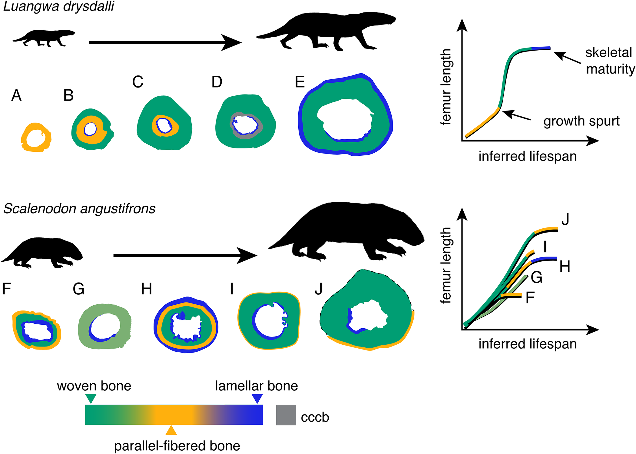
Comparative schematic of changes in bone tissue composition as femur size increases in Luangwa and Scalenodon (silhouettes not to scale). Luangwa (A–E) has juvenile growth characterized by parallel-fibered bone (A) that is quickly built upon by woven bone (B, C), interpreted as a growth spurt. The incipient slower-growing tissue is later remodeled or replaced (D), and at large size, an external fundamental system (EFS) of lamellar bone (E) indicates skeletal maturity. In contrast, Scalenodon (F–J) does not show stereotyped changes in bone tissue composition with increasing size. The earliest record of growth is a woven-parallel complex throughout all sampled elements, but many individuals show different proportions of peripheral parallel-fibered tissue (F, H, I, J), with only H indicating skeletal maturity, whereas G shows continued deposition. Regions of endosteally deposited lamellar tissue are common for both taxa, indicating expansion and remodeling of the medullary cavity. Silhouette modified from G. Ugueto’s unpublished illustrations.

Comparison with Nonmammalian Cynodont Bone Histology
The bone histology of L. drysdalli is unusual among cynodonts that have been thin sectioned due to a growth spurt in early ontogeny. By contrast, S. angustifrons bone histology is similar to that of some other cynognathians and traversodontids that show rapid bone growth that is not interrupted (i.e., lacks lines of arrested growth or annuli). Interestingly, cynognathians represent the first group of cynodonts to demonstrate physiological differences related to growth strategy driven by dietary niche (Botha and Chinsamy Reference Botha and Chinsamy2000). For example, the femora of carnivorous Cynognathus are composed of sustained rapidly deposited woven-parallel tissue, whereas the co-occurring and similarly sized herbivore Diademodon has growth marks that interrupt rapidly deposited tissue (Botha and Chinsamy Reference Botha and Chinsamy2000). It is possible that a different diet, such as omnivory, could explain the growth spurt seen in Luangwa. As suggested by Kemp (Reference Kemp1980a), the wear facets and occlusion pattern of Luangwa from the Ntawere Formation of Zambia indicate that there is no reason to assume an exclusively herbivorous diet. Furthermore, aggregations of Scalenodon skeletons are interpreted to have accumulated around shrinking ponds, reflecting seasonally drought-stressed environments (Smith et al. Reference Smith, Sidor, Angielczyk, Nesbitt and Tabor2017). That Luangwa is also found in these same deposits but in smaller numbers could indicate that their diet did not force aggregations around vegetated ponds.
Other gomphodont species that have been histologically sampled include Andescynodon, which is characterized by lamellar tissue throughout the middle and outer cortex of an isolated femur (Chinsamy and Abdala Reference Chinsamy and Abdala2008). Andescynodon is an early-diverging traversodontid from South America with a small-medium body size (estimated skull length = 90 mm) and unusually slow-growing bone histology compared with other traversodontids. Medium-bodied species from South America include Massetognathus pascuali (maximum skull = 205 mm in length; Chinsamy and Abdala Reference Chinsamy and Abdala2008), where small femora show slow-growing tissue. However, a recent report by Garcia Marsà et al. (Reference Garcia Marsà, Agnolín and Novas2022) revealed uninterrupted rapid growth in distinctly longer M. pascuali femora than those sampled by Chinsamy and Abdala (Reference Chinsamy and Abdala2008). This result indicates intraspecific variation in growth and a decoupling between element size and inferred age in Massetognathus, similar to what is seen in S. angustifrons.
Comparative histological data of other traversodontids from southern African and South American Anisian–Carnian deposits show that acyclical, moderately to rapidly deposited tissue characterizes growth throughout early ontogeny (e.g., Traversodon, Protuberum, Exaeretodon, and Scalenodontoides) (de Ricqlès Reference de Ricqlès1969; Botha-Brink et al. Reference Botha-Brink, Abdala, Chinsamy and Chinsamy-Turan2012; Veiga et al. Reference Veiga, Botha-Brink and Soares2018). However, this comparative sample does not provide insights into traversodontid life history or degrees of developmental plasticity, as only immature tissue has been sampled thus far. From the results presented here, the bone histology of S. angustifrons reflects the highest degree of intraspecific variation in growth dynamics for the clade. This may also be echoed in M. pascuali (Carnian), but a larger sample size is needed to confirm this. In contrast, Luangwa long bone histology provides the most compelling evidence of attenuating growth in traversodontids. However, Luangwa is distinct, because it shows a life history marked by a growth spurt that continued until skeletal maturity was reached. Interestingly, Thrinaxodon also shows a growth spurt in its femoral histology (Botha and Chinsamy Reference Botha and Chinsamy2005). These authors illustrate that the smallest Thrinaxodon femur has parallel-fibered bone toward the outer cortex, whereas a larger and presumably more mature individual shows a woven-parallel complex as the dominant tissue type. Although not described in detail, it appears that Thrinaxodon has the same growth curve inferred for Luangwa, with slower growth during early stages of development, a rapid increase from a growth spurt later in ontogeny, and a decrease in growth at large size (Botha and Chinsamy Reference Botha and Chinsamy2005). Critically, Thrinaxodon is one of the few cynodonts for which ontogenetic information is available. That a similar life history characterized by a multiphased growth rate and determinate growth is seen in Thrinaxodon and Luangwa suggests that this pattern is not the result of environmental fluctuations, but rather inherent biological factors that were either convergent or ancestral in eucynodonts. It is equally parsimonious for the similar life histories to be driven by convergence or ancestry, and continued work with a broader histological sample of nonmammalian cynodonts can shed light on this question. Mounting evidence from this and previous work describing the highly variable tissue types that characterize different nonmammalian species indicates that growth dynamics and developmental patterns were labile in the mammalian stem.
Developmental Plasticity and Its Role in the Evolution of Mammalian Life Histories
Mammals evolved from small-bodied probainognathians during the Late Triassic. The prevailing hypothesis for mammalian life history evolution is that highly variable growth rates were lost in the immediate ancestors of mammals due to small body size and canalization of other physiological traits related to mammalian endothermy and maternal care (Farmer Reference Farmer2000, Reference Farmer2020; Chinsamy and Hurum Reference Chinsamy and Hurum2006; Kemp Reference Kemp2006; Hurum and Chinsamy-Turan Reference Hurum, Chinsamy-Turan and Chinsamy-Turan2012; Veiga et al. Reference Veiga, Botha-Brink and Soares2018). When this loss occurred, or if it happened multiple times in separate lineages, is currently unknown. Pieces of evidence to support the loss of developmental plasticity in stem mammals comes from different examples of stereotyped growth across the skeleton when compared with ancestral nonmammalian cynodonts (Hurum Reference Hurum, Chinsamy-Turan and Chinsamy-Turan2012). For example, cranial allometry and diphyodonty in mammaliaforms indicates stereotyped (i.e., constrained) development of the skull (O’Meara and Asher Reference O’Meara and Asher2016). Multiple histological analyses of mammaliaform hard tissues reveal disparate life histories with little to no evidence of highly developmentally plastic growth. Slow reptilian-like growth characterizes Morganucodon (Newham et al. Reference Newham, Gill, Brewer, Benton, Fernandez, Gostling and Haberthür2020), whereas a placental-like life history is seen in multituberculates and early placental mammals (e.g., Funston et al. Reference Funston, dePolo, Sliwinski, Dumont, Shelley, Pichevin and Cayzer2022; Weaver et al. Reference Weaver, Fulghum, Grossnickle, Brightly, Kulik, Mantilla and Whitney2022). However, the degree of developmental plasticity among out-group members of Mammaliaformes are not as clear due to limited fossils. One previous histological analysis of small-bodied prozostrodont cynodonts (e.g., Brasilitherium and Brasilodon) show a mosaic of woven, parallel-fibered, and lamellar tissue (Botha-Brink et al. Reference Botha-Brink, Soares and Martinelli2018), unlike the compositional changes in bone tissue reflecting small-bodied marsupial and placental life histories outlined in Weaver et al. (Reference Weaver, Fulghum, Grossnickle, Brightly, Kulik, Mantilla and Whitney2022). However, additional histological analyses are needed to test for intraspecific variation in growth in these and other Late Triassic cynodonts.
Considering the broader context of cynodont life histories known from the bone histology of at least 27 species, patterns emerge that afford a reappraisal of the evolution of stem-mammalian growth strategies and the role that developmental plasticity has on our understanding of mammal origins. The life histories inferred from Permo-Triassic cynodonts indicate relaxed developmental patterns during a time when the assembly of the mammalian body plan began to evolve (Ruta et al. Reference Ruta, Botha-Brink, Mitchell and Benton2013). Eucynodont species that survived the end-Permian mass extinction or diversified in the earliest Triassic have variable growth patterns and life histories. For example, the bone histology of Galesaurus and Thrinaxodon reveals histovariation across the skeleton throughout ontogeny (de Ricqlès Reference de Ricqlès1969; Botha and Chinsamy Reference Botha and Chinsamy2005; Butler et al. Reference Butler, Abdala and Botha-Brink2019). Thrinaxodon bone histology documents a growth spurt from two femora at 42% and 97% maximum known size. This growth spurt is not seen in forelimb elements but could have been resorbed from medullary expansion (author’s personal observations). By contrast, Galesaurus shows high incipient rates of growth and a prolonged phase of slow growth that begins in elements that are approximately 65% maximum length (Butler et al. Reference Butler, Abdala and Botha-Brink2019). A midcortical annulus in Thrinaxodon marks the transition in bone mineral matrix and the beginning of the growth spurt. Galesaurus shows a peripheral growth mark in the context of slow, parallel to lamellar bone tissue, suggesting that growth continued over a prolonged period in this taxon. The different life histories seen in Thrinaxodon and Galesaurus may have afforded differential survival during fluctuating climates of the Early Triassic (Botha-Brink et al. Reference Botha-Brink, Abdala, Chinsamy and Chinsamy-Turan2012) or the ability for Thrinaxodon to reach the Antarctic portion of southern Pangea (where Galesaurus has yet to be found; Sidor et al. Reference Sidor, McIntosh, Gee, Hammer, Makovicky, Smith, Smith, Tabor, Whitney and Woolley2023).
Few non-traversodont gomphodontian species have been thin sectioned (e.g., Diademodon, Trirachodon, and Langbergia), but they provide evidence that the stock lineage that gave rise to traversodontids had an incremental life history with seasonally arrested growth (Botha and Chinsamy Reference Botha and Chinsamy2000, Reference Botha and Chinsamy2004; Botha-Brink et al. Reference Botha-Brink, Abdala, Chinsamy and Chinsamy-Turan2012). In contrast to the life history of small-bodied forms like Thrinaxodon and Galesaurus, larger-bodied gomphodonts may have survived in Induan and Olenekian postextinction ecosystems by their ability to rebound to relatively rapid growth rates after seasonal stress.
During the Middle–Late Triassic, experimentation and mosaicism of the mammalian phenotype was at its peak among cynodonts. Evolutionary rates were high among cynognathians (Ruta et al. Reference Ruta, Botha-Brink, Mitchell and Benton2013), and multiple skeletal systems indicate relaxed evolutionary trends, as evidenced in forelimb posture and function (e.g., Lungmus and Angielczyk Reference Lungmus and Angielczyk2019) and mosaic evolution of craniodental systems (LeBlanc et al. Reference LeBlanc, Brink, Whitney, Abdala and Reisz2018). From an osteohistological perspective, the results presented here show that cynodont life-history evolution was similarly relaxed during this interval of synapsid evolution, and interspecific variation in bone tissue composition is interpreted to have resulted from intrinsic differences in growth rates (i.e., growth spurt in Luangwa not seen in Scalenodon), whereas intraspecific variation in bone tissue composition from elements of the same size in Scalenodon is interpreted as developmental plasticity. However, this is still an interpretation, and not the mechanism by which intraspecific variation arose. This raises the interesting issue of whether the intraspecific variation in growth seen in Scalenodon can be interpreted as (1) the outcome of natural selection on the ability to modulate growth rates in response to environmental conditions or (2) the inherent life-history trade-offs that occur between individuals of a population that natural selection then acts on. I cannot address this with confidence, because it is difficult to reconcile what mechanisms underlie the variation in growth seen in Scalenodon. I favor the interpretation of developmental plasticity as a result of differing environmental conditions, because multiple individuals at the same size/length show different developmental stages in bone tissue, suggesting that extrinsic factors influenced the allocation of resources differently. We will never know the effective environment that acted on each individual fossil that we histologically sample, but we can come as close as possible to isolating potential mechanisms that drive variation and natural selection by controlling confounding variables that the fossil record imbues. I have controlled for many of the factors that impact growth in this sample, including: phylogenetic affinity, similar body size, similar inferred ecology, and spatial and temporal resolution. I cannot, however, say with confidence that any of the individuals that were histologically sampled lived at the same time, as this level of accuracy is next to impossible to support in the fossil record. What we are left with is a sample that captures variation at the level of the individual, but our interests are in the larger evolutionary context of how growth dynamics evolved in the mammalian stem lineage. Therefore, from the available evidence at the level of the individual, the results presented here show variation that I interpret as intrinsic and extrinsic factors that impacted growth and development in the mammalian stem lineage. Indeed, and regardless of the mechanisms that underlie their pattern, the innate differences in life history among Scalenodon and Luangwa highlight that growth plasticity is itself labile in the deep mammalian stem. Moreover, that contemporary traversodontids show disparate degrees of intraspecific variation in growth dynamics does not support a gradual or stepwise loss of development plasticity. Considering this, we should expect developmental plasticity to persist near the common ancestor of mammals and between species that are phylogenetically and temporally close to that ancestor.
Assessing Histological Developmental Plasticity in the Fossil Record
Variability in development is well demonstrated in extinct vertebrates, particularly in early-diverging dinosaurs and their closest relatives (e.g., early sauropodomorphs, silesaurids) (Griffin and Nesbitt Reference Griffin and Nesbitt2016a; Chapelle et al. Reference Chapelle, Botha and Choiniere2021; Barta et al. Reference Barta, Griffin and Norell2022). However, no prior analyses have focused on developmental plasticity in the synapsid lineage. Well-sampled groups like dinosaurs provide examples of anomalously high variation in postnatal development using a combination of morphological and histological datasets (e.g., Griffin and Nesbitt Reference Griffin and Nesbitt2016a; Chapelle et al. Reference Chapelle, Botha and Choiniere2021; Barta et al. Reference Barta, Griffin and Norell2022). From a histological perspective, the same high level of variation is seen in S. angustifrons.
Some of the methods used to assess dinosaur polymorphic development, such as ontogenetic sequence analysis, are not available for S. angustifrons, as many of the thin-sectioned elements are incompletely preserved; however, some femora appear more robust than others of the same size. Despite this limitation, evidence for intraspecific variation in cynodont bone tissue composition and size is available from bone histology presented here. This result demonstrates that polymorphic growth is present among Triassic dinosauriforms and cynodonts but does not imply that the mechanisms that resulted in polymorphic growth are the same for both lineages. Further, highly polymorphic growth may be plesiomorphic for Amniota and may remain labile until it is advantageous to delay or accelerate development in response to environmental conditions. From foundational studies of life-history trade-offs (e.g., Stearns Reference Stearns1983, Reference Stearns1989) it follows that resource allocation will differ between individuals within a population and those individuals that grow more slowly do not benefit from delaying maturity indefinitely. Therefore, the ability to allocate resources efficiently and grow plastically is advantageous and may underlie the life histories of many extinct species whose bone histology is incompletely known. Reconciling whether intraspecific variation in growth is the result of individual variation or in response to different environmental conditions that imposed different resource allocation during growth in the fossil record requires histological sampling from highly resolved spatial and temporal intervals to afford observations of intrinsic and extrinsic factors that impact growth.
Comparative Growth Rates in Lifua Member Terrestrial Vertebrates
Histological analyses of archosaurs from the same middle–upper Lifua Member study area afford the rare opportunity to compare the growth, life history, and degrees of developmental plasticity from a contemporaneous assemblage of terrestrial fauna. Asilisaurus kongwe and Stenaulorhynchus stockleyi were previously histologically analyzed to reveal opposite ends of the growth rate spectrum. Stenaulorhynchus is characterized by slow incremental growth (Werning and Nesbitt Reference Werning and Nesbitt2016) akin to living heterothermic ectotherms, and Asilisaurus is characterized by uninterrupted woven and parallel-fibered bone (Griffin and Nesbitt Reference Griffin and Nesbitt2016b) similar to the bone tissue types suggestive of elevated metabolic rates (e.g., Huttenlocker and Farmer Reference Huttenlocker and Farmer2017). As discussed earlier, Asilisaurus also demonstrates highly polymorphic growth, indicating that size and age are poorly correlated, as in S. angustifrons (Griffin and Nesbitt Reference Griffin and Nesbitt2016a).
By contrast, the tibia and femur of one individual of S. stockleyi indicate that skeletal maturity was reached before death via slow and incremental growth (Werning and Nesbitt Reference Werning and Nesbitt2016). Little insight is gained from this comparison, because S. stockleyi has a much slower rate of growth that arrested regularly throughout ontogeny, whereas A. kongwe, and S. angustifrons show variable proportions of sustained rapid growth throughout early ontogeny. The growth spurt present in Luangwa provides some evidence to suggest that cynodonts were able to grow faster than their dinosauriform contemporaries, but additional work comparing the proportions of different tissue types from a broader taxonomic sample of contemporary dinosaur and mammal “precursors” is needed to investigate whether there are quantitative differences in growth rates.
Histological comparison of Lifua Member fauna also contextualizes whether there is a taphonomic or biologic bias on mature tissue types, as very few individuals recovered from this study area represent skeletally mature individuals (see Kulik Reference Kulik2023). That the bone histology of S. stockleyi represents a skeletally mature individual (Werning and Nesbitt Reference Werning and Nesbitt2016) indicates that there is not a taphonomic bias removing large and mature cynodont and silesaur elements from these localities. However, it is possible that young rhynchosaurs and old cynodonts and dinosaurs lived in a different environment than what is preserved in the Lifua Member. From the available fossil record, rhynchosaur elements are substantially larger and more robust than cynodont and silesaur remains (e.g., femur length = 182 mm, midshaft circumference = 102 mm; Werning and Nesbitt Reference Werning and Nesbitt2016), yet all three taxa co-occur in middle–upper Lifua localities. Therefore, it is likely that a biological bias such as mortality rates drives the size spectrum and low recovery rate of skeletally mature individuals. Interestingly, high mortality rates are hypothesized to underlie the development of highly plastic growth curves in modern population dynamics (Marty et al. Reference Marty, Dieckmann, Rochet and Ernande2011). Poor environmental conditions or density-dependent mortality could explain the advantage of developmentally plastic growth seen in Scalenodon and Asilisaurus, but its absence in Luangwa is less clear. Scalenodon represents the most abundant cynodont recovered from middle–upper Lifua Member localities and could have experienced higher mortality rates due to smaller range sizes, competition, or predation from other larger archosaurs. While Asilisaurus is not as numerically abundant, competition with other archosaurs could underlie increased mortality rates.
An alternative interpretation of these results, and the one that is favored for dinosaurs (Griffin and Nesbitt Reference Griffin and Nesbitt2016a), is that individual variation in growth and development is high in early-diverging members and is eventually lost in crownward members. While a loss of polymorphic development has been demonstrated in dinosaurs, the same cannot be said for cynodonts. From the available cynodont histological analyses and the results presented here, high intraspecific variability in growth is labile among early-diverging members of Traversodontidae but its presence in Scalenodon and absence in Luangwa does not indicate a progressive loss of developmental plasticity. Considerably more questions than answers are put forth with this result. Is high developmental plasticity widespread in Cynodontia? Is developmental plasticity more labile in certain lineages (i.e., Traversodontidae) than others (i.e., Probainognathia)? Or is plasticity only selected for in some early-diverging members? The available histological data preclude confident speculations. Nonetheless, from the results presented here, there are many opportunities to test for the constraint or lack thereof in stereotyped changes in bone tissue with increasing size and further explore the role of flexible growth rates in the origin of cynodont clades, including mammals.
Acknowledgments
I would like to thank my Ph.D. advisor C. A. Sidor and committee members G. Wilson Mantilla, S. Santana, and T. Popowics for their help and support throughout the development of the dissertation chapter this article is based on. L. Marilao is thanked for her assistance in making thin sections and K. Schoenberger is thanked for helping with data collection. S. Powers and J. Ricks are thanked for their efforts to catalog and inventory the Tanzanian and Zambian fossil collections temporarily housed at the Burke Museum. Fossils were curated, prepared, and photographed by M. Rich, C. Abramson, G. Livingston, B. Crowley, K. Abrams, K. Anderson, B. Masek, and other volunteers from the Burke fossil lab. Scientific illustrations were drawn by C. Shin with support from the University of Washington Walker Family and Hoag Awards. Fossils were collected with help from the 2007 and 2012 field crews to Tanzania and supported from NGS 7787-05, NGS 8962-11, and NSF EAR-1337569 (to C. A. Sidor), NSF DBI-0306158 (to K. D. Angielczyk). I also thank B. Gee, M. Whitney, and Y. Haridy for their advice, histology brainstorming sessions, and thoughtful feedback throughout the various stages of this project and its many drafts. C. Griffin and an anonymous reviewer are thanked for their helpful comments that improved the quality of this article.
Competing interests
The author declares no competing interests.
Data Availability Statement
Supplementary files of high-resolution thin-section images are available from the MorphoSource digital repository under project ID 636652: https://www.morphosource.org/projects/000636652?locale=en.