


Introduction
Mud shrimps of the genus Acutigebia Sakai, Reference Sakai1982 (Decapoda: Gebiidea: Upogebiidae) are distinguished from other genera of the family by a rostrum without inferior spines separated by broad U-shaped notch from prominent paired anterior lobes and by the presence of a row of denticles (crista dentata) on the mesial surface of the third maxilliped. Acutigebia currently comprises six species distributed from the eastern Indian Ocean to the western Pacific (Liu and Liu, Reference Liu and Liu2013; Poore and Ahyong, Reference Poore and Ahyong2023). Most species of Acutigebia exhibit restricted geographic ranges: A. laticauda Liu and Liu, Reference Liu and Liu2013 and A. serrifera Liu and Liu, Reference Liu and Liu2013 are known only from the Xisha Islands in the South China Sea; A. simsoni (Thomson, Reference Thomson1893) from Australia; A. kyphosoma Sakai, 1993 only from northwestern Australia; and A. danai (Miers, Reference Miers1876) only from New Zealand (Miers, Reference Miers1876; Thomson, Reference Thomson1893; Sakai, Reference Sakai1993, Reference Sakai2006; Liu and Liu, Reference Liu and Liu2013). A. trypeta (Sakai, Reference Sakai1970) has been recorded from Japan and Australia, representing the widest distribution within the genus (Sakai, Reference Sakai1970, Reference Sakai1984). In addition to these six species, an unnamed species, Upogebia (Upogebia) sp. α De Man (Reference Man1928), collected from a depth of 31 m off the west coast of Gebé Island, Indonesia, was included in Acutigebia by Sakai (Reference Sakai1982, Reference Sakai2006).
Recent surveys along the Pacific coast of Japan have revealed an undescribed species of Acutigebia, representing the second record of this genus in the country. Because this species closely resembles A. simsoni, this study compares the morphology and genetics of the two species and provides information on the coloration and habitat of the new species.
Material and methods
All Japanese specimens used in this study were preserved in 70% ethanol. They are deposited in the Coastal Branch of the Natural History Museum and Institute, Chiba, Japan (CMNH-ZC), Kanagawa Prefectural Museum of Natural History, Japan (KPM–NH), and Tokushima Prefectural Museum, Japan (TKPM-AR). In addition, we examined the specimens of A. simsoni deposited in Museums Victoria (NMV). Carapace length (cl), measured from the tip of the rostrum to the midpoint of the posterodorsal margin of the carapace, was used as an index of body size.
To complement the morphological comparisons, we attempted to sequence the mitochondrial 16S rRNA marker from four paratype specimens of the new species and four specimens of A. simsoni of which only one was successful. In addition, three mitochondrial markers (12S rRNA, COI, and H3) were sequenced from one paratype specimen of the new species and one specimen of A. simsoni. Partial fragments of three mitochondrial genes (12S, 16S rRNA, and COI) and one nuclear gene (histone 3 [H3]) were amplified using the primers and protocols detailed in Supplementary Table S1. The sequencing was outsourced to Eurofins Genomics K.K. Potential contamination was assessed using the Basic Local Alignment Search Tool. The 16S rRNA gene sequences were aligned using ClustalW (Thompson et al., Reference Thompson, Higgins and Gibson1994) implemented in MEGA 11 (Tamura et al., Reference Tamura, Stecher and Kumar2021) and deposited in GenBank (Table 1). Pairwise genetic distances (p-distance) based on the Kimura 2-parameter model (Kimura, Reference Kimura1980) were calculated in MEGA 11 to evaluate genetic divergence among available congeners.
List of specimens of Acutigebia used for source of partial 16S rRNA gene in this study

Table 1 Long description
The table links each listed species to a specimen voucher number and a corresponding GenBank accession number. Acutigebia succinacia sp. nov. appears four times with vouchers KPM-NH 5771, 5739, 5740, and 5741, paired with accessions LC922686, LC922688, LC922689, and LC922687. Acutigebia simsoni appears once with voucher NMV J56040 and accession LC922690. The entries indicate multiple sequenced specimens for the new species compared with a single reference specimen for A. simsoni. No additional metadata such as gene region, collection locality, or sequencing method is provided, so comparisons are limited to specimen identifiers and accession links.
Results and discussion
Order Decapoda Latreille, Reference Latreille1802
Infraorder Gebiidea de Saint Laurent, Reference Saint Laurent1979
Family Upogebiidae Borradaile, Reference Borradaile1903
Genus Acutigebia Sakai, Reference Sakai1982
[New Japanese name: Shinobi-anajako-zoku]
Upegebia (Acutigebia) Sakai, Reference Sakai1982: 69. – Sakai, Reference Sakai1987: 306 (list).
Acutigebia Sakai, Reference Sakai1993: 105. – Poore, Reference Poore1994: 105 (key). – Davie, Reference Davie2002: 480. – Sakai, Reference Sakai2006: 18. – Poore, 2004: 190. – Poore and Ahyong, Reference Poore and Ahyong2023: 243.
Type species. Gebia danai Miers, Reference Miers1876 (by original designation).
Acutigebia simsoni (Thomson, Reference Thomson1893)
Acutigebia simsoni (Thomson, Reference Thomson1893). (A–E) NMV J1482, female (cl 15.2 mm); (F–I) NMV J24119, female (cl 9.4 mm). (A, F) Anterior carapace, antennule, antenna (lateral); (B, G) Anterior carapace (dorsal); (C) Right antennule; (D, H) Telson, left uropod; (E, I) Right pereopod 1 (lateral).

Figure 1 Long description
The image shows detailed illustrations of Acutigebia simsoni anatomy labeled A to I. A and F depict the lateral view of the anterior carapace, antennule and antenna. B and G show the dorsal view of the anterior carapace. C illustrates the right antennule. D and H display the telson and left uropod. E and I present the right pereopod 1 in lateral view. The scale indicates 2 millimeters for A to E and F to I.
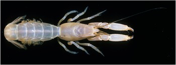
Acutigebia simsoni (Thomson, Reference Thomson1893), habitus. Colour photo of live specimen, NMV J24119. Photo Michael Marmach, © Museums Victoria/CC BY.

Gebia simsoni Thomson, Reference Thomson1893: 49, pl. 1 figs 3–5.
Upogebia simsoni. – Fulton and Grant, Reference Fulton and Grant1902: 61, pl. 5 figs 5 and 6. – McNeill, Reference McNeill1926: 305. – Hale, Reference Hale1927a: 85. – Hale, Reference Hale1927b: 309. – McNeill and Ward, Reference McNeill and Ward1930: 362.
Upogebia (Upogebia) simsoni. – Borradaile, Reference Borradaile1903: 543. – De Man, Reference Man1927: 24. – De Man, Reference Man1928: 28 (list), 38, 40, 52. – Poore and Griffin, Reference Poore and Griffin1979: 301, fig. 52.
Upogebia (Acutigebia) simsoni. – Sakai, Reference Sakai1982:71, fig. 14c–e.
Acutigebia simsoni. – Davie, Reference Davie2002: 480. – Sakai, Reference Sakai2006: 19. – Poore, 2004: 190, figs 53a, b, c, 54d, 55a, pl. 12f. – Gowlett-Holmes, Reference Gowlett-Holmes2008: 217 (colour figs).
Not Upogebia simsoni. – Dworschak, 1992: 225–226 [probably A. danai (Miers, Reference Miers1876)].
Material examined. Australia, Tasmania: King Island, Naracoopa, 39.92°S, 144.13°E, 08.03.1960 (MRG stn), NMV J1482 (female, 15.2 mm). Gordon Jetty, 43.2333°S, 147.25°E, 28.04.1985 (stn TAS-46), NMV J16595 (female, 8.2 mm; juvenile, 2.8 mm). J.M. Moverley Tasmanian Coastal Survey, 1995–1996 stations: Norfolk Bay, Lime Bay, 42°57.2′S, 147°42.5′E, 3–4 m, Zostera bed, 13.04.1995 (stn JMTAS-224), NMV J77222 (female, 6.7 mm); 23.02.1996 (stn JMTAS-239), NMV J77223 (juvenile, 3.8 mm). Spring Bay, 42°34′S, 147°53.5′E, 2–3 m, Zostera bed, 15.08.1995 (stn JMTAS-275), NMV J77224 (female, 5.4 mm); 15.08.1995 (stn JMTAS-276), NMV J77225 (2 juveniles, 6.1, 7.9 mm).
Victoria: Cape Paterson, Eagles Nest, 38.6708°S, 145.671°E, 28.03.2012 (stn BMS-001), NMV J56040 (female, 4.4 mm). Shoreham, 38.43°S, 145.05°E, 10.11.1902 (Fulton colln), NMV J16585 (2 females, 11.0, 12.3 mm; ovigerous female, 11.9 mm). Shoreham, Honeysuckle Point, 38.43°S, 145.05°E, 7.04.1983, NMV J1481 (female, 11.8 mm). Flinders, Ocean Beach, 38.48°S, 145.02°E, 1969 (MSG stn 76/01/69), NMV J16596 (ovigerous female, 15.3 mm). Flinders, Black Head, 38.48°S, 145.03°E, 08.08.1901 (Fulton colln), NMV J16586 (ovigerous female, 12.5 mm). Phillip Island, Cat Bay, 38.50°S, 145.138°E, 10.01.1970 (MRG stn 02/70), NMV J16721 (ovigerous female, 13.7 mm). Portland Bay, reef below lighthouse, 38.37°S, 141.603°E (stn CRUST 141), 26.02.1992, NMV J40256 (male, 8.3 mm [with pair of epicaridean isopods attached to pleopods, Bopyridae: Phyllodurus sp. now registered as NMV J77221]), NMV J24118 (female, 7.0 mm). Portland Bay, reef 400 m off lighthouse, 38.37°S, 141.607°E (stn CRUST 142), 27.02.1992, NMV J24119 (female, 9.4 mm).
Australia: Locality unknown, M.E. Lowe colln (donated by Kane), NMV J16587 (female, 11.1 mm [with pair of epicaridean isopods attached to pleopods, Bopyridae: Phyllodurus sp. now registered as NMV J77220]).
Diagnosis
Rostrum about quarter times as long as distance between tip of rostrum and cervical groove. Carapace width about two-fifths times as long as distance between tip of rostrum and cervical groove. Gastric lateral carinae parallel anteriorly. Anterolateral lobe longer than wide at base, 0.45 as long as rostrum. Telson 1.5 times as wide as long; posterior margin widely concave. Eyestalk half as long as rostrum. Antennal peduncle article 1 lower margin without spine. Pereopod 1 merus lower margin with 1 or 2 basal teeth plus 2 proximal spines; propodus lower margin serrate at midpoint; dactylus lower margin smooth, narrowing unevenly halfway along, dactylus upper margin carinate with denticles, dactylus lateral face carinate, denticulate.
Colour
Lateral carapace and ridges yellow–orange; dorsally with silvery gloss.
Distribution
East coast of Tasmania (type locality), Victoria, New South Wales, Queensland, Northern Territory, South Australia (Thomson, Reference Thomson1893; Fulton and Grant, Reference Fulton and Grant1902; McNeill, Reference McNeill1926; Hale, Reference Hale1927b; McNeill and Ward, Reference McNeill and Ward1930; Poore and Griffin, Reference Poore and Griffin1979). The 1929 record from New Zealand (Dworschak, 1992) is likely to be a misidentification of A. danai. A. simsoni has not been recorded in New Zealand since.
Acutigebia succinacia sp. nov
[New Japanese name: Kohaku-shinobi-anajako]
Acutigebia succinacia sp. nov., habitus. (A, G, H) Paratype, KPM-NH 5738, female (cl 8.1 mm); (B, I, J) Paratype, KPM-NH 5736, female (cl 6.3 mm); (C, D) Holotype, KPM-NH 5735, male (cl 7.0 mm); (E, F) Paratype, CMNH-ZC 03047, male (cl 7.6 mm). (A, B) In aquarium; (C, E, G, I) Lateral view; (D, F, H, J) Dorsal view.

Figure 3 Long description
The image A showing a close-up photograph of a single Acutigebia succinacia sp. nov. on a flat surface, with a dark background. The body is oriented left to right, with long antennae extending to the left and multiple legs along the underside. The label A is in the upper left. The image B showing a close-up photograph of a single Acutigebia succinacia sp. nov. on a flat surface, with a dark background. The body is oriented left to right, with long antennae extending to the left and multiple legs along the underside. The label B is near the upper left of this sub-image. The image C showing a close-up photograph of a single Acutigebia succinacia sp. nov. in lateral view on a dark background. The body is oriented left to right, with long antennae extending to the left. The legs are visible along the underside. The label C is at the left. The image D showing a close-up photograph of a single Acutigebia succinacia sp. nov. in dorsal view on a dark background. The body is oriented left to right. Two long antennae extend to the left. Multiple legs extend outward from both sides. The label D is at the left. The image E showing a close-up photograph of a single Acutigebia succinacia sp. nov. in lateral view on a dark background. The body is oriented left to right, with long antennae extending to the left. The legs are visible along the underside. The label E is at the left. The image F showing a close-up photograph of a single Acutigebia succinacia sp. nov. in dorsal view on a dark background. The body is oriented left to right. Two long antennae extend to the left. Multiple legs extend outward from both sides. The label F is at the left. The image G showing a close-up photograph of a single Acutigebia succinacia sp. nov. in lateral view on a dark background. The body is oriented left to right, with long antennae extending to the left. The legs are visible along the underside. The label G is at the left. The image H showing a close-up photograph of a single Acutigebia succinacia sp. nov. in dorsal view on a dark background. The body is oriented left to right. Two long antennae extend to the left. Multiple legs extend outward from both sides. The label H is at the left. The image I showing a close-up photograph of a single Acutigebia succinacia sp. nov. in lateral view on a dark background. The body is oriented left to right, with long antennae extending to the left. The legs are visible along the underside. The label I is at the left. The image J showing a close-up photograph of a single Acutigebia succinacia sp. nov. in dorsal view on a dark background. The body is oriented left to right. Two long antennae extend to the left. Multiple legs extend outward from both sides. The label J is at the left. A scale bar at the bottom right reads 5 mm.
Acutigebia succinacia sp. nov. (A–C) Holotype, KPM-NH 5735, male (cl 7.0 mm). (A) Anterior carapace, antennule, antenna (lateral); (B) Anterior carapace (dorsal); (C) Telson, left uropod.

Figure 4 Long description
The image A shows a lateral view of a Acutigebia succinaciasp. nov.'s anterior carapace, antennule and antenna. The image B shows a dorsal view of the anterior carapace with detailed surface texture. The image C shows the telson and left uropod, highlighting the structure and segmentation. Each diagram includes a scale bar indicating 1 millimeter for reference.
Acutigebia succinacia sp. nov. (A–G) Holotype, KPM-NH 5735, male (cl 7.0 mm). (A) Right pereopod 1 (lateral); (B) Right maxilliped 3 (inner, lateral); (C) Right maxilliped 3 (outer lateral); (D) Right pereopod 2 (lateral); (E) Right pereopod 3 (lateral); (F) right pereopod 4 (lateral); (G) Right pereopod 5 (lateral).

Figure 5 Long description
Illustrations of Acutigebia succinacia sp. nov. anatomical parts labeled A to G, each with a scale bar indicating 1 millimeter. A shows the right pereopod 1 in lateral view. B depicts the right maxilliped 3 from an inner lateral perspective. C illustrates the right maxilliped 3 from an outer lateral view. D presents the right pereopod 2 in lateral view. E shows the right pereopod 3 in lateral view. F displays the right pereopod 4 in lateral view. G illustrates the right pereopod 5 in lateral view. Each part is detailed with distinct features and proportions, providing a comprehensive view of the crustacean's anatomy.
Collection site of Acutigebia succinacia sp. nov. (A) Intertidal rocky shore, Tsumeki-zaki, Shimoda city, Shizuoka Prefecture, Japan; (B) Same, individual of Acutigebia succinacia sp. nov. [specimen not collected].

Figure 6 Long description
The image A showing a wide view of a rocky shore covered with many rounded stones of different sizes. A small object lies among the stones. The image B showing a close-up view into a wet rock crevice. A single Acutigebia succinacia sp. nov. lies along the crevice, with its body aligned lengthwise in the narrow gap between rocks.
[urn:lsid:zoobank.org:act:666BFE8C-97B2-4608-A5E8-1E4198720CD3]
Type material
Holotype: Japan, Shizuoka Prefecture, Shimoda city, Tsumeki-zaki, 34°39.32′N 138°59.04′E, coll. S. Kyotani, by hand, 13 January 2025, KPM-NH 5735 (male, cl 7.0 mm).
Paratypes: Shizuoka Prefecture: Collected with holotype, KPM-NH 5736 (female, cl 6.3 mm); CMNH-ZC 03047 (male, cl 7.6 mm); TKPM-AR 3624 (female, cl 7.6 mm), coll. S. Kyotani. Collected with holotype, TKPM-AR 3623 (female, cl 7.2 mm), coll. R. Yamashita. Type locality, KPM-NH 5737 (female, cl 7.9 mm), coll. S. Kyotani, 17 November 2024; KPM-NH 5738 (female, cl 8.1 mm), coll. T. Nagasaka; KPM-NH 5771 (male, cl 7.5 mm, DNA voucher), coll. S. Kyotani, 7 December 2025.
Shikinejima Island, Tokyo: Ishijirokawa beach, 34°19.24′N 139°13.22′E, hand net, coll. S. Kyotani, 21 November 2025, KPM-NH 5739 (male, cl 6.4 mm, DNA voucher).
Wakayama Prefecture: Arita, Kushimoto town, 33°28.56′N 135°44.48′E, coll. T. Nagasaka, hand net, 12 October 2025, KPM-NH 5740 (female, cl 7.7 mm, DNA voucher).
Kagoshima Prefecture: Shibushi city, Shibushi town, detailed locality is not disclosed, coll. A. Mizuno, by hand, 29 April 2025, KPM-NH 5741 (female, cl 8.5 mm, DNA voucher).
Other material: Shizuoka Prefecture: Type locality, KPM-NH 5742–5743 (2 juveniles, cl 3.0-4.4 mm), coll. T. Nagasaka; KPM-NH 5744–5745 (1 male, 1 juvenile, cl 4.5, 5.4 mm), coll. R. Yamashita; CMNH-ZC 03046 (female, cl 8.0 mm), coll. S. Kyotani, 17 November 2024; CMNH-ZC 03048 (male, cl 6.7 mm), coll. R. Yamashita, 7 January 2026.
Wakayama Prefecture: Collected with KPM-NH 5740, TKPM-AR 3625 (2 males, 1 female, cl 5.8–6.8 mm), coll. T. Nagasaka.
Diagnosis
Rostrum about quarter times as long as distance between tip of rostrum and cervical groove. Carapace width about two-fifths times as long as distance between tip of rostrum and cervical groove. Gastric lateral carinae parallel anteriorly. Anterolateral lobe broadly triangular, about as wide as long, 0.4 as long as rostrum. Telson 1.4 times as wide as long; posterior margin convex. Eyestalk half as long as rostrum. Antenna article 1 lower margin with spine. Pereopod 1 merus lower margin with 2–4 proximal spines; propodus lower margin with 5–11 spines at midpoint; dactylus lower margin smooth, dactylus upper margin carinate with denticles, dactylus lateral face smoothly carinate.
Description (male holotype)
Carapace width 0.45 times as long as distance between tip of rostrum and cervical groove. Rostrum triangular, 1.4 times as long as wide at base, tapering evenly, 0.3 times as long as long as distance between tip of rostrum and cervical groove, with apical tooth and about 15 small tubercles on submedian gastric ridge on either side; dorsal surface of rostrum and anterior midgastric region with few tubercles. Anterolateral lobes 0.4 times as long as rostrum, well separated from rostrum, inner margins diverging, lateral margin and lateral carinae parallel, with about 12 evenly spaced teeth. Eyestalk about half the length of rostrum, reaching just beyond end of lateral lobe, unarmed.
Antennular peduncle unarmed.
Antennal peduncle article 1 with one lower spine; article 2 cylindrical with free sclerite distally, upper part bearing dichotomously branched scaphocerite; article 3 with one lower spine; article 4 and 5 unarmed; dense setae on upper margin of article 4 and 5.
Maxilliped 3 ischium crista dentata comprising about 20 denticles; merus shorter than ischium, twice as long as wide, with 7 teeth on lower margin; carpus unarmed; about 0.8 times as long as merus; propodus 1.7 times as long as wide, about 1.2 times as long as carpus, unarmed, lower margin rounded; dactylus unarmed, about 0.8 times as long as propodus; exopod of 3 articles, extending halfway along merus.
Pereopod 1 basis lower margin with tooth. Ischium lower margin with 1 tooth. Merus about 2.3 times as long as wide, lower margin with 4 proximal teeth, upper margin, convex, with 1 subdistal tooth. Carpus half as long as merus, triangular, lower margin with 1 distal tooth, upper margin with 2 distal teeth. Propodus cylindrical, barrel-shaped, about 1.7 times as long as wide, lower margin with 5 teeth at midpoint, dorsal margin, carinate, with 1 distal tooth; fixed finger tapering distally, as long as wide at base, slightly curved, with notch below base of dactylus, with row of 5 small teeth on proximal inner edge. Dactylus tapering distally, about 0.75 times as long as propodus, with rows of setae extending to the tip along each face, upper and lower margins; upper margin denticulate, carinate; lateral face with smooth carina. The texture of the tip resembles a claw, with the most distal portion appearing black.
Pereopod 2 ischium unarmed. Merus about 3.5 times as long as wide, lower margin with 3 teeth and row of long setae. Carpus about 0.4 times as long as merus, lower margin with 1 tooth. Propodus about twice as long as wide, about 1.5 times as long as carpus, unarmed, upper margin rounded; Dactylus as long as propodus.
Pereopod 3 unarmed. Merus about 3.3 times as long as wide. Carpus upper margin lobe-like, about half as long as merus. Propodus almost as long as carpus, about 2.2 times as long as wide. Dactylus about 1.1 times as long as propodus, widest at midpoint.
Pereopod 4 unarmed; similar to but narrower than pereopod 3; ratio of lengths of merus, carpus, propodus and dactylus 2.3:1:1.2:1.5.
Pereopod 5 unarmed. Dactylus curved, with rounded apex; ratio of lengths of merus, carpus, propodus and dactylus 1.9:1.3: 1.7:1.
Pleonal tergites smooth. Telson 1.4 times as wide as long; posterior margin convex; 0.7 times as long as pleonite 6. Uropodal exopod paddle-shaped, 1.1 times as long as wide; endopod subtriangular, anterior margin straight, apex rounded, posterior margin with strong lobe, almost twice as long as wide.
Variability
This species exhibits individual variation in the number of teeth or spines on several body parts. The lower margin of the pereopod 1 merus bears 2–4 proximal spines; the lower margin of the pereopod 1 propodus bears 5–11 spines at the midpoint; the lower margin of the pereopod 2 merus bears 1–3 teeth. Additionally, the fixed finger of pereopod 1 ordinarily bears a row of 3–5 teeth, whereas in specimen KPM-NH 5738 the fixed finger lacks teeth entirely; the upper margin of pereopod 2 propodus unarmed, whereas in specimen KPM-NH 5741 has 1 tooth on both pereopods 2.
Size
Largest male cl 7.6 mm; largest female cl 8.5 mm
Colour pattern in life
Bright ambient light (Figure 3C–F): Entire body light yellow to light reddish yellow, tip of pereopod 1 dactylus blackish.
Dark ambient light (Figure 3G, H): Cornea greyish white, pereopods 2–5 yellowish white, remaining body regions strong reddish orange, tip of pereopod 1 dactylus blackish.
After moulting (Figure 3I, J): Entire body whitish translucent, the interior of the carapace light yellow, tip of pereopod 1 dactylus greyish.
Distribution
Presently known only from Pacific coast of Japan, Honshu and Kyushu: Shimoda city, Shizuoka Prefecture, Honshu (type locality); Shikinejima Island, Izu Islands, Tokyo; Kushimoto town, Wakayama Prefecture, Honshu; Shibushi city, Kagoshima Prefecture, Kyushu.
Ecology
In the type locality, A. succinacia sp. nov. was collected from burrows beneath boulders buried in intertidal muddy sand substrate (Figure 6A, B). At other sites, A. succinacia sp. nov. was collected from beneath buried boulders in the subtidal zone.
Etymology
This specific epithet refers to the body colouration, which ranges from reddish orange to light yellow and resembles the colour of amber (succinacia amber-coloured in Latin).
Remarks
The new species described in this study is assigned to Acutigebia based on the following features: triangular rostrum with an apical tooth (Figure 4A), and carapace bearing lateral lobes that are well separated from the rostrum (Figure 4B; Poore and Ahyong, Reference Poore and Ahyong2023). Among the six valid species of Acutigebia, A. succinacia sp. nov. most closely resembles A. simsoni in overall morphology, sharing the following features: (1) anterolateral lobes of carapace arranged parallel to each other (Figure 4B); (2) a row of teeth on the lower margin of the pereopod 1 propodus (Figure 5A); (3) several teeth on the pereopod 1 merus (Figure 5A). In contrast, comparison of the two species reveals a stable morphological difference in the antennal peduncle: in A. succinacia sp. nov., the lower margin of article 1 with a spine (Figure 4A), whereas in A. simsoni the lower margin of antennal article 1 is unarmed (Figure 1A, F). Additionally, the shape of the lateral lobes differs between the two species: in A. succinacia sp. nov., the lateral lobe is broadly triangular and about as wide as long (Figure 4B), whereas in A. simsoni the lateral lobe is longer than wide at the base (Figure 1B, G). Moreover, the coloration of A. succinacia sp. nov. and A. simsoni is similar. Fulton and Grant (Reference Fulton and Grant1902) described the coloration of A. simsoni as ‘Cream yellow shading to pale pink’. The strong reddish orange coloration observed in this study in dim light was not noted in Fulton and Grant (Reference Fulton and Grant1902), though the tendency for the colour to shift from yellowish to reddish is similar. Whereas the colour image of A. simsoni (Figure 2) and in Poore and Ahyong (Reference Poore and Ahyong2023) appear considerably whiter, indicating that the colour pattern of A. simsoni requires further investigation.
Liu and Liu (Reference Liu and Liu2013) noted that A. serrifera is morphologically similar to A. simsoni. They compared the merus of pereopod 1 noting that A. serrifera bears a row of small spines or teeth (actually denticles) on the lower margin, and carpus of pereopod 2 unarmed (Liu and Liu, Reference Liu and Liu2013: Figure 3A, C). A. succinacia sp. nov. differs from A. serrifera by the presence of 2–4 teeth on proximal lower margin of merus of pereopod 1 and the presence of 1 tooth on distal lower margin of carpus of pereopod 2 (Figure 5A, D).
In Japan, Acutigebia was previously represented only by A. trypeta (Sakai, Reference Sakai1970, Reference Sakai2006). A. succinacia sp. nov. is clearly distinguished from A. trypeta by the following features: (1) the lower margin of the pereopod 1 propodus is unarmed in A. trypeta (Sakai, Reference Sakai1970: fig. 2Bf, g), whereas A. succinacia sp. nov., bears a row of spines at the midpoint of the lower margin of the pereopod 1 propodus (Figure 5A); (2) in A. trypeta, the central portion of the lateral margin of the telson protrudes and becomes markedly narrower posteriorly (Sakai, Reference Sakai1970: fig. 2Ac), whereas in A. succinacia sp. nov., the central portion of the lateral margin of the telson is smooth and tapers gradually towards the posterior section (Figure 4C). Furthermore, the orientation of the lateral lobes may also be significant in distinguishing the two species: in A. trypeta, the lateral lobes converge anteriorly (Sakai, Reference Sakai1970: fig. 2Ab), whereas in A. succinacia sp. nov., they are arranged parallel to each other (Figure 4B).
Previous literature has reported several habitats for species of Acutigebia. Fulton and Grant (Reference Fulton and Grant1902) described A. simsoni as burrowing beneath stones on muddy substrates below the low water line. Sakai (Reference Sakai1970) noted that A. trypeta lives in narrow tunnels inside large coral blocks situated in the mid-intertidal zone. Liu and Liu (Reference Liu and Liu2013) recorded A. laticauda and A. serrifera living within narrow tunnels of coral reef. In this study, A. succinacia sp. nov. was collected from beneath buried boulders in the intertidal to subtidal zones. Especially at the type locality, specimens of A. succinacia sp. nov. were observed from the intertidal zone, where they burrowed within muddy sand substrates (Figure 6B). This habitat bears a striking resemblance to the record of A. simsoni by Fulton and Grant (Reference Fulton and Grant1902) among previous studies.
Genetic differences in the 491 bp 16S rRNA gene sequences between four individuals of A. succinacia sp. nov. and one of A. simsoni obtained in this study are shown in Table 2. The p-distance within the four paratype specimens of A. succinacia sp. nov. ranged from 0.00% to 0.62%, whereas the p-distance between these and A. simsoni ranged from 6.34% to 6.60%. The range of interspecific K2P genetic divergence varies considerably between species, and examples are known where the divergence is under 4% in some interspecific pairs (e.g., Cabezas et al., Reference Cabezas, Macpherson and Machordom2009; Wang et al., Reference Wang, Gan and Li2017; Komai, Reference Komai2022; Jiang et al., Reference Jiang, Zhou, Ma, Wang, Xie and Chen2025). In light of these results, the interspecific divergence obtained in this study provides genetic evidence supporting the distinct species status of A. succinacia sp. nov. and A. simsoni. The nucleotide sequences (12S rRNA, COI, and H3 obtained in this study) of two specimens have been deposited in GenBank (A. succinacia sp. nov., KPM-NH 5771: 12S rRNA, LC922885; COI, LC923594; H3, LC923593, and A. simsoni, NMV J56040: 12S rRNA, LC922886).
K2P genetic divergence of partial 16S rRNA gene sequence (491 bp) between A. succinacia sp. nov. and A. simsoni
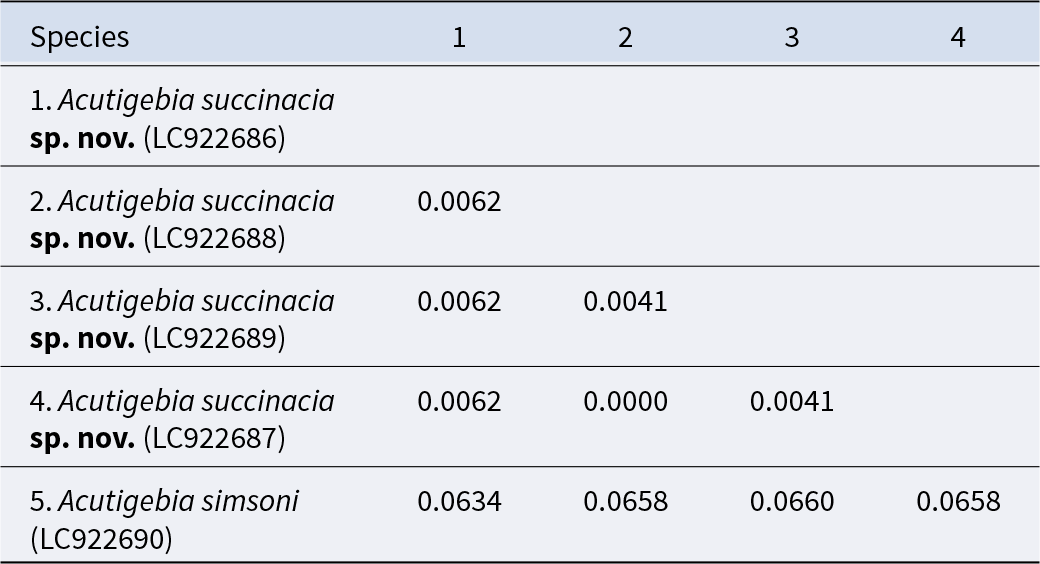
Table 2 Long description
The table reports pairwise genetic distances among five specimens, with values shown for each comparison. Four entries are Acutigebia succinacia sp. nov. (LC922686, LC922688, LC922689, LC922687) and one is Acutigebia simsoni (LC922690). Within A. succinacia, distances are very small, ranging from zero to 0.0062. The smallest within-species distance is zero between A. succinacia LC922688 and LC922687, indicating no difference for the measured marker. Other A. succinacia comparisons are 0.0041 between LC922689 and LC922688, 0.0041 between LC922687 and LC922689, and 0.0062 between LC922686 and each of LC922688, LC922689, and LC922687. In contrast, A. simsoni shows much larger distances from all A. succinacia samples, ranging from 0.0634 to 0.0660, with the largest being 0.0660 versus A. succinacia LC922689. Blank cells reflect that only one triangle of the distance matrix is populated, so each pair is listed once.
Key to species of Acutigebia
1. Pereopod 1 propodus lower margin unarmed.…………………2
– Pereopod 1 propodus lower margin serrate or with teeth………4
2. Pereopod 1 dactylus with subdistal tooth on lower margin, with serrate upper margin ………………… A. trypeta (Sakai, Reference Sakai1970)
– Pereopod 1 dactylus without subdistal tooth on lower margin, with smooth upper margin ……………………………………3
3. Rostrum about one quarter length of rostrum to cervical groove. Telson square, about as wide as long ………………………………… A. kyphosoma (Sakai, Reference Sakai1993)
– Rostrum about one sixth length of rostrum to cervical groove. Telson rectangular, wider than long …………………………… A. laticauda Liu and Liu, Reference Liu and Liu2013
4. Gastric lateral carinae diverging anteriorly. Pereopod 1 propodus lower margin with sharp teeth along most of length; dactylus lateral face carinate, denticulate ……… A. danai (Miers, Reference Miers1876)
– Gastric lateral carinae converging anteriorly or parallel. Pereopod 1 propodus lower margin denticulate along some of length; dactylus lateral face smoothly carinate or denticulate proximally ……………………………………………………..5
5. Gastric lateral carinae weakly converging anteriorly …………………………… A. serrifera Liu and Liu, Reference Liu and Liu2013
– Gastric lateral carinae weakly parallel anteriorly …………………………………………………………………6
6. Antenna article 1 lower margin with spine. Lateral lobe broadly triangular, about as wide as long ……… A. succinacia sp. nov.
– Antenna article 1 lower margin without spine. Lateral lobe longer than wide at base ………………… A. simsoni (Thomson, Reference Thomson1893)
Supplementary material
The supplementary material for this article can be found at https://doi.org/10.1017/S0025315426101350.
Acknowledgements
We are grateful to Aritomo Mizuno (Okinawa Prefecture), Tadanosuke Nagasaka (Saitama city, Saitama Prefecture), and Ryunosuke Yamashita (Tokyo University of Marine Science and Technology) for providing specimens; Koki Horikawa, Daiju Ogushi, and Naoto Takiyama (Tokyo University of Marine Science and Technology) for sampling; Dr Junji Okuno (Natural History Museum and Institute, Chiba), Dr Takehiro Sato (Kanagawa Prefectural Museum of Natural History), and Dr Yuya Suzuki (Tokushima Prefectural Museum) for registering type specimens, Dr Yumi Henmi (Kyoto University), Dr Tomoyuki Komai (Natural History Museum and Institute, Chiba), Dr Hiroki Nakajima (Yokosuka city Museum), and Dr Koetsu Kon (Tokyo University of Marine Science and Technology) for helpful advice. We thank Peter Dworschak (Natural History Museum, Vienna) and Kareen Schnabel (Earth Sciences New Zealand, Wellington) for discussion about the identity of the specimen in Vienna.
Author contributions
S.K. designed the study, carried out the genetic analysis and described the new species. G.C.B.P examined the Australian species. S.K. and G.C.B.P. wrote the paper together.
Funding
Nothing.
Competing interests
The authors declare none.
Data availability
The corresponding author can provide the data on request.




 Open access

