

Virchow-Robin space (VRS) is a subpial space surrounding the perforating arteries and arterioles of the brain, and dilated VRS (dVRS) appears as small and sharply delineated structures of cerebrospinal fluid signal intensity that follow the orientation of the perforating vessels and run perpendicular to the brain surface.Reference Heier, Bauer, Schwartz, Zimmerman, Morgello and Deck 1 - Reference Udaka, Sawada and Kameyama 3 The presence of dVRS has been reported in both normal persons and those with various disorders such as hypertension, depression, and dementia but the clinical implications of dVRS remain controversial.Reference Maclullich, Wardlaw, Ferguson, Starr, Seckl and Deary 2
Because the striatum (STR) is a critical region in the pathogenesis of parkinsonism, many previous studies have explored the role of dVRS in STR on parkinsonian patients and have yielded conflicting results.Reference Mancardi, Romagnoli, Tassinari, Gandolfo, Primavera and Loeb 4 - Reference Zijlmans, Daniel, Hughes, Revesz and Lees 10 Some authors suggest that dVRS is either a single pathologic feature or a clinical modifier in parkinsonian patients.Reference Fenelon, Gray, Wallays, Poirier and Guillard 7 , Reference Laitinen, Chudy, Tengvar, Hariz and Bergenheim 8 , Reference Mestre, Armstrong and Walsh 11 The dopamine transporter (DaT) is abundantly expressed in the striatal terminals of dopaminergic neurons and is well correlated with striatal dopamine concentrations.Reference Hersch, Yi, Heilman, Edwards and Levey 12 Dopamine transporter positron emission tomography (DaT-PET) is a useful diagnostic tool to investigate the dopaminergic innervation of STR in patients with parkinsonism.Reference Rinne, Ruottinen, Bergman, Haaparanta, Sonninen and Solin 13 , Reference Frost, Rosier and Reich 14
Although DaT-PET alterations can result from structural lesions, such as an infarct to the STR, the relationship between dVRS and DaT-PET remains undetermined.Reference Vaamonde, Flores, Gallardo and Ibanez 15 There are two types of dVRS. Type 1 dVRS presents with normal surrounding tissue, and type 2 dVRS presents with surrounding rarefication and abnormal gliosis.Reference Mancardi, Romagnoli, Tassinari, Gandolfo, Primavera and Loeb 4 The role of dVRS on DaT-PET likely differs based on the type of dVRS.
Based on the hypothesis that dVRS plays a modifying role in parkinsonism, we aimed to investigate the influence of dVRS in STR on the clinical features and neuroimaging of parkinsonian patients.
Methods
Participants
Patients were recruited from those who visited our clinic for evaluation of parkinsonism. The evaluation was done by standard protocol of our Parkinson registry, including neurological evaluation of parkinsonism, cognitive function testing and brain magnetic resonance imaging (MRI). Dopamine transporter-PET was done in those with an uncertain diagnosis. Because only a few patients exhibited dVRS in STR on their MRI, we included all patients from our registry that underwent both 1.5T MRI and DaT-PET and exhibited dVRS on their MRI, regardless of clinical diagnosis.
Parkinsonism was defined as the presence of at least two of four cardinal signs (rest tremor, rigidity, bradykinesia, and loss of postural reflexes). Clinical diagnosis of Parkinson Disease (PD) was made using the UK Parkinson’s Disease Society Brain Bank Clinical Diagnostic Criteria (UKPDS).Reference Gibb and Lees 16 Diagnosis of Multiple System Atrophy (MSA), Progressive Supranuclear Palsy (PSP), and Frontotemporal Dementia (FTD) were made using the consensus clinical criteria for each diagnosis.Reference Gilman, Wenning and Low 17 - Reference Neary, Snowden and Gustafson 19 A diagnosis of Vascular Parkinsonism (VaP) was made in patients with symmetric parkinsonism with predominance in the lower body and in the absence of typical features suggestive of other parkinsonian disorders. Reference Zijlmans, Daniel, Hughes, Revesz and Lees 10 Those with a history of stroke within one year from the time of diagnosis and those with new vascular lesions on brain MRI were excluded.
Demographic data and vascular risk factors, including hypertension, diabetes, hyperlipidemia, smoking, heart disease and history of stroke, were assessed using medical records. Motor and cognitive statuses were evaluated by Hoehn and Yahr stage (HY stage), Korean Mini-Mental Status Examination (K-MMSE), Korean version of Montreal Cognitive Assessment (MoCA-K), and Frontal Assessment Battery (FAB).
This study was approved by the Institutional Review Board of Kyung Hee University Hospital. All participants gave informed consent before the study.
Neuroimaging
Virchow-Robin space in STR was defined as the presence of small (less than three mm) isointense signals with cerebrospinal fluid on both T1 and T2 weighted images.Reference Heier, Bauer, Schwartz, Zimmerman, Morgello and Deck 1 - Reference Udaka, Sawada and Kameyama 3 Dilated Virchow-Robin space was evaluated on axial MRI slices in which both the caudate nucleus (CN) and the putamen could be best visualized. Each STR was divided into three segments: CN, anterior putamen (AP), and posterior putamen (PP). Anterior putamen and PP were separated by an imaginary line from the genu of internal capsule, which was perpendicular to the anterior–posterior commissure line. The severity of dVRS was assessed in each segment using a visual semi-quantitative scale (none=0, mild=1, moderate=2, and severe=3) and a quantitative scale by counting the actual number of dVRS [0 (none), 1-9=1 (mild), 10-19=2 (moderate), and ≥20=3 (severe)]Reference Maclullich, Wardlaw, Ferguson, Starr, Seckl and Deary 2 , Reference Doubal, MacLullich, Ferguson, Dennis and Wardlaw 20 , Reference Klarenbeek, van Oostenbrugge, Lodder, Rouhl, Knottnerus and Staals 21 The sum of the six segment scores generated the dVRS score.
Dopamine transporter-PET was performed using 18F-fluorinated N-3-fluoropropyl-2beta-carboxymethoxy-3-beta-(4-iodophenyl) notropane (18F-FPCIT), as reported previously.Reference Hwang, Hong, Ahn, Yi, Lee and Kim 22 The DaT-PET findings in the six segments (CN, AP, and PP, bilaterally) were blindly evaluated by an expert in nuclear medicine (IKH). The findings on DaT-PET were qualitatively designated as normal (=0) or abnormal (=1) in each segment, and scores were then summed to generate a DaT-PET score.
Anterior-posterior gradient (APg) was assessed by comparing the severity of dVRS and DaT-PET abnormality (ab-DaT-PET) in the anterior and posterior segments. Anterior-posterior gradient was determined to be present if dVRS in the CN was less severe than in the putamen and if dVRS in the AP was not more severe than in the PP. With regards to DaT-PET, APg was present if the abnormality was absent in the CN and found in both the AP and PP or in only the PP.
The correlation between dVRS and ab-DaT-PET was designated in each segment as concordant if dVRS was present with ab-DaT-PET or as discordant if dVRS was either present with normal DaT-PET or absent with ab-DaT-PET. A concordant segment was assumed to represent pathologic dVRS in STR. Concordance rate (CR) was calculated [CR=number of concordant segments/number of concordant and discordant segments] to assess its correlation with clinical and neuroimaging scales. Patients were divided into two groups according to the mean CR value (one with CR≥mean, and the other with CR<mean).
Leukoaraiosis (LA) was defined as the presence of hyperintense lesions in the periventricular or subcortical regions on fluid-attenuated inversion recovery MRI sequences. We evaluated LA severity based on a visual rating scale (LA score).Reference Wahlund, Barkhof and Fazekas 23 LA score was designated as mild (1-4) or severe (>4). We defined silent infarctions (SIs) as focal hyperintensities of three mm or larger on T2-weighted images (T2WIs) in patients without relevant history or neurologic deficit.Reference Zhu, Dufouil, Tzourio and Chabriat 24 SIs showed hypointensity on T1-weighted images. Cerebral microbleeds (CMBs) were defined as small (less than five mm in diameter), rounded, homogeneous, and hypointense signals on the gradient echo or T2-weighted images. We differentiated CMBs from perforating vessels and iron deposition by reviewing serial images.Reference Greenberg, Vernooij and Cordonnier 25
Statistical Analysis
Groups were compared using the Student’s t test, Mann-Whitney U test or Chi-square (Fisher’s exact) test. Agreement between semi-quantitative and quantitative scales on dVRS was assessed using the intraclass correlation coefficient (ICC). Correlations between neuroimaging and clinical scales were analyzed by Spearman’s rho. The cutoff p value was set at 0.05. All statistical analyses were conducted using the SPSS 19.0 package for Windows (IBM Corp. Armonk, NY, USA).
Results
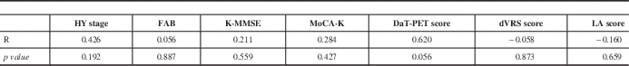
Eleven patients were included in this study. Clinical diagnoses and major neuroimaging findings are summarized in Table 1. The mean CR was 0.39. Demographics, clinical scales and neuroimaging findings are compared between those with CR ≥0.39 and CR <0.39 (Table 2).
Clinical diagnosis and analysis of magnetic resonance imaging and dopamine transporter positron emission tomography

Abbreviations: dVRS, dilated Virchow-Robin Space; CN, caudate nucleus; AP, anterior putamen; PP, posterior putamen; DaT-PET, dopamine transporter positron emission tomography; CR, concordance rate; A-P, anterior-posterior; MRI, magnetic resonance imaging; LA, leukoaraiosis; FTD, Frontotemporal Dementia; R, right; L, left; B, bilateral; NA, not applicable; PSP, Progressive Supranuclear Palsy; VaP, Vascular Parkinsonism; PD, Parkinson Disease; MSA, Multiple System Atrophy
* In Case 8, right caudate nucleus and putamen was not evaluated due to adjacent cerebral infarction.
Comparison between the patients with concordance rate ≥0.39 and <0.39

Data expressed by median (range)
Abbreviations: CR, concordance rate; HY stage, Hoehn and Yahr stage; K-MMSE, Korean mini mental status examination; MoCA-K, Montreal Cognitive Assessment Korea; FAB, frontal assessment battery; DaT-PET, dopamine transporter positron emission tomography; dVRS, dilated Virchow-Robin Space; LA, leukoaraiosis
Representative concordant and discordant cases are shown in Figure 1. We observed dVRS in 42 of 63 segments and ab-DaT-PET in 33 of 63 segments. Three segments from case eight were excluded from the analysis due to a lesion associated with adjacent ischemic stroke (Table 1).
Concordance between dopamine transporter scan (DaT-PET) abnormality and dilated Virchow-Robin space (dVRS). (A) A concordant case. DaT-PET shows abnormal findings and brain magnetic resonance imaging (MRI) shows moderate to severe degree of dVRS in the bilateral caudate nucleus, anterior and posterior putamen. (B) A discordant case. DaT-PET shows normal findings, whereas brain MRI shows variable dVRS in the caudate nucleus and putamen.

Anterior-posterior gradient of dVRS and ab-DaT-PET was present in five cases (nine sides; Table 1). Anterior-posterior gradient was common in those with PD or MSA. In one case with PD and another with MSA, APg of dVRS was concordant with that of ab-DaT-PET. In VaP, there was only one case (two sides) with APg of dVRS and another (one side) with APg of ab-DaT-PET.
The conformity between scoring scales for dVRS was excellent (ICC=0.964, p value <0.001). The quantitative dVRS scale was evaluated on only one axial slice because the same dVRS could be counted repeatedly in more than one slice. In addition, an accurate count of dVRS could be limited by imaginary segmentation of STR. Thus, we used a visual semi-quantitative scale for further statistical analysis.
There was no significant correlation between the presence of dVRS and ab-DaT-PET. No neuroimaging or clinical scales were significantly correlated with CR (Table 3). There were no significant differences in clinical features and neuroimaging findings between those patients with CR ≥0.39 and CR <0.39 (Table 2). Leukoaraiosis, mild hydrocephalus, CMBs, and cerebral infarctions were variably present in brain MRI scans of certain individuals (Table 2). The dVRS score was strongly correlated with MoCA-K and FAB and moderately correlated with K-MMSE; however, it was not correlated with age, LA score, or HY stage (Figure 2).
Correlations between dilated Virchow Robin space (dVRS) score and clinical and neuroimaging findings. There is strong correlation between the dVRS score and Frontal Assessment Battery (FAB) and between the dVRS score and Korean version of Montreal Cognitive Assessment (MoCA-K), and moderate correlation between the dVRS score and Korean Mini-Mental Status Examination (K-MMSE).

Correlation coefficient between concordance rate and clinical or neuroimaging scales
Abbreviations: R, correlation coefficient; HY stage, Hoehn and Yahr stage; K-MMSE, Korean mini mental status examination; MoCA-K, Montreal Cognitive Assessment Korea; FAB, frontal assessment battery; DaT-PET, dopamine transporter positron emission tomography; dVRS, dilated Virchow-Robin Space; LA, leukoaraiosis
Discussion
In our study, we observed the presence of dVRS in various parkinsonian disorders, including PD. Although PD is considered to be a presynaptic disorder that leaves the STR least affected, in a pathologic study, 18 of 76 (23.7%) patients with pathologically confirmed PD and 3 of 24 cases (12.5%) with other etiologies had striatal pathology, including dVRS; however, the clinical implications of dVRS are obscure.Reference Hughes, Daniel, Kilford and Lees 6
In a previous study, the globus pallidus ipsilateral to the limbs exhibiting more severe motor symptoms showed more dVRS, suggesting a pallidotomy-like effect of dVRS on the contralateral limbs.Reference Laitinen, Chudy, Tengvar, Hariz and Bergenheim 8 There was a pathology-proven case of axial-dominant parkinsonism with isolated dVRS in STR that showed no response to levodopa.Reference Fenelon, Gray, Wallays, Poirier and Guillard 7 These studies suggested that dVRS could affect parkinsonian symptoms. However, in a recent case series of PD with large dVRS, dVRS was not associated with ab-DaT-PET but was correlated with atypical clinical features.Reference Mestre, Armstrong and Walsh 11 In our study, the severity of parkinsonism, evaluated by HY stage, was not correlated with the dVRS score, excluding a significant role of dVRS in clinical parkinsonism.
Because type 2 dVRS is associated with greater pathologic change, type 2 was suggested to be associated with clinical parkinsonism, whereas type 1 was a feature of normal aging.Reference Mancardi, Romagnoli, Tassinari, Gandolfo, Primavera and Loeb 4 , Reference Murrow, Schweiger, Kepes and Koller 5 , Reference Fenelon, Gray, Wallays, Poirier and Guillard 7 Recently, dVRS was suggested to be a marker of inflammation.Reference Wuerfel, Haertle and Waiczies 26 Thus, type 2 dVRS may affect dopaminergic terminals in STR, resulting in decreased 18F-FPCIT uptake in DaT-PET, while type 1 dVRS may simply push dopaminergic terminals aside, leaving 18F-FPCIT uptake unaffected. To test this hypothesis, we studied the relationship between dVRS and ab-DaT-PET. First, the correlation between the presence of dVRS and ab-DaT-PET was studied, which was found to be insignificant. Second, we studied whether APg of dVRS coincided with APg of ab-DaT-PET. Concurrence between APg of dVRS and ab-DaT-PET was observed in only two (three sides) out of four patients (eight sides) with PD, suggesting a minimal pathologic contribution of dVRS to ab-DaT-PET. Moreover, CR, a representation of type 2 dVRS, was not correlated with clinical parkinsonism. Thus, all the results showed that dVRS did not play an important role in ab-DaT-PET and clinical parkinsonism, suggesting that the majority of observed dVRS may be type 1.
Dilated VRS severity was reported to be correlated with cognitive dysfunction.Reference Maclullich, Wardlaw, Ferguson, Starr, Seckl and Deary 2 Our study also showed a significant correlation between dVRS and cognitive function tests. Frontal function was shown to be best correlated with dVRS severity (Spearman’s rho between FAB and the dVRS score=−0.868, p<0.01). Our results reaffirm the role of STR in frontal function via the frontal-STR loop.Reference Alexander, DeLong and Strick 27
In this study, we demonstrated the universality of dVRS in various parkinsonian disorders, the discrepancy between dVRS and ab-DaT-PET, and the possible cognitive contribution of dVRS. Our study is limited by the small number of subjects, heterogeneous clinical diagnoses and the inability to discriminate between two types of dVRS. Further studies using more sophisticated MRI techniques in a larger number of patients are necessary for a more comprehensive understanding of the role of dVRS.
Disclosures
Dokyung Lee does not have anything to disclose. Il Ki Hong does not have anything to disclose. Tae-Beom Ahn does not have anything to disclose.


