

From their very first appearance in the fossil record, trilobites possessed elaborate compound eyes (Lindström Reference Lindström1901; Clarkson et al. Reference Clarkson, Levi-Setti and Horváth2006; Schoenemann et al. Reference Schoenemann, Pärnaste and Clarkson2017a; Schoenemann Reference Schoenemann2021). There are two principal types of trilobite eyes. The first is the so-called holochroal eye, which is in shape and internal structure very similar to the eyes of modern-day active insects and crustaceans. It is a so-called apposition compound eye (Schoenemann et al. Reference Schoenemann, Pärnaste and Clarkson2017a). Apposition compound eyes of trilobites consist of a sensory and a dioptric apparatus (lens and crystalline cone) (Schoenemann et al. Reference Schoenemann, Pärnaste and Clarkson2017a; Scholtz et al. Reference Scholtz, Staude and Dunlop2019; Schoenemann & Clarkson Reference Schoenemann and Clarkson2020, Reference Schoenemann and Clarkson2021). The crystalline cone is a cellular, clear structure giving way to focus light on a central light-guiding structure, the rhabdom, which is part of the sensory cells encompassing it. This entire system is termed an ommatidium. Pigment cells isolate the units optically from each other (Land & Nilsson Reference Land and Nilsson2012). By that, the inputs of the visual field of each ommatidium are summed up to one signal, and the image seen by the arthropod consists of ‘dots’, each one produced by an individual ommatidium. Similar to the pixels of a computer graphic, the vision is mosaic-like in total (Müller Reference Müller1826, Exner Reference Exner1891). Consequently, the resolution of vision here depends mainly on the number of facets and the aperture of each ommatidium.
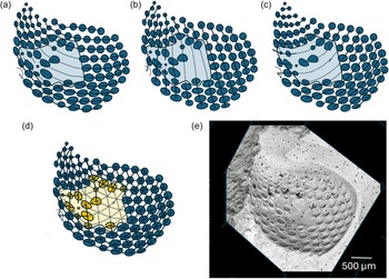
The second compound eye system of trilobites is that of the schizochroal eyes, which can be found only among trilobites of the phacopid suborder Phacopina. Schizochroal eyes seem to have developed during the Ordovician and reached their full expression during the Devonian, before the demise of the group during the great mass extinction at the end of that geological period (Clarkson et al. Reference Clarkson, Levi-Setti and Horváth2006). Equipped with their schizochroal eyes, phacopine trilobites possessed a hyper-compound eye (Schoenemann et al. Reference Schoenemann, Clarkson, Bartels, Südkamp, Rössner and Ryck2021), so far unique among arthropods. Here, the individual visual units were fewer in number than in holochroal eyes, each covered by a wide lens, generally much larger than the lenses of holochroal eyes, some up to more than 1 mm in diameter, as in Drotops megalomanicus Struve Reference Struve1990. Below the large lenses lie sophisticated systems consisting of one or more concentric rings of minuscule ommatidia, each cupped by a tiny lens. The small ommatidia are grouped around a central structure of still unknown function (Schoenemann et al. Reference Schoenemann, Clarkson, Bartels, Südkamp, Rössner and Ryck2021). While the functioning of this hyper-compound eye system is still unknown, it offers perspectives on the possible pooling of signals, functioning as a kind of amplifier, or a kind of sharing function among the ommatidial members of the system.
An eye of the Ordovician phacopine trilobite, Achatella sp. described herein shows evidence of a healed injury, probably caused by a macrophagous predatory attack and can provide information on the genetic program of compound-eye formation, at least in phacopine trilobites.
1. The visual surface of schizochroal eyes
The visual surface, and thus the visual field of the schizochroal eye is oriented anteriorly, anterior-laterally, and just slightly backward. The palpebral lobe covers the upper side of the eye. In contrast to, for example, many Silurian phacopine trilobites with relatively large but densely packed lenses (Fig. 1a–d), in Devonian phacopine trilobites, each large lens is separated at half a lens-diameter distance from its neighbour horizontally, while the vertical distance of the lenses is smaller (Schoenemann Reference Schoenemann2021, Reference Schoenemann2025). Horizontally, the visual surface is curved, offering a field of view often of ∼140°, while vertically, the visual surface is quite steep, just slightly tilted backwards, allowing the lower lenses protruding (Fig. 1a–d), providing some visual perception of the water-column above the trilobite. Due to the higher vertical resolution, movements in the vertical direction can be perceived better than in the horizontal direction (Fordyce & Cronin Reference Fordyce and Cronin1989, Reference Fordyce and Cronin1993), but the visual field horizontally is much wider than vertically. Intriguingly, in many phacopines the lenses protrude quite markedly from the visual surface, with the consequence that each lens has an almost hemispherical surface and a wide range (about 180°) to capture light (Fig. 1b), and so these interspaces may be covered optically (Schoenemann Reference Schoenemann2007). Clarkson & Levi-Setti (Reference Clarkson and Levi-Setti1975) showed that the large lenses of phacopine trilobites, in fact, are lens systems. The subunits of the schizochroal eye lenses have different refractive indices, resulting in a complex function of the optical system as a whole. This was suggested to be a system to correct spherical and chromatic aberration, typical of all thick lenses (Clarkson & Levi-Setti Reference Clarkson and Levi-Setti1975). It has been shown more recently that the schizochroal eye actually is a highly specialized system – it is a hyper-compound eye, where below each of the large lenses a small individual compound eye is located (Schoenemann et al. Reference Schoenemann, Clarkson, Bartels, Südkamp, Rössner and Ryck2021)
Aspect and field of view of the typical phacopine compound eye and the injured compound eye of Achatella sp. (a) Drawing of a typical phacopine compound eye (in Acaste downingiae (Salter)), lateral view (Clarkson Reference Clarkson1966, Fig. 1a). (b) Hemispherical lens protruding from the visual surface, gathering light from ∼180°. (c) Drawing of a typical phacopine compound eye (in Acaste downingiae (Salter)) as seen from above (Clarkson Reference Clarkson1966, Fig. 1b). Note that each lens can gather light from above. (d) Visual surface of c seen from the side and from above. (e) Eye with intact visual surface of Achatella sp. RGM 1495535 from the same location as f–m. (f) Injured and healed visual surface of the injured compound eye of Achatella sp. RGM 1495534, The Haerst, Zwolle, NL. (g) Injured specimen with interpretative arrows and lines. Blue line: dent in the visual surface, yellow arrows: restored lenses, smaller than in the normal pattern; black arrows: restored lenses larger than in the normal pattern; red arrow: tiny lens, minuscule but in the correct position. (h) Injured specimen with interpretative markings. Blue: lenses which are smaller; yellow: lenses which are larger than in the normal pattern; green: lenses with irregular shapes; blue line: dent in the visual surface. (i) Small tubercles not just between the lenses, but also above some of the marginal lenses. (j) Example of a part of the intact visual surface. (k) Red rectangle indicates the position of image in i, blue rectangle indicates the position of j. (l) Injured and restored part of the visual surface. (m) Affected region, marked in blue.

The geometry of the schizochroal eye was nicely described by Clarkson (Reference Clarkson1966) who subsequently (Reference Clarkson and Martinsson1975) discussed how it developed during the ontogeny of the trilobite. Starting from the upper side of the eye with a generative zone, during growth the lenses were ordered in a logarithmic spiral, and with every moulting, one row of lenses was added, while the generative zone moved downwards until the final size of the eye was reached. The size of the lenses increased with each moulting. In addition, the spirals grew anteriorly, adding small new lenses. Consequently, a characteristic lens pattern evolved, constant for each species and sometimes even within a family or superfamily (Clarkson Reference Clarkson and Martinsson1975).
While in earlier schizochroal eyes such as in the Silurian Acaste (Fig. 1a–d) the lenses are generally more closely spaced than those in Devonian phacopines, it is remarkable that in the eye of the Ordovician Achatella sp., the more advanced mode is already represented. As Figure 1e shows, Achatella sp. possessed remarkably protruding compound eyes, probably indicating a function of elaborated visual orientation and visually guided behaviour.
2. Analyses of the injured eye of Achatella sp.
The isolated eyes of the Ordovician trilobite Achatella sp. (Fig. 1e–m) were found in a glacial erratic boulder of silicified carbonate from the Upper Sandbian Stage (Haljala/Keila regional stages (CIII–DII)), originating from Northern Scandinavia and the Baltic area (see section 6, Materials and Methods) at The Haerst, near Zwolle, The Netherlands. Although the upper part is missing, the left eye of Achatella sp. in Figure 1e shows the characteristic pattern of a schizochroal compound eye. The visual surface of a second eye (Fig. 1f–m), however, is obviously disturbed. One may argue that the smooth areas within the visual surface are an unprepared matrix covering the visual surface, and the intact lenses lie below. However, there are clearly smaller and larger lenses of irregular size in this area (Fig. 1f–h, l, m), and there is a a conspicuous, linear dent (blue dashed line on Fig. 1g, h). This suggests a healed eye injury, which needs to be described and is interpreted as a healed, restored injury, probably caused by a macrophagous predatory attack. Furthermore, it can provide information on the genetic program of compound-eye formation, at least in phacopine trilobites.
For analysis, each lens in the interpretative graphics was represented by a blue spot of equivalent size, as it was not covered by the matrix, and the visual surface was visible (Fig. 2). The oval shape of lenses in the graphic results from the photograph’s parallax. Then, the centres of the lens representants were connected, indicating the principles of order within the visual surface.
Organisation of the injured and restored eye of Achatella sp. (a) Horizontal rows of lenses, maintained after healing, indicated by the dotted line. (b) Vertical rows of lenses, maintained after healing, indicated by the dotted line. (c) Oblique arrangement of the lenses maintained after healing indicated by the dotted line. (d) All restored lenses (yellow) are still in the correct place (yellow: affected region). (e) Anterior view of the investigated damaged eye of Achatella sp. (RGM 1495534) for comparison.

Figures 1m and 2e show clearly that in the anterior part of the eye, the typical regular lens arrangement pattern, as described above, is still intact, whereas in the central area, it is not, as indicated by the pale blue areas in Figures 2a–c. Several features characterize this destroyed part of the visual surface: 1. There is an indented line across the upper part of the eye (blue dashed line, Fig. 1g, h). 2. Some lenses are smaller than expected by the regular pattern (see anterior part of the eye), indicated by yellow arrows and blue transparent circles (Fig. 1h). 3. Some lenses are larger than expected (indicated by dark green arrows (Fig. 1g) and yellow transparent circles (Fig. 1h). 4. The general pattern of vertical and horizontal order of the lenses is retained (Fig. 3a–e), even if a substituted lens is very small (red arrow, Fig. 1g), 5. Some of the lenses adjacent to the indented line are not round but of irregular shape. (Fig. 1h in green).
There are lines of tubercles of unknown function within the cuticular interspaces between the lenses (Fig. 1i). Remarkably, the same tubercles occur in a few lenses adjacent to the smooth areas. If these tubercles are built on the lenses and they possibly were of similar character as those within the interspaces, the lenses may not have been transparent or fully functional.
3. Healed injuries in trilobites
Healed trilobite injuries are frequently reported, mainly caused by macrophagous predatory attacks on the trilobite body (for an overview, see, e.g., Owen Reference Owen1985; Rudkin Reference Rudkin1985; Babcock Reference Babcock1993; Bicknell & Pates Reference Bicknell and Pates2020; Bicknell et al. Reference Bicknell, Smith, Howells and Foster2022). They must be distinguished from other malformations, such as those caused by teratological reasons, damage during moulting or other activities and also from characteristic swellings, often indicating parasitic violations, genetic malformations or the misformation of individual lenses in the compound eyes (e.g., in Owen Reference Owen1985, Bicknell et al. Reference Bicknell, Smith and Hopkins2024, Reference Bicknell, Smith, Amati and Hopkins2025).
Healed injuries are mainly reported from the lateral areas of the trilobite thorax and pygidium, more rarely from the cephalon or the rachis, indicating that the latter were more often lethal than the former (Owen Reference Owen1985). This is understandable if we assume that the heart of trilobites, as typical for all tetraconates, was an elongated tube and lay dorsally along the rachis. The open circulatory system of these animals inevitably leads to bleeding to death in the event of an injury, especially if this is located near the heart, and if the immune system does not act quickly enough closing the wound. While evidence of injuries of the cephalon are rare, a good example of a complicated laceration of an eye and its healing was described for the trilobite Telephina intermedia (Thorslund Reference Thorslund1935) by Schoenemann et al. (Reference Schoenemann, Clarkson and Høyberget2017b). In this specimen, the indented area inside the visual surface (Schoenemann et al. Reference Schoenemann, Clarkson and Høyberget2017b, Fig. 1e, f) and the imperfect restoration of obviously lost lenses into the now smooth and structureless area of the eye surface indicate an incompletely healed destruction of the visual surface. Other reports of injured cephala were provided by Babcock (Reference Babcock1993), Fatka et al. (Reference Fatka, Budil and Grigar2015), Pates & Bicknell (Reference Pates and Bicknell2019) and Bicknell et al. (Reference Bicknell, Smith and Hopkins2024).
4. Interpretation of the injured eye
Healing processes, as regarded here and by other authors, reflect the existence of functioning immune and repair mechanisms, which probably evolved before or during the early Cambrian (e.g., Morris & Jenkins Reference Morris and Jenkins1985; Jago & Haines Reference Jago and Haines2002; Fatka et al. Reference Fatka, Budil and Grigar2015; Pates et al. Reference Pates, Bicknell, Daley and Zamora2017; Bicknell & Pates Reference Bicknell and Pates2020; Bicknell et al. Reference Bicknell, Smith, Howells and Foster2022). Zong (Reference Zong2021) also described the difficulties and dangers that may arise from exoskeleton irregularities due to the healing processes. Since, in the case of the eye of Achatella sp., the visual surface is closed, and there is a complete, albeit altered lens pattern, the animal to which this eye belonged, must already have finished at least one moult after the lesion. It is an interesting question, whether the lesion occurred during a moult, which made it ‘easier’ to be repaired during an ongoing process of forming an eye, or whether it was possible to repair the leak during ‘normal’ life of the trilobite, in the time between two moults. Unfortunately, we probably cannot solve this question.
The trilobite survived the injury, and the way the repair was carried out provides information about the genetic program that determines the formation of the pattern of a visual surface in a schizochroal compound eye. We interpret the linear furrow as a line of indenting and tearing of the visual surface. Below that, due to a greater injury, lies a smooth lensless area, framed in its upper part by some lenses of irregular size. Inside the smooth area, most original lenses are missing, just some new lenses were installed, and intriguingly, the vertical and horizontal order of the new lenses is retained, even after the relatively great destruction of the visual surface. The size of the restored lenses is often smaller or larger than originally. There are smaller lenses at the lower margin of the injured zone, indicating where the injury finishes.
The entire lesion has a dimension of about 750 × 750 µm. The irregular shape and the wide flat area filled with homogeneous replacement tissue suggest a hole closed by a successful healing process. Such a hole could have been caused by a sharp ‘tool’, such as the sharp beak of a small cephalopod, as has been suggested in different trilobites (Schoenemann et al. Reference Schoenemann, Clarkson and Høyberget2017b, Fatka et al. Reference Fatka, Budil and Mikuláš2022). Orthocerids are among the most important predators of the Ordovician period and were equipped with a pointed, parrot-like beak, and certainly represent serious predators of trilobites. Beaks of fully grown orthocerid, however, may usually be too large to injure a small trilobite eye, as here, some millimetres in size. Crustaceans of that time, bottom dwellers such as ostracodes or phyllocarids might have left larger bite marks, or had a lifestyle other than predatory (Brahm & Geiger Reference Brahm and Geiger1966; Williams et al. Reference Williams, Perrier, Bennett, Hearing, Stocker and Harvey2015).
An unsuccessful attack by a eurypterid might be considered. Eurypterids appeared at the beginning of the Ordovician (Wang et al. Reference Wang, Braddy, Botting and Zhang2023) but so far it seems that the Baltic Ordovician is devoid of eurypterids; they appear there in the Silurian in the fossil record (Tetlie Reference Tetlie2007). Perhaps, however, the injured trilobite eye is a first sign of their existence during the Baltic Ordovician. The scale of the injury would be compatible with the fact that among the earliest eurypterids were some smaller forms, such as Orcanopterus manitoulinensis Stott et al. 2005, Brachyopterus stubblefieldi Størmer 1951, Onychopterella augusti Braddy, Aldridge & Theron 1995, and Paraeurypterus anatoliensis Lamsdell, Hosgör & Selden Reference Lamsdell, Hoşgör and Selden2013 (P. Selden pers. communication 2025). An abrasion of the visual surface by feeding from conspecifics shortly after moulting in a soft-shelled stage is rather unlikely due to the linear dent in the visual surface. Thus, the origin of the lesion remains unresolved.
Finally, considering these findings, we can find a hierarchic genetic program for establishing an ordered visual surface, at least for the schizochroal eyes of trilobites.
The first superordinate instance is the vertical and diagonal arrangement of the lenses, as established by the mode of growing. It is retained/repeated during growth, while at each new moulting, a new row of lenses and anterior lenses are added. The pattern continues during each moult and is retained and enforced during the restoration of an extended injury. It must be connected with each genetic command to establish a lens. The second instance is the size of the lenses. The appropriate size, as expected by the regularity of the lens pattern within the visual surface, is not always retained. Some of the lenses, especially along the tearing line and the outer margin of the lesion, are too small – probably because of a non-intact generative equipment, and the restoration probably depends on the possibilities and conditions of the restoration site. Significantly, some of the lenses restored in the more central part of the now smooth, injured area are larger than would be expected by order of the original pattern, and may indicate that the system has some capacity for regulation regarding the light-gathering capacity (large lenses with a higher light gathering capacity ‘trying’ to compensate for the loss of the other lenses).
5. Discussion
It is possible to indirectly determine which area of the eye was injured and destroyed, as indicated by the blue zone in Figure 1m. The findings suggest implications about the genetic program of compound eye-formation, at least here in the schizochroal eye of Achatella sp. As long as the system is able to replace missing lenses with even the smallest lenses, the pattern of order as described above is stable and primary to the other steps of formation. It even overcomes the severe destruction of the visual surface. Apparently, the shape of the lenses is not as fixed as the general order. Even the smallest replaced lenses are round. So, this part of the program also seems to be stable in principle. At the margin of the injury, however, the lenses show an irregular shape. As in other arthropods, the lenses probably were built by a more or less liquid/gel-determined preliminary stage, a process that finally results in an optimal relation between volume and contents – a sphere, and thus forming a functional lens. Some of the replaced lenses along the indented line (Fig. 1g, h: light blue line) are of irregular shape; they were probably disturbed mechanically during growth by the external remains of the destroyed cuticle close to the newly growing lenses not allowing the development of perfectly round structures. So here, we probably have more structural reasons for an imperfect lens shape rather than a failure of the genetic program. Most remarkable is the difference in size of the restored lenses. Adjacent to those that are too small compared to the original pattern, probably because the conditions of growth here were insufficient after destruction, are others that are too large. One may suggest that the larger ones compensate for the loss of captured light in the smaller ones. This implies that the amount of light-capturing area is also part of the genetic program. It seems that the amount of light captured is aimed at, and the genetic program tries to regulate misfits. Logically, this part of the genetic program is subordinate to the others (position and shape of the lenses).
Finally, it is clear that the system is not always able to regenerate the original lenses if the lesion is too extensive (smooth area). It builds properly round lenses and not irregular, random systems, if not located directly at the margin of the injury. So, the program to form round lenses still exists in a hierarchical manner. The superordinate is the position within the visual surface; the shape of the lenses is not genetically determined, but controlled by the physical conditions during their establishment, and the size of the lenses again is genetically determined, because the system probably ‘tries’ to compensate for lost light-gathering surfaces by establishing larger lenses.
6. Materials and methods
The specimens are deposited at the Naturalis Diversity Center, Darwinweg 2, 2333 CR Leiden, NL. RGM 1495534 the injured eye, RGM 1495535 the intact eye. They were found at a sandpit in the middle of the Netherlands near the town of Zwolle, called The Haerst (see Fig. 3a; Van Uum Reference van Uum2003).
(a) Map of the Pleistocene (Weichselian) of The Netherlands (Van Uum Reference van Uum2003) showing The Haerst (red star) from where the erratic block yielding the injured eye of Achatella sp. was obtained. (b) Chart showing the stratigraphical position of the beds from which the erratic block yielding the injured eye of Achatella sp. was derived within the Upper Sandbian Stage (Haljala Substages Idavere CIII–Johvi DI) (adapted from Rhebergen Reference Rhebergen2009). (c) The Eridanos river system (Rhebergen Reference Rhebergen2009).

Many boulders from this pit, up to 20cm across, are angular (transported in ice) and some were rounded by river transport. To obtain sand and gravel, a boat with a long pipe that can be lowered to depths of up to 30 metres is located in the sand pit. Jets and a basket are mounted at the end of the pipe. Jets stir up the sand above the bottom, where the fossil-bearing blocks lie. The pipe sucks the sand through the basket, which filters out waste. Sand and gravel are transported through a long tube to the shore, where the material is mechanically cleaned and sorted.
Among the gravel are Ordovician limestone erratics (in Dutch called ‘Baksteenkalk’). These boulders are most greyish in colour and can be placed in the Upper Sandbian Stage (Haljala/Keila stages (CIII–DII) (Fig. 3b). They are of entirely or partially leached, silicified carbonate, with a Late Ordovician fossil fauna (van Keulen & Rhebergen Reference van Keulen and Rhebergen2017). It is generally assumed that they were frozen in the river ice of the Eridanos (Fig. 3c), which flowed from the north of Scandinavian into the North Sea (Overeem et al. Reference Overeem, Weltje, Bishop-Kay and Kroonenberg2001). The ice thawed, and the boulders fell out into its delta. The North Sea coastline lay along the current Dutch-German border one million years ago. Late Pliocene and Early Pleistocene ice age glaciers slid over the delta, covering the north of the Netherlands and pushed a lot of material to the south. After retreating, thus thawed, vast amounts of sand and gravel were left behind in deep glacier tongues (Fig. 3a; Van Uum Reference van Uum2003).
Because the fossils are completely dissolved the empty space shows both sides of each fossil after breaking the boulder. These, often razor-sharp external moulds, hold much important information about the fossil’s appearance.
The boulder containing the injured specimen also contains several ostracods: Bolbihithis, Tetrada, and Rakverella; the trilobites Chasmops (a pygidium and a librigena) and Harpidella (a cranidium); several brachiopods and crinoid stem ossicles; Mastopora concava and tiny single-celled Leiosphaeridia-like algae; Machaeridia plates; chitinozoa and the bryozoans Corynotrypa, Diplotrypa and Graptodictya.
The equipment used for making images is a Wild M400 Heerbrugg photomacroscope with a mounted Canon EOS 50D camera and Helicon Focus/Remote software for stacking images. Air tools and sharp needles to break away bits of surrounding material. To strengthen soft material, Paraloid B72 TM (thermoplastic acrylic resin) is dissolved in acetone.
Acknowledgements
We thank handling editor Alan Owen, Peter Sheldon and an anonymous reviewer for their valuable comments that improved the manuscript, and Paul A. Selden for his supportive discussions about eurypterids.
Competing interests
The author(s) declare none.


 Open access