Introduction
Language, considered as the most unique function of cognition in human beings, is conducted by complicated networks including multiple brain regions across both hemispheres. Aphasia refers to the impairment and loss of language function resulting from brain injury. It primarily manifests as difficulties in language generation, naming and comprehension. Reference Jiménez de la Peña, Gómez Vicente, García Cobos and Martínez de Vega1 The profound effects of aphasia extend to impede patients’ learning and occupational capabilities, significantly diminishing their overall quality of life.
Since the 19th century, researchers have delved into the study of aphasia. The initial identification of the Broca and Wernicke areas marked the pioneering understanding of distinct functional language regions in the brain, associated with language production and comprehension, respectively. Reference Mesulam, Thompson, Weintraub and Rogalski2 In the early 20th century, Marin and Head Reference Rutten3 proposed a shift in perspective, suggesting that language involves a comprehensive brain network rather than being confined to specific regions. Moving into the 21st century, Hickok and Poeppel Reference Hickok4 introduced a dual-stream model of language processing. According to this model, language undergoes joint processing through two anatomically separate and functionally specific pathways – the ventral stream and the dorsal stream. The ventral stream, encompassing the superior and middle temporal lobes and the anterior frontal lobe, plays a role in language recognition. On the other hand, the dorsal stream, consisting of the posterior frontal lobe, the dorsal aspect of the temporal lobe and the tectum, transforms the auditory signals into articulatory representations, ultimately expressed through the vocal tract.
With the developments of modern technologies, functional neuroimaging techniques, such as functional MRI (fMRI), positron emission tomography (PET) and magnetoencephalography, are extensively utilized in research on human cognition and language functions. The widespread adoption of fMRI has provided researchers with a visual means to unravel language processing, language recovery and the neural plasticity mechanisms involved in aphasic rehabilitation. Reference Crosson, McGregor and Gopinath5 The blood-oxygen-level-dependent (BOLD) signal is increasingly used to examine the neural correlates of aphasia in fMRI studies. Reference Crinion, Holland, Copland, Thompson and Hillis6 The elevation of the BOLD signal is associated with an increase in neuronal activity in a corresponding brain region. However, most previous fMRI studies have focused on the brain regions that are co-activated between aphasic patients and healthy controls or the brain regions that are activated only in aphasic patients. The main objective of those studies was to determine the compensatory brain regions after aphasia, which can then be stimulated or inhibited (e.g., by invasive or noninvasive therapies) to promote the recovery of language function. However, many overlook the brain regions that showed reduced activation in aphasic patients. In the present study, we determined the brain regions with reduced activation during fMRI language tasks in aphasic patients. For brain regions with reduced activation, normal activation is observed in the healthy control group during task-based fMRI. However, in aphasic patients, these brain regions exhibit weaker activation compared to the control group. We examine task-state fMRI studies that utilize overall group analysis to compare activation patterns between aphasic patients and normal controls during fMRI language tasks. These studies reported brain regions with reduced activation in aphasic patients compared to controls, along with the specific coordinates of peak activation in these regions. Through activation likelihood estimation (ALE) meta-analysis, we identify brain regions with consistently reduced activation in aphasic patients during language tasks. The discovery is beneficial for further understanding the language network and providing potential avenues for functional recovery in post-aphasic individuals.
Materials and methods
Literature search strategy
We searched the PubMed, Web of Science and Springer databases from December 1964 to June 2022. Terms relating to aphasia (Aphasia) and neuroimaging (functional MRI or fMRI or functional imaging) were used.
Inclusion and exclusion criteria of literature
After reading the titles, abstracts and full text of the articles, we applied the following inclusion criteria: (1) Patients with secondary aphasia. (2) Studies on secondary aphasia using task-based fMRI. (3) Whole-brain voxel-based analysis and overall group analysis were utilized. (4) Peak activation coordinates (MNI or Talairach) were reported. (5) The literature was in English.
Exclusion criteria were as follows: (1) Peak coordinates of fMRI were not reported in the literature or supplementary materials. (2) The coordinates were obtained using region of interest (ROI) and small volume correction (SVC). (3) Studies did not compare activation patterns between aphasic patients and healthy controls. (4) Subjects with primary aphasia. (5) Each study had less than seven subjects or subjects younger than 18 years of age. (6) The study did not have a normal control group. (7) Publication was a review, case report, systematic review, animal study or irrelevant literature.
Data extraction
Two researchers independently extracted the data, including articles title, first author, publication date, the number of aphasia patients and normal healthy controls, aphasia type, lesion location, fMRI tasks, stimuli of fMRI tasks, coordinate type (MIN or Talairach), coordinate source and peak coordinate of healthy control M aphasia patients during fMRI tasks. When two researchers disagreed, the decision was made through discussion. If the problem could not be resolved after discussion, a third researcher with higher seniority was invited to intervene and evaluate the results.
ALE meta-analysis
Ginger ALE (http://www.brainmap.org/ale) was used to perform meta-analysis. It’s a coordinate-based meta-analysis software that calculates the activation likelihood for each voxel in the brain under certain conditions. Coordinates reported in the included literature represent the maximally activated voxels, but the actual activated brain region comprises many voxels. Therefore, the activation sites from individual experiments are reconstructed using the 3D Gaussian smoothing distribution provided in Ginger ALE. Voxels closer to the peak activation point are assigned higher activation probabilities, while those farther away have lower probabilities. To compute commonalities across multiple experiments, Ginger ALE calculates activation distribution maps for each experiment and then combines these maps to determine the activation likelihood for each voxel across experiments.
First, we organized all three-dimensional (X, Y, Z) coordinates into a txt file. Ginger ALE is based on MNI or Talairach space coordinates. Due to the different standard spaces used in the included literature, we should standardize the coordinate spaces. Talairach coordinates need to be converted to MNI coordinates using the Lancaster (SPM) algorithm provided in the Ginger ALE. The optimal threshold method in Ginger ALE software was adopted, namely, the cluster-level inference method. The parameter setting was as follows: cluster-lever FWE p = 0.05, the number of displacement tests was selected as 1000, the minimum cluster size was 200 cubic mm3 and the obtained results were presented in an excel file and a “nii” format files contained ALE image. We utilized Mango software (https://mangoviewer.com/) to read “nii” format files and merge this file with the standard brain map, which can generate ALE images.
Results
Data description
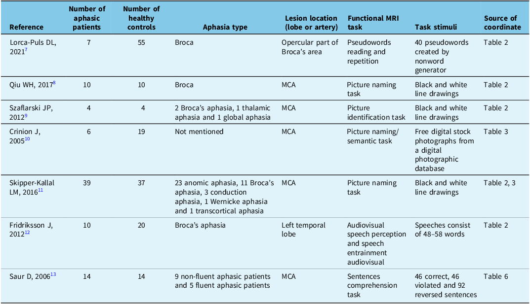
A total of 6806 relevant articles were retrieved from PubMed, Web of Science and Springer databases. After reading the titles and abstracts, 213 articles met the inclusion criteria, and 86 duplicate articles were excluded. We read the full text of all included articles, of which 120 were excluded for the following reasons: subjects were primary aphasia patients (n = 35), the subjects were under the age of 18 (n = 2), total subjects of studies were fewer than seven (n = 7), there was no normal control group (n = 22), the study used ROI or SVC research methods (n = 15), MNI or Talairach coordinates of fMRI tasks were not reported (n = 22) or studies did not compare activation patterns between aphasic patients and healthy controls (n = 17). The remaining seven articles were included in the analysis. Both the literature inclusion process and the reasons for exclusion are detailed in a flowchart (Figure 1). The specifics of the included studies are outlined in Table 1. Reference Lorca-Puls, Gajardo-Vidal and Team7–Reference Saur, Lange and Baumgaertner13 From the included literature, we compiled fMRI results from 90 patients with secondary aphasia and 159 normal healthy controls, extracting a total of 53 task-based fMRI coordinates.
Flowchart of the selection process for included papers. The flowchart shows the whole searching and selecting process of all literatures from December 1964 to June 2022. Ultimately, activation foci from seven studies were included in the ALE meta-analysis.

Basic information of included studies

Table contains all features of included references publication date and first author, the number of aphasic patients and healthy controls, aphasia type, lesion location, the type of fMRI task, task stimuli and source of coordinate.
MCA = middle cerebral artery.
ALE meta-analysis
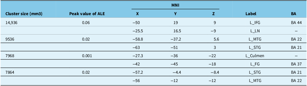
We conducted a comprehensive analysis of fMRI data from secondary aphasia patients and healthy controls during language tasks. The ALE analysis revealed brain regions with reduced activation in secondary aphasia patients, including the left inferior frontal gyrus (IFG), left middle temporal gyrus (MTG), left superior temporal gyrus (STG), left fusiform gyrus (FG), left lentiform nucleus and culmen of the cerebellum. The ALE image illustrating these findings is presented in Figure 2. The results of the ALE meta-analysis, MNI coordinates and Brodmann areas partitions are presented in Table 2.
Results of ALE meta-analysis. ALE maps of brain regions with reduced activation in aphasic patients during fMRI language tasks. Left IFG, left MTG, left STG, left FG, left lentiform nucleus and culmen of the cerebellum showed consistent reduced activation. ALE single dataset analyses thresholder at p < 0.001 uncorrected voxel-wise, FWE p < 0.05 cluster wise, 1000 permutations. IFG = inferior frontal gyrus; MTG = middle temporal gyrus; STG = superior temporal gyrus; FG = fusiform gyrus; LN = lentiform nucleus.

Abnormal brain regions with reduced activation in aphasia patients

The table shows the clusters with reduced activation in aphasic patients during task−state fMRI studies. For each cluster, we provided the cluster size, MNI coordinates, anatomical markers (Harvard−Oxford atlas) and BA partition.
MNI = Montreal Neurological Institute; L = left brain hemisphere; IFG = inferior frontal gyrus; LN = lentiform nucleus; MTG = middle temporal gyrus; STG = superior temporal gyrus; FG = fusiform gyrus; BA = Brodman’s partition.
Discussion
In this study, our aim was to pinpoint brain regions with reduced activation in aphasic patients during fMRI language tasks. Across the seven studies we reviewed, overall group analyses were conducted to compare activation patterns between normal controls and aphasic patients, identifying brain regions that showed reduced activation in aphasic patients during language tasks compared to controls. Notably, these brain regions with reduced activation in fMRI did not correlate with damaged brain regions observed in structural MRI. Furthermore, as highlighted by Turkeltaub, interpreting these brain regions with reduced activation in fMRI as damaged is problematic, as overall group analysis methods can exclude the effects derive from damaged brain regions. Reference Skipper-Kallal, Lacey, Xing and Turkeltaub11 The brain regions with reduced activation in aphasic patients can be explained by the following hypotheses. First, language functions are governed by the language networks, and stroke-induced damage disrupts these networks. Reference Price, Warburton, Moore, Frackowiak and Friston14 The brain regions with reduced activation fail to receive outgoing signals from the damaged brain areas, leading to reduced activation in these brain regions and impacting language production and comprehension. Second, in aphasic patients with left-hemisphere strokes, compensatory activation increases in homologous brain regions in the right hemisphere. This signal from homologous brain regions can be transmitted via the corpus callosum to the left hemisphere, inhibiting activation in brain regions of the left hemisphere, which manifests as reduced activation in task-based fMRI. Reference Qiu, Wu and Yang8 Across our seven included studies, aphasia was consistently post-stroke, including both hemorrhagic and ischemic strokes, with most patients in the chronic phase (≥6 months post-onset). Stroke-affected vessels primarily involved the left middle cerebral artery, and the types of aphasia included Broca’s aphasia, Wernicke’s aphasia, conduction aphasia and transcortical sensory aphasia.
The ALE meta-analysis indicated that the left IFG, left STG, left MTG, left FG, left lentiform nucleus and the culmen of the cerebellum show reduced activation in aphasic patients during fMRI language tasks. The left IFG plays a critical role in semantic recognition and decision-making. Winhuisen et al. Reference Winhuisen, Thiel and Schumacher15 used repetitive transcranial magnetic stimulation (TMS) combined with positron emission tomography (PET) to identify language functional areas in post-stroke aphasic patients, assessed at 10 days and 8 weeks post-stroke. They targeted the left IFG with repetitive TMS, temporarily disrupting its function. Then, they found that inhibitory stimulation of the left IFG at both time points impaired language function and worsened performance in verb generation tasks. Other researchers also revealed a positive correlation between activation levels in the left IFG and performance in language tasks. Reference Saur, Lange and Baumgaertner13 Our ALE meta-analysis suggests that aphasic patients exhibit significantly reduced activation in the left IFG compared to normal controls during language tasks, particularly concentrated in the triangular part of the left IFG as indicated by ALE images. Functional MRI Reference Rolheiser, Stamatakis and Tyler16 and diffusion tensor imaging (DTI) Reference Frey, Campbell, Pike and Petrides17 studies have confirmed the crucial role of the functional connectivity between the triangular part of the left IFG and the temporal lobe in semantic and lexical processing. In this study, most aphasic patients resulted from left middle cerebral artery stroke, leading to temporal lobe damage and disruption of fiber connections between the temporal lobe and the triangular part of the left IFG. This disruption causes reduced activation levels in the left IFG, particularly in the triangular part, which impacts the patients’ language functions.
The language function is governed by language networks. Task-based fMRI have shown significant activation of the left IFG during semantic tasks, suggesting its vital role in semantic processing. Similarly, a neuroimaging study reveals the co-activated brain regions associated with semantics, particularly the posterior MTG (pMTG). Reference Kuperberg, Sitnikova and Lakshmanan18 It is hypothesized that pMTG serves as a “convergence point” in semantic decision-making, integrating semantic information from the frontal and temporal lobes. Our findings also indicate reduced activation in the left IFG and pMTG, further substantiating their correlation. Davey et al. Reference Davey, Cornelissen and Thompson19 included a group of healthy participants and used inhibitory TMS to suppress pMTG function. They found that participants performed worse on object identification tasks. Simultaneously, Whitney et al. Reference Whitney, Kirk, O’Sullivan, Lambon Ralph and Jefferies20 targeted the left pMTG and triangular part of the left IFG as sites for inhibitory stimulation in a study involving 16 healthy individuals. After stimulation, participants completed a semantic decision task. The study revealed that participants whose pMTG and IFG were inhibited showed prolonged response times and increased error rates, particularly when exposed to task stimuli with weak semantic associations. In addition, we observed a close association between reduced activation of the STG and language function impairment. Schwarz et al. Reference Schwarz, Lizarazu, Lallier and Klimovich-Gray21 analyzed cortical responses to phonological and lexical-semantic processing using magnetoencephalography. The results indicate that the peaks of both phonological and lexical-semantic effects are located in the STG, demonstrating a significant correlation between left STG and symptoms of dyslexia in patients. Additionally, there is a piece of evidence suggesting that long-term language training can enhance left STG activity and is associated with improved infant reading skills. Conversely, reduced activation in STG corresponds to varying degrees of impairment in patients. Reference Simos, Fletcher and Bergman22
The left FG is pivotal in the process of language comprehension. It serves as part of the convergence zone, primarily responsible for integrating multimodal information and storing/retrieving language information. Reference Binder, Frost, Hammeke, Cox, Rao and Prieto23 Functional MRI studies have confirmed this view, showing the involvement of the left FG in processing various modalities of information. Sharp et al. Reference Sharp, Scott and Wise24 conducted PET on chronic aphasic patients and healthy controls during language tasks. Their study found significant activation in bilateral FG in healthy controls and aphasic patients during language tasks. However, when comparing activation levels between aphasic patients and controls, they observed the left FG activation was notably reduced in aphasic patients compared to controls. The ROI-based analysis ulteriorly demonstrated a significant correlation between reduced left FG activation in aphasic patients and restricted language function. Reference Galton, Patterson and Graham25 Damage to the left FG impairs semantic processing. Performance of semantic memory tasks positively correlates with cortical volume of the left FG in aphasia patients. Significant semantic memory deficits were observed in patients with herpes simplex encephalitis extending to the left FG. Reference Schmolck, Kensinger, Corkin and Squire26 Consistent with the results of our ALE analysis, reduced activation in the left FG has implications for patients’ language function.
Language, as one of the most complex cognitive functions, relies not only on the intricate cortex but also on subcortical structures and the cerebellum. Bilateral lateral cerebellar regions are considered to be involved in language production. Reference Yuan, Li and Du27 The cerebellum serves as a crucial forward-driving node in the reading network, playing a role in word processing. The traditional “lesion-deficit” hypothesis posits that cerebellar dysfunction can lead to impairments in speech monitoring, thereby affecting language function. Reference Sihvonen, Virtala, Thiede, Laasonen and Kujala28 In that study, we found that language impairment correlates with reduced activation in the culmen of the cerebellum. Functional imaging evidence supports this view, showing increased activation in the culmen of the cerebellum during writing tasks involving higher complexity or creativity. Therefore, reduced culmen activation in aphasic patients correlates with disorder in processing complex texts. In addition, subcortical structures such as the lentiform nucleus, caudate nucleus and thalamus are implicated in language processing. According to our research, reduced activation in the lentiform nucleus may impact language function. Newberg et al. Reference Peres, Moreira-Almeida, Caixeta, Leao and Newberg29 conducted PET on patients with left-hemisphere stroke-induced aphasia. Their findings indicate a positive correlation between activated levels in the left lentiform nucleus and speech fluency in aphasic patients. While direct damage to brain regions is a primary cause of language decline, reduced activation in undamaged brain areas is also significant for poorer language performance among aphasic patients. Understanding these brain regions with reduced activation not only enhances our comprehension of language networks but also holds the potential for advancing aphasia treatment strategies.
The processes of language production and comprehension are facilitated by the coordinated efforts of various brain regions, as evidenced by functional and structural imaging studies. Conditions such as stroke can result in multiple forms of language impairment. First, damage can lead to gray matter injury in specific areas, resulting in dysfunction of language functions. Second, injury can affect white matter fiber bundles. When white matter is damaged, the transmission of signals involved in language processing is interrupted, leading to aphasia. The superior longitudinal fasciculus (SLF) and arcuate fasciculus (AF) are the primary fiber bundles of the dorsal stream of the language dual-stream model. Previous research has often treated these two bundles as an entity without detailed differentiation. More recently, the two tracts have been deemed distinct, although AF is still widely considered as one of the branches of the SLF. Reference Shekari and Nozari30 The AF, connecting the frontal and temporal lobes, is one of the most extensively studied neural pathways involved in language processing. Research on post-stroke aphasia indicates a significant correlation between the integrity of the left AF and auditory comprehension and naming abilities in chronic stroke survivors. Reference Yu, Jiang and Sun31 In comparison, the SLF is considered a complex fiber bundle, composed of three or more branches that connect regions of the frontal, parietal and temporal lobes. McKinnon et al. Reference McKinnon, Fridriksson and Basilakos32 conducted language task tests on chronic stroke aphasia patients, revealing associations between semantic and phonological task accuracy and axonal loss in the SLF.
The ventral stream of the dual-stream model, such as the inferior longitudinal fasciculus (ILF) and uncinate fasciculus (UF), also play crucial roles in language processing. The ILF connects the occipital and temporal lobes, originating from the occipital pole and projecting to the temporal gyri and fusiform gyrus. Zhao et al. Reference Zhao, Thiebaut de Schotten, Altarelli, Dubois and Ramus33 conducted DTI on 32 patients with reading disorder and 32 normal controls. The results indicated a significant reduction in leftward lateralization of the ILF in individuals with reading disorders. Meanwhile, studies have linked lesions in the left ILF to deficits in semantic word mapping in patients with aphasia. Reference Faulkner and Wilshire34 In addition, the UF connects the anterior temporal lobe to the orbitofrontal cortex and is involved in semantic processing. Decreased fractional anisotropy values in the UF have been identified as significant predictors in patients with semantic dementia. Reference Bouchard, Wilson, Laforce and Duchesne35 Furthermore, in primary progressive aphasia patients, the integrity of the UF correlates significantly with fluency in naming and category decision tasks. Reference Bouchard, Wilson, Laforce and Duchesne35 Overall, our study identifies brain regions with reduced activation in aphasia patients, including the left IFG, left STG, left MTG, left FG, left lentiform nucleus and the culmen of the cerebellum. These regions are interconnected by fiber bundles such as the AF, SLF, ILF and UF. Damage to these fiber tracts may impair fiber connection between brain regions, resulting in reduced activation in these brain regions and thereby affecting language production and comprehension.
Our result from ALE analysis has stipulated that the left IFG, left STG, left MTG, left FG, left lentiform nucleus and the culmen of the cerebellum exhibit reduced activation and hold significant implications for further understanding of language networks and recovery of language functions in aphasic patients. Currently, treatment options for aphasia remain exceedingly limited, and conclusive findings regarding treatment effectiveness are lacking. Speech and language therapy represents the most fundamental and widely utilized approach but provides modest benefits. In recent years, noninvasive brain stimulation (NIBS) techniques have been extensively applied to restore language function following aphasia. TMS and transcranial direct current stimulation (tDCS) are the primary NIBS modalities employed, both of which modulate neural activity by inducing currents within the central nervous system. Shah-Basak et al. Reference Shah-Basak, Sivaratnam and Teti37 and de Jongh et al. Reference de Jongh, de Munck, Baayen, Jonkman, Heethaar and van Dijk38 suggested that targeting the perilesional areas with NIBS can enhance functional recruitment, aiding in the recovery of language functions in aphasic patients. In these studies, the efficacy of NIBS targeting structurally damaged brain regions is constrained. However, our research identifies brain regions showing reduced activation in aphasic patients during fMRI language tasks, which still respond to NIBS interventions. Thus, activating NIBS can potentially increase activation in these regions, facilitating language function recovery. Griffis et al. Reference Griffis, Nenert, Allendorfer and Szaflarski39 targeted the left IFG for TBS therapy and assessed treatment effects with fMRI, finding increased activation in the left IFG and improved performance in language tasks. Fridriksson et al. Reference Fridriksson, Rorden, Elm, Sen, George and Bonilha40 divided post-stroke aphasic patients into experimental and sham stimulation groups, targeting the posterior temporal lobe for tDCS, and observed a significant improvement in naming task accuracy in the experimental group after 3 weeks of treatment. These studies collectively demonstrate that NIBS for these brain regions with reduced activation led to conspicuous enhancement in language function among aphasic patients, offering new possibilities for aphasia treatment.
Limitation
This study has several limitations. First, our study just included seven task-based fMRI studies due to the limited prior research on brain regions with reduced activation. Second, these regions identified by ALE meta-analysis could provide new therapeutic targets for NIBS, but the efficacy requires further exploration. These findings also highlight our future research direction. We intend to conduct multicenter studies using task-based fMRI in aphasic patients to further validate brain regions of reduced activation and analyze the feasibility of these regions as targets for NIBS.
Acknowledgments
We thank Liwen Bianji (Edanz) (www.liwenbianji.cn) for editing the English text of a draft of this manuscript.
Author contributions
Xin-ming Yu and Wen-ming Lv contributed equally to this work.
(1) Research Project: (A) Conception, (B) Organization, (C) Execution.
(2) Statistical Analysis: (A) Design, (B) Execution, (C) Review and critique.
(3) Manuscript: (A) Writing of the first draft, (B) Review and critique.
XM Y: 1C, 2B, 2C and 3A.
WM L: 1C and 3B.
QW Y: 1C and 2B.
XZ K: 1C and 2C.
XL L: 1A, 1C, 2C and 3B.
LW Z: 1A, 1B, 2A, 2C and 3B.
Funding statement
This work was supported by the China Postdoctoral Science Foundation (no. 2022M711742 to Xiaoliang Liu), Youth Science Foundation Cultivation and Support Program of Shandong First Medical University (no. 202221-050 to Xiaoliang Liu), Shandong Provincial Natural Science Foundation (no. ZR2023QH032 to Xiaoliang Liu) and Shandong Provincial Natural Science Foundation (no. ZR2023ZD17 to Liangwen Zhang).
Competing interests
The authors declare that they have no known competing financial or personal relationships that could have appeared to influence the work reported in this paper.