Introduction
Meige syndrome (MS) is a focal movement disorder characterized by orofacial dystonia and involuntary eyelid movements involving the orbicularis oculi muscles and periocular muscles.Reference LeDoux1 The average age of MS onset is approximately 50–60 years, and this disorder is more common in females. The etiology and pathophysiological mechanism underlying MS are poorly understood. Altered synaptic plasticity and cortical hyperexcitability may contribute to the development of MS.Reference Simonyan, Cho, Hamzehei Sichani, Rubien-Thomas and Hallett2 Deep brain stimulation (DBS) has emerged as a safe and effective treatment option for patients with MS; however, the mechanism underlying the effect of DBS on MS remains unclear. The key to optimal DBS application may lie in differential brain structural and functional alterations.Reference Hao, Lv and Zheng3,Reference Hao, Zheng and Zhang4 Recently developed image analysis techniques, such as voxel-based morphometry (VBM) and surface-based morphometry (SBM), have been shown to be effective at detecting subtle anatomical alterations in the brain.Reference Goto, Abe and Hagiwara5 Surface-based methods have advantages over VBM because of their superior ability to measure cortical characteristics, including cortical thickness and folding patterns, and their lower level of bias during the image registration and alignment processes.Reference Ghosh, Kaur and Shah6
In addition to motor symptoms, nonmotor symptoms, including emotional disorders and sleep disorders, have been found to be comorbidities in MS patients.Reference Defazio, Hallett, Jinnah, Conte and Berardelli7 Sleep disorders are likely to occur intrinsically rather than secondary to motor symptoms, as they are not effectively alleviated by botulinum toxin injection.Reference Ray, Kutty, Pal and Yadav8 However, no existing study has investigated the mechanism of sleep disorders in MS patients.
Therefore, we aimed to identify structural alterations in MS brains, to examine structural differences between MS patients with sleep disorders and those without sleep disorders using SBM, to clarify the pathogenic mechanism of MS and to elucidate the effect of DBS on MS patients.
Methods
Participant information
Clinical data from 42 right-handed patients with primary MS were consecutively collected in the Department of Neurosurgery of Peking University People’s Hospital from January 2020 to June 2023. The diagnosis of MS was made by an experienced chief neurosurgeon based on the previously established diagnostic criteria. The exclusion criteria were as follows: (1) had neurological diseases other than MS; (2) had severe psychotic disorders or cognitive impairment; (3) had metabolic diseases, such as diabetes, hyperthyroidism or hypothyroidism; (4) had a positive urine toxicology or pregnancy test; (5) used psychotropic drugs in the past 2 months; (6) had other serious systemic diseases; and (6) had MRI contradictions. In addition, 30 healthy, right-handed, age- and sex-matched individuals without MS were selected as healthy controls (HCs). All subjects provided written informed consent, and this study was approved by the Ethical Review Committee of Peking University People’s Hospital.
Clinical assessment
The baseline data included age, sex, education level, assessment of dystonia and sleep-related clinical symptoms. Symptom severity was assessed by an experienced neurosurgeon who was not involved with the study, thus ensuring objectivity and accuracy. The severity of motor symptoms was assessed using the Burke–Fahn–Marsden Dystonia Rating Scale for motor symptoms (BFMDRS-M) (scores ranged from 0 to 40, with higher scores indicating more severe symptoms).Reference Burke, Fahn, Marsden, Bressman, Moskowitz and Friedman9 The severity of sleep disorders was assessed using the Pittsburgh Sleep Quality Index (PSQI),Reference Buysse, Reynolds Iii, Monk, Berman and Kupfer10 a scale that differentiates “poor” sleep from “good” sleep by measuring the following aspects: subjective sleep quality, sleep latency, sleep duration, habitual sleep efficiency, sleep disturbances, usage of sleeping medications and daytime dysfunction over the last month. A score of 5 or greater indicates the presence of sleep disorders. In this study, patients were divided into a sleep disorder group (SD, PSQI score of 5 or greater) and a normal sleep group (NS, PSQI score of less than 5).
MR data acquisition
T1-weighted thin-slice structural images of the brain were acquired with the following parameters: repetition time = 4.9 ms, echo time = 2.0 ms, field of view = 240 × 240 mm,Reference Simonyan, Cho, Hamzehei Sichani, Rubien-Thomas and Hallett2 flip angle = 15°, slice thickness = 1 mm, in-plane resolution = 1 × 1 mmReference Simonyan, Cho, Hamzehei Sichani, Rubien-Thomas and Hallett2 and 170 slices. A body coil was used for transmission, and a normal quadrature coil was used for reception. All MR data were collected by an experienced radiologist on a Discovery 750 3.0T MRI (GE Healthcare, Waukesha, WI).
SBM processing
Imaging data were processed using SPM12-v7771 software and the CAT12.7-Beta (r1615) toolbox, both of which were installed in the MATLAB 2017b operating environment.Reference Christian, Robert, Paul, Florian and Eileen11 In the initial step of voxel-based processing, T1 images were first screened for quality, artifacts and potential abnormalities. Anatomical images that met the criteria were then converted and reoriented on the anterior commissure-posterior commissure (AC-PC) line. Reoriented images were subjected to preliminary denoising, coarse affine bias correction and registration, followed by skull stripping, whole-brain signal calibration, redenoising and rapid registration with locally adaptive segmentation. Preprocessed images were then fitted to a DARTELReference Ashburner12 template and segmented. The Desikan-Killiany 40 (DK40) atlas was chosen as the reference atlas.
Cortical thickness
Cortical thickness was estimated using the automatic surface preprocessing algorithm in the CAT12 toolbox by reconstructing the central surface of the left and right hemispheres through a projection-based thickness calculation method. This algorithm also estimates the white matter (WM) distance using tissue segmentation and projects the local maxima (which are equivalent to cortical thickness) to other gray matter (GM) voxels. The WM distance defines this adjacent relationship.Reference Dahnke, Yotter and Gaser13 A 15-mm full-width-at-half-maximum (FWHM) smoothing kernel was applied.
Gyrification
To calculate the local gyrification index (GI) maps, the absolute mean curvature approach was utilized. A mesh of the central surface, located between the GM/CSF and the GM/WM boundary, was constructed through cortical surface extraction.Reference Dahnke, Yotter and Gaser13 We then calculated the local absolute mean curvature of the ventral surface by averaging the mean curvature values from each vertex point within 3 mm of a specific position. To smooth the GI maps, a 20-mm FWHM kernel was applied.
Cortical complexity (fractal dimension)
The cortical complexity, that is, fractal dimension, was determined using the spherical harmonic reconstructions method, as detailed in a previous study.Reference Yotter, Nenadic, Ziegler, Thompson and Gaser14 This technique yields a consistent number of vertices across all reconstructed surfaces, thereby minimizing the effects of individual vertex alignment, resampling and interpolation, thus leading to more precise reconstructions. After generating individual vertexwise fractal dimension maps, a 20-mm FWHM kernel was applied for smoothing.
Sulcus depth
The sulcus depth was determined by calculating the Euclidean distance between the central surface and its convex hull, which was further computed from the surface and smoothed with a 20-mm FWHM kernel.Reference Dahnke, Yotter and Gaser13 The surface data were visually inspected for artifacts and homogeneity, and the overall image quality was checked to ensure compliance with the statistical quality control criteria.
Statistical analyses
The data are presented as the mean ± standard deviation (SD) unless otherwise indicated. Demographic and clinical data, such as age, sex, BFMDRS-M scores and PSQI scores, were analyzed using either the chi-square test (for categorical variables) or the independent samples t test or ANOVA, as appropriate (for continuous variables). P < 0.05 was considered to indicate statistical significance. Two-tailed t tests were used to compare cortical thickness, fractal dimension, gyrification and sulcus depth across different brain regions between groups, and data were corrected using the familywise error rate (FWE) for multiple comparisons to control for type I errors. A threshold of p < 0.001 (FWE uncorrected) or p < 0.05 (FWE corrected) for statistical significance was applied for regions in the DK40 cohort. Statistical analyses were conducted with SPSS statistical software (version 24.0; IBM Corp.).
Results
Demographic and clinical characteristics
A total of 42 patients (13 males and 29 females) with MS were included in this study; their ages ranged from 35 to 69 years, with an average age of 56.90 ± 8.17 years. The years of education ranged from 5 to 24 years, with an average of 11.62 ± 4.79 years. The disease durations of MS ranged from 1 to 12 years, with an average of 3.99 ± 3.23 years. The BFMDRS-M scores ranged from 4.0 to 17.0, with an average of 8.76 ± 4.40. The PSQI scores of the MS patients ranged from 0 to 15 points, with an average of 5.24 ± 4.32 points. Additionally, a total of 30 HCs (11 males and 19 females) were included in this study, with an average age of 54.63 ± 7.74 years. The years of education ranged from 6 to 21 years, with an average of 12.23 ± 4.76 years. The BFMDRS-M and PSQI scores of the MS group were significantly higher than those of the HC group (p < 0.05). There were no significant differences in age, sex or years of education between the MS patients and HCs (Table 1).
Demographics and clinical information of MS group and HC group

MS = Meige syndrome; HC = healthy control.
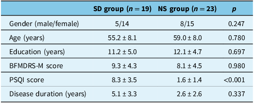
After dividing patients into groups based on their PSQI scores, there were 19 patients in the SD group and 23 patients in the NS group. There were no significant differences in age, sex or BFMDRS-M scores between the SD and NS groups (p > 0.05) (Table 2).
Demographics and clinical information of SD group and NS group

SD = sleep disorder; NS = normal sleep.
Surface-based morphometry metrics
Comparison between MS patients and healthy controls
Cortical thickness
A comparison of cortical thickness measurements between MS patients and HCs revealed significant increases in MS patients in bilateral posterior cingulate, isthmus cingulate, caudal anterior cingulate and left medial orbitofrontal and rostral anterior cingulate regions (Table 3, Figure 1). Significant decreases in cortical thickness among MS patients were detected in bilateral postcentral, precentral, superior frontal, paracentral, superior parietal, lateral occipital, pericalcarine, lingual, cuneus, parahippocampal, entorhinal, fusiform, inferior temporal and left inferior parietal, insula, temporal pole, middle temporal, The banks of the superior temporal sulcus (BANKSSTS) and right precuneus regions (pFWE > 0.05) (Table 3, Figure 2).
Regions with increased cortical thickness in Meige syndrome than in healthy control, displayed from the left, bottom and right surfaces. The color scale represents the range of t values.

Regions with decreased cortical thickness in Meige syndrome than in healthy control, displayed from the left, bottom and right surfaces. The color scale represents the range of t values.

Between-group comparison of cortical thickness

BANKSSTS = The banks of the superior temporal sulcus; MS = Meige syndrome; HC = healthy control.
Gyrification
Gyrification measurements indicated significant decreases in the right fusiform, parahippocampal, inferior temporal (pFWE > 0.05) and right precentral, postcentral, lingual, fusiform, pars triangularis, lateral occipitofrontal, insula and left superior temporal and BANKSSTS (p < 0.001, uncorrected) regions among MS patients. Additionally, a significant increase was observed in the right medial orbitofrontal region (p < 0.001, uncorrected) (Table 4).
Between-group comparison of gyrification index and cortical complexity

BANKSSTS = The banks of the superior temporal sulcus; MS = Meige syndrome; HC = healthy control.
Cortical complexity
MS group showed significant decrease in cortical complexity in the right lingual, parahippocampal, isthmus cingulate, precuneus (pFWE < 0.05) and left parahippocampal (p < 0.001, uncorrected) regions. Additionally, the MS group showed an increase in left superior parietal and precuneus regions (p < 0.001, uncorrected) (Table 5).
Between-group comparison of cortical complexity

MS = Meige syndrome; HC = healthy control.
Sulcus depth
The comparison of the sulcus depth between the MS group and the HCs revealed a decrease in left inferior temporal, middle temporal and fusiform regions among the MS patients (p < 0.001, uncorrected) (Table 6).
Between-group comparison of sulcus depth

MS = Meige syndrome; HC = healthy control.
Comparison between the SD group and NS group
The comparison of cortical thickness measurements and gyrification measurements between the SD and NS groups did not reveal any significant differences.
The comparison of cortical complexity between the study groups revealed increased complexity in the right insula and decreased complexity in the left lateral occipital and right pars triangularis regions of the SD group (p < 0.001, uncorrected) (Table 7). The comparison of sulcus depth measurements revealed that the SD group showed a decrease in the right inferior parietal region (p < 0.001, uncorrected) (Table 8).
Comparison of cortical complexity between SD group and NS group

SD = sleep disorder; NS = normal sleep.
Comparison of sulcus depth between SD group and NS group

SD = sleep disorder; NS = normal sleep.
Discussion
The etiology of primary MS has not been fully elucidated. In the present study, we observed significant differences in cortical thickness, cortical complexity and gyrification across the whole brain between MS patients and HCs, with the most significant differences observed in the central cortical regions. Additionally, substantial differences were observed in occipital and temporal lobes, suggesting widespread alterations in cortical characteristics associated with MS. To our knowledge, this is the first SBM-based study of MS.
The present study found cortical thinning prominently in the bilateral postcentral, precentral, superior frontal and paracentral regions. The reduced cortical thickness found in these areas accentuates abnormalities in the sensorimotor network among MS patients. The postcentral gyrus, which houses the primary somatosensory cortex, serves as a primary receptor for general bodily sensations receiving sensory data from the body from thalamic radiation. Previous studies have consistently demonstrated structural and functional alterations in the primary somatosensory cortex in individuals with various forms of movement disorders.Reference Defazio, Berardelli and Hallett15,Reference Wu, Zhang and Li16 Martino et al.Reference Martino, Di Giorgio and D’Ambrosio17 analyzed whole-brain GM volume in patients with primary blepharospasm and found decreased GM volume in the primary sensory cortex (S1). Furthermore, functional imaging studies have reported hyperactivity in primary sensory cortices in patients with primary dystonia, including blepharospasm and cervical dystonia.Reference Nguyen, Kelly and Glickman18,Reference Feng, Yin and Wang19 Given that sensory symptoms such as dry eye, burning sensations and photophobia are frequently reported in MS patients, structural alterations may contribute to the impairment of sensorimotor integration. The precentral gyrus includes the motor cortex and part of the premotor cortex, which are differentially involved in the initiation and subsequent execution of movements. VBM-based studies of idiopathic blepharospasm have shown increased GM in the bilateral precentral gyri with hypoactivity.Reference Zhang, Huang, Li, Shang and Yang20 Yang et al.Reference Yang, Liu and Lv21 observed significant increases in cerebral blood flow in the middle frontal gyrus and bilateral precentral gyrus among MS patients. The findings of our study are also consistent with previous research using transcranial magnetic stimulation, which revealed abnormal cortical activities in the motor cortex in several forms of dystonia.Reference Sommer, Ruge, Tergau, Beuche, Altenmüller and Paulus22 The superior frontal and paracentral areas are functionally connected to other frontal and parietal regions, which are also involved in motor functioning and subserve spatial attention. Importantly, the supplementary motor area (SMA) occupies one-third of the superior frontal gyrus and is considered to be crucial in planning and coordinating complex movements. Xu et al. reported greater GM volume in the bilateral SMA in patients with blepharospasm than in hemifacial spasm patients and HCs, suggesting that SMA plays a role in the genesis of dystonic symptoms.Reference Xu, Luo and Peng23 Functional research has also shown altered excitability of the SMA in other forms of dystonia.Reference Huang, Zhang, Li, Shang and Yang24 Taken together, these findings suggest that abnormalities in the SMA may be associated with the loss of control of undesired involuntary movements in MS patients.
A significant decrease in cortical thickness was also observed bilaterally in the fusiform, lingual, parahippocampal, lateral occipital, inferior temporal, cuneus and pericalcarine regions as well as in adjacent areas. The fusiform gyrus is integral to visual processing, particularly emotional perception and stimulus-response tasks. Structural and functional alterations of this region have been identified in several forms of dystonia, including blepharospasm, cervical dystonia and embouchure dystonia.Reference Zhang, Huang, Li, Shang and Yang20,Reference Prell, Peschel and Köhler25,Reference Haslinger, Noé and Altenmüller26 The inferior temporal gyrus is a visual associative region that receives input from the occipital lobe and connects comprehensive information to the prefrontal cortex to achieve higher-level functions. The inferior temporal gyrus, along with the lateral occipital area, facilitates comprehensive visual information processing. Lesions affecting this region may lead to visual impairments observed in MS patients. The lingual gyrus is situated on the posteroinferior portion of the medial surface of the occipital lobe and, as it extends anteriorly, merges with the parahippocampal gyrus at the tentorial surface of the temporal lobe. The parahippocampal gyrus has significant interconnectivity to other cortical limbic structures as well as the amygdala, with many recognized functions, including episodic memory, visuospatial processing, complex emotive processing and cognitive function.Reference Aminoff, Kveraga and Bar27,Reference Echávarri, Aalten and Uylings28 Taken together, these findings suggest that cortical thinning in these areas is associated with motor deficits in the eye as well as nonmotor symptoms, such as anxiety and depression, in MS patients.
Increases in cortical thickness were found bilaterally in the posterior cingulate, isthmus cingulate and caudal anterior cingulate regions as well as in the medial orbitofrontal and rostral anterior cingulate regions of the left hemisphere. The cingulate cortex, interconnected with the limbic system, plays roles in attentional processes, memory retrieval and pain modulation.Reference Coghill and Squire29,Reference Pflugshaupt, Nösberger, Gutbrod, Weber, Linnebank and Brugger30 Multimodal imaging studies have revealed structural and functional alterations of the anterior, isthmus and posterior cingulate areas in MS and other dystonias.Reference Huang, Zhang, Li, Shang and Yang24,Reference Jochim, Li and Gora-Stahlberg31,Reference Hanekamp and Simonyan32 The medial orbital gyrus is implicated in emotional processing and reward responses, while increased cortical thickness may indicate compensatory mechanisms following sensory circuit imbalances.Reference Rolls33
The gyrification index is one of the most commonly used metrics for measuring cortical folding complexity. In this study, we found a significant decrease in the gyrification index in the fusiform, parahippocampal and inferior temporal regions of the right hemisphere, consistent with our findings of cortical thickness alterations. Abnormal folding patterns in the fusiform gyrus have been associated with impairments in visual attention and cognitive control in depression. Chen et al.Reference Chen, Liu and Zuo34 reported a decreased gyrification index in the right fusiform gyrus in patients with major depressive disorder, which may indicate a correlation between structural changes in the brain and psychological symptoms in MS patients. The parahippocampal gyrus is a part of the default mode network and is involved in pain processing, memory and the stress response and plays roles in the pathophysiological process of migraine, depression, anxiety and tinnitus.Reference Yin, Lan and Tian35,Reference To, Song, Mohan, De Ridder, Vanneste, Langguth, Kleinjung, De Ridder, Schlee and Vanneste36 In a study of patients with blepharospasm, Hou et al.Reference Hou, Zhang and Wei37 demonstrated altered functional connectivity of the default mode network, pointing toward its potential role in the pathogenesis of dystonic symptoms. The inferior temporal cortex is crucially involved in the visual recognition of objects, which could be related to motor symptoms of the eye in MS patients.
The evaluation of cortical complexity revealed significant increases in the left lingual, parahippocampal, isthmus cingulate and precuneus cortexes in the MS group. Increased cortical complexity in the left superior parietal gyrus and reduced sulcal depth in the left inferior temporal gyrus, middle temporal gyrus and fusiform gyrus were also observed. The lingual gyrus and parahippocampal gyrus are implicated in visual and cognitive functions, with structural abnormalities potentially underpinning nonmotor symptoms in MS. The precuneus, crucial for memory and environmental perception, exhibits functional abnormalities in movement disorders and depression.Reference Reetz, Lencer and Hagenah38,Reference Cheng, Rolls, Ruan and Feng39 In addition, the precuneus has been proven to be extensively connected with the SMA,Reference Leichnetz40 suggesting its role in MS symptomatology.
Additionally, comparisons between MS patients with and without sleep disorders revealed significant cortical indicator changes in the inferior parietal, insula, lateral occipital and pars triangularis regions. These regions are critical for motor control, cognition, vision and language functions. Studies have highlighted increased functional connectivity in the insula among individuals with sleep disorders,Reference Cheng, Rolls, Ruan and Feng39 Moreover, Kim et al.Reference Kim, Lee and Lee41 reported that the activation level of the right insula correlates with the severity of both sleep and depressive disorders, confirming the association of this brain region with sleep disorders. Calcium imaging in the cortical layers of mouse brains revealed spatiotemporal patterns of global cortical activity related to sleep stages, with significant activation observed in the occipital cortical areas, particularly during rapid eye movement sleep.Reference Wang, Fei and Liu42 Cortical-level brain activity participates in the control of sleep stages, while neurotransmitter systems involving interactions among the brainstem, hypothalamus, basal forebrain, thalamus and cortex predominantly regulate the modulation of sleep and wakefulness.Reference Brown, Basheer, McKenna, Strecker and McCarley43 However, this study did not detect structural changes in these brain regions, underscoring the need for further research with larger cohorts to explore the underlying mechanisms of sleep disorders in MS patients.
Notably, although the detailed cortical metric SBM can be extracted from the MS brain, it tends to attain less accuracy over certain subcortical regions. For example, it is highly important that alterations in the basal ganglia and the associated areas and circuits are accounted for. However, due to the limitations of mixed dystonia forms and relatively small sample sizes, previous studies have produced inconsistent results, and the associations of structural alterations in the basal ganglia of MS patients with disease development and treatment response remain unclear.Reference Martino, Di Giorgio and D’Ambrosio17,Reference Pantano, Totaro and Fabbrini44 . In addition, although functional changes in certain brain regions, such as the basal ganglia, are essential for the development and maintenance of muscle tone disorders, they may not necessarily lead to anatomical changes. As a result, further research on a larger scale and with different modalities, such as fMRI, is needed.
Conclusions
In summary, the findings of the present study strongly suggested that MS patients have cortical structural abnormalities compared with HCs. The alterations in cortical metrics are predominant across the postcentral, precentral, superior frontal and paracentral regions as well as the occipital and temporal lobes, thus revealing the pathophysiology of motor and nonmotor symptoms underlying MS. No significant difference was detected when comparing MS patients with and without sleep disorders. Future studies with larger sample sizes and multiple modalities are needed to detect small but significant differences in cortical parameters between groups of patients with and without sleep symptoms.
Data availability statement
The data that support the findings of this study are available on request from the corresponding author. The data are not publicly available due to privacy or ethical restrictions.
Author contributions
Y.T.: Conceptualization, methodology, software, data curation, investigation, writing – original draft preparation. R.L.: Supervision, validation, writing – reviewing and editing.
Funding statement
This study was funded by grants from Peking University People’s Hospital (2017-T-01) and partly supported by the Beijing Municipal Health Commission (SHOUFA-202224085).
Competing interests
The authors declare no conflict of interest.