

Introduction
Marine organisms increasingly suffer from environmental stress caused by pollution, overfishing, and coastal degradation. The sea floor acts as a final repository for various pollutants, while surrounding waters facilitate exposure to toxic substances, particularly heavy metals. Although national and local authorities are responsible for addressing environmental decline, experts recommend integrated approaches for more thorough and realistic evaluations of contaminated habitats (Matranga et al., Reference Matranga and Yokota2012). The impact of human activities and the number of stressors have surged in recent decades due to rising demands for natural resources and population growth, especially in coastal areas (Halpern et al., Reference Halpern, Frazier, Afflerbach, Lowndes, Micheli, O’Hara, Scarborough and Selkoe2019).
Aquatic sediments are significant reservoirs for contaminants, notably heavy metals, which comprise over 90% of the total heavy metal content in aquatic ecosystems (Simpson & Batley, Reference Simpson and Batley2016; Chapman et al., Reference Chapman, Wang, Janssen, Goulet and Kamunde1998; Luoma & Rainbow, Reference Luoma and Rainbow2008; Benson et al., Reference Benson, Asuquo, Williams, Essien, Ekong, Akpabio and Olajire2018; Farhat, Reference Farhat2019). Research indicates that heavy metal accumulation in sediments can disrupt metabolism, cause DNA damage, and potentially result in lethal outcomes for organisms through direct ingestion, skin contact, or indirectly via the food chain (Valavanidis et al., Reference Valavanidis, Vlahogianni, Dassenakis and Scoullos2006; Amiard et al., Reference Amiard, Amiard-Triquet, Barka, Pellerin and Rainbow2006). Among heavy metals, cadmium (Cd) is particularly insidious due to its high bioavailability in anoxic sediments, lack of known biological role, and prevalence in the Persian Gulf, where industrial effluents and shipping activities elevate sediment concentrations to levels exceeding global averages (e.g., up to 11 µg/l in Qeshm Island sediments; local monitoring data). Cd was selected for this study as a model pollutant because of its documented bioaccumulation in benthic invertebrates, interference with calcium metabolism, and understudied chronic effects on echinoderm physiology in semi-enclosed basins like the Gulf (Segar, Reference Segar1971; Nriagu, Reference Nriagu1990; Farhat, Reference Farhat2019). Due to insufficient anthropogenic recycling (Nriagu, Reference Nriagu1990), chronic cadmium issues persist in the sediments of industrialized nations (Bryan & Langston, Reference Bryan and Langston1992). Coastal areas near urban centres, heavily impacted by human activity, typically show cadmium pollution, with up to 90% of coastal sediment cadmium originating from anthropogenic sources (Förstner, Reference Förstner1980). Thus, sediments can act as cadmium reservoirs long after initial contamination (Tessier & Campbell, Reference Tessier and Campbell1987; Salomons et al., Reference Salomons, de Rooij, Kerdijk and Bril1987). Elevated metal concentrations emphasize sediments’ role as direct toxins for aquatic life. Beyond its harmful effects on the ecosystem and soft tissues, cadmium also disrupts calcite deposition in calcium-carbonate-dependent organisms, weakening their skeletal structures (Roesijadi, Reference Roesijadi1996; Fichet et al., Reference Fichet, Radenac and Miramand1998). Youness et al. (Reference Youness, Mohammed and Morsy2012) suggest that this happens due to cadmium replacing calcium, leading to the development of fragile skeletal structures that once relied on robust calcium carbonate frameworks (Wang et al., Reference Wang, Zhu, Shi, Weng, Jin, Kong and Nordberg2003; Youness et al., Reference Youness, Mohammed and Morsy2012).
Regeneration is a vital biological process that enables organisms to recover from injuries, replace lost body parts, and maintain their ecological roles. In marine ecosystems, echinoderms such as brittle stars exhibit remarkable regenerative abilities, which are essential for their survival and ecological functions, including sediment mixing and nutrient cycling (Candia Carnevali, Reference Candia Carnevali2006; Ben Khadra et al., Reference Ben Khadra, Sugni, Ferrario, Bonasoro, Oliveri, Martinez and Carnevali2018). This process is particularly important in dynamic marine environments where physical damage and predation are common. However, exposure to heavy metals like cadmium poses a significant threat to regeneration by disrupting cellular mechanisms, inducing oxidative stress, and interfering with calcium metabolism, which is critical for tissue repair and skeletal integrity (Fichet et al., Reference Fichet, Radenac and Miramand1998; Wang et al., Reference Wang, Zhu, Shi, Weng, Jin, Kong and Nordberg2003; Youness et al., Reference Youness, Mohammed and Morsy2012). The impairment of regeneration not only affects individual survival but also has cascading effects on population dynamics and ecosystem stability. Understanding how cadmium and other pollutants impact regeneration in echinoderms is crucial for assessing the resilience of marine biodiversity in polluted environments and for developing effective conservation strategies.
The alteration of contaminated sediments by specific organisms influences pollutant mobility, but our understanding of cadmium’s effects on infaunal organisms, particularly echinoderms in anoxic sediments, is limited. Factors such as sediment binding, metal loads, temperature, salinity, pH, and redox potential affect the bioavailability and toxicity of contaminants (Förstner, 1984; Bryan & Langston, Reference Bryan and Langston1992). Despite their ecological significance, echinoderms are underrepresented in ecotoxicological studies, which mostly focus on anecdotal records of heavy metal presence in certain species (e.g., Segar, Reference Segar1971; Leatherland and Burton, Reference Leatherland and Burton1974; Stenner and Nickless, Reference Stenner and Nickless1975), acute toxicity (Eisler, Reference Eisler1971; Ahsanullah, Reference Ahsanullah1976), or cadmium uptake (den Besten et al., Reference den Besten, Valk, Van Weerlee, Nolting, Postma and Everaarts2001). The chronic effects of heavy metals on echinoderm physiology and ecology remain poorly understood.
Brittle stars, being benthic organisms in close contact with contaminated sediments, are particularly vulnerable to heavy metal accumulation. Their regeneration process involves reconstructing lost tissues or body parts due to trauma or self-amputation (Mattson, Reference Mattson1976; Goss, Reference Goss2013; Ben Khadra et al., Reference Ben Khadra, Sugni, Ferrario, Bonasoro, Oliveri, Martinez and Carnevali2018). This regeneration can occur at various life stages and biological levels (Daviddi, Reference Daviddi2014; Ben Khadra et al., Reference Ben Khadra, Ferrario, Di Benedetto, Said, Bonasoro, Candia Carnevali and Sugni2015), generally decreasing in extent as organisms become more complex (Alvarado, Reference Alvarado2000). Invertebrates typically exhibit greater regenerative capabilities than vertebrates, with some able to fully regenerate from body fragments or reproduce asexually (Thouveny and Tassava, Reference Thouveny and Tassava1997; Candia Carnevali, Reference Candia Carnevali2006; Agata and Inoue, Reference Agata and Inoue2012). Evidence suggests that regeneration enhances their adaptation and survival in marine ecosystems (Ben Khadra et al., Reference Ben Khadra, Sugni, Ferrario, Bonasoro, Oliveri, Martinez and Carnevali2018). The overall regeneration process is divided into three phases: repair, early regenerative, and advanced regenerative, which may vary between and within classes (Ben Khadra et al., Reference Ben Khadra, Sugni, Ferrario, Bonasoro, Oliveri, Martinez and Carnevali2018) and could be affected by heavy metal presence. To address these gaps, this study examines Cd’s ecotoxicological impacts on Ophiocoma scolopendrina arm regeneration, using histological analysis to quantify delays and anomalies in a polluted Gulf context.
To address these concerns about heavy metals threatening marine life, this study aims to investigate the effects of cadmium on the macrobenthic community in the Persian Gulf. We selected O. scolopendrina as our model organism, as it is prevalent in the southern Qeshm Island region of Iran and directly exposed to polluted sediments. Documenting the arm regeneration patterns in brittle stars provides insight into their adaptability in a unique marine environment with higher temperatures and salinity than other seas. The Persian Gulf’s distinct ecosystem and physicochemical parameters can influence the physiology and adaptability of its organisms. Rising pollution levels, especially heavy metals (noted globally and in the Persian Gulf), raise concerns about local biota. Notably, no previous studies have examined the effects of heavy metals on O. scolopendrina’s regenerative capacity, either globally or in the Persian Gulf. Thus, our research aims to elucidate the histological impacts of this environmental stressor on their regeneration abilities. Additionally, beyond species-specific responses, our findings will contribute to understanding the broader ecological impacts of heavy metal pollution on marine biodiversity in the underrepresented Persian Gulf region.
Materials and methods
Animal collection, maintenance, and regeneration tests
Non-regenerating adult O. scolopendrina specimens (disc diameter ∼14 mm) were collected on 26 September 2019, from the intertidal zone of Qeshm Island (26°55′31.23″ N, 56°14′18.71″ E) (Figure 1). They were transported to the University of Hormozgan laboratory in Bandar Abbas, Iran, where they acclimatized for 12 days in aerated aquaria with seawater at 25°C and 39 ppt salinity. Water parameters were continuously monitored, and the animals were fed twice a week with Microvore Microdiet (Brightwell Aquatics) (Ferrario et al., Reference Ferrario, Khadra, Czarkwiani, Zakrzewski, Martinez, Colombo, Bonasoro, Carnevali, Oliveri and Sugni2018).
Sampling site in Qeshm Island (Iran) and our experimental model (O. scolopendrina).

Figure 1 Long description
A) A thematic location map showing sampling sites on and around Qeshm Island (Iran). A legend titled “Legend” includes the label “Sampling site.” Sampling-site markers appear as small circular points placed mainly along the island’s coastline, with additional points on nearby small islands and along the adjacent mainland coast across the water. Place labels visible on the map include “Qeshm Island,” “Qeshm,” “Dargahan,” “Khamir,” “Bandar Abbas,” “Hormuz,” “Larak,” and “Kish.” A north arrow is shown at the lower-right of the map. B) A close-up view of a small, star-shaped marine organism with a central disc and five long, thin arms. A scale bar at the lower-left reads “1 cm.”.
Traumatic arm amputation was conducted using a scalpel, amputating a maximum of two arms per animal at 1 cm from the disc. The animals were first anesthetized in a 3.5% MgCl2 (6H2O) solution mixed in a 1:1 ratio with filtered seawater and distilled water (Blowes et al., Reference Blowes, Egertová, Liu, Davis, Terrill, Gupta and Elphick2017; Ferrario et al., Reference Ferrario, Khadra, Czarkwiani, Zakrzewski, Martinez, Colombo, Bonasoro, Carnevali, Oliveri and Sugni2018). They were then divided into two experimental groups, each with three replicates (six aquaria total, each containing seven brittle stars; n = 21 per group). One group was allowed to regenerate in control aquaria, maintaining the same chemical and physical seawater parameters as the acclimatization aquaria, while the other group was placed in treated aquaria with a 50 µg/l CdCl2 solution (Bachoo, Reference Bachoo2002) for specific durations: 24 hours, 72 hours, and 7, 14, 21, 35, and 42 days post-amputation (p.a.). The 50 µg/l Cd concentration was selected as sublethal to simulate elevated pollution scenarios, based on prior dose–response data in echinoids showing no mortality at this level while inducing physiological stress (Bachoo, Reference Bachoo2002), and considering ambient Cd levels of 11 µg/l at the sampling site. This concentration allows detection of regenerative delays without confounding lethality. Regeneration time points were chosen to capture key phases (repair: 24–72 hours; early: 1 week; intermediate: 2 weeks; advanced: 3–6 weeks) as established in histological studies of related ophiuroids (e.g., Amphiura filiformis and Ophioderma longicaudum; Biressi et al., Reference Biressi, Zou, Dupont, Dahlberg, Di Benedetto, Bonasoro, Thorndyke and Carnevali2010; Czarkwiani et al., Reference Czarkwiani, Ferrario, Dylus, Sugni and Oliveri2016), enabling direct comparisons while accounting for potential delays under Gulf conditions (higher salinity/temperature). Regenerating arms were collected using a scalpel following the same anesthetization process for subsequent analysis.
Light microscopy/histological analysis
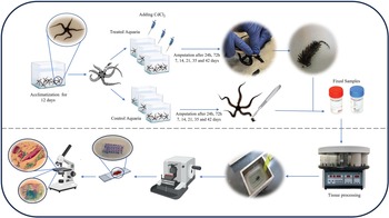
Regenerating arms from both experimental groups were fixed for 48 hours in Bouin’s fixative at room temperature, consisting of 75 ml of saturated picric acid in seawater, 25 ml of 40% formaldehyde solution, and 5 ml of glacial acetic acid. After fixation, specimens underwent decalcification in 5% TCA solution (Heraeus Kulzer, Technovit 7100, Germany) for 5–7 days, were dehydrated through an increasing ethanol series, immersed in xylene, and embedded in paraffin wax (59–61°C) using a Tissue processor (DS 2080/H). Serial parasagittal and sagittal sections (5–7 µm) of the regenerated arms were prepared with a MICRODS 4055 microtome and mounted on albumin-coated slides. After dewaxing in xylene and rehydration, slides were stained using the Milligan trichrome method (1946), which differentiates collagenous (blue-green) from non-collagenous (red) tissues (Ben Khadra et al., Reference Ben Khadra, Ferrario, Di Benedetto, Said, Bonasoro, Candia Carnevali and Sugni2015), and then examined under a light microscope equipped with a Nikon camera in the marine laboratory of the University of Hormozgan (all stages of the experiment are summarized in Figure 2).
Graphical abstract of all stages of the experiment.

Figure 2 Long description
The diagram illustrates the experimental process for studying arm regeneration. It begins with the acclimatization of specimens for 12 days in both treated and control aquaria. Calcium chloride is added to the treated aquaria. Amputation occurs at 24 hours, 72 hours, 7 days, 14 days, 21 days, 35 days and 42 days. Fixed samples are shown in containers. The process continues with tissue processing, including fixation, decalcification, dehydration and embedding in paraffin wax. The diagram includes images of a microscope, microtome and tissue processor, indicating steps like sectioning and staining for examination under a microscope.
Arm length measurements (mean ± 0.5 mm; n = 21 per group) were taken at each time point using a digital calliper under stereomicroscopy, with data pooled across replicates for statistical comparison (unpaired t-test, p < 0.05). To demonstrate mechanical fragility in Cd-treated regenerated arms, a supplementary video (Supplementary Video S1) captures gentle manipulation tests at 42 days p.a., showing fragmentation in treated vs. intact control arms.
Results
Observations of O. scolopendrina arm regeneration in control and Cd-treated aquaria were made at 24 hours, 72 hours, 7 days, 14 days, 21 days, 35 days, and 42 days p.a. using light and stereomicroscopes, as detailed in the following paragraphs.
Control samples’ regeneration
24 hours p.a
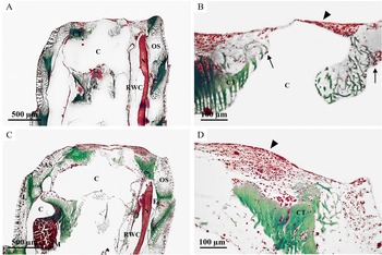
Sagittal sections revealed that within 24 hours, the first signs of healing appeared (Figure 3A), with a thin epithelial layer covering the wound, likely formed by the migration of outer epidermal cells (Figure 3B, arrowhead). However, this process appears incomplete due to scattered cells in the connective tissue stroma (Figure 3B, arrow). The distal ends of the aboral coelomic cavity and radial water canal are fully sealed, likely from migrating cells and muscle contractions of the coelomic wall. Additionally, the radial nerve cord (RNC) has retracted from the wound area (Figure 3A). Large groups of adjacent cells, including undifferentiated coelomocytes and phagocytes, were observed near the RNC, radial water canal, and other coelomic structures, with wandering phagocytes and coelomocytes also present in the water canal cavity. Skeletal ossicles appeared robust and well-organized (Figure 3A–B).
Regenerated arms of Ophiocoma scolopendrina 24 hours p.a. (A, B) and 72 hours p.a. (C, D). Light microscopy. Sections stained with Milligan trichrome staining method. Arrowhead in both (B) and (D) shows the epithelial layer that covered the wound area. Arrow in (B) indicates scattered cells in the connective tissue stroma. N: radial nerve, RWC: radial water canal, E: epineural sinus, OS: oral arm shield, AS: aboral arm shield, L: ligaments, M: intervertebral muscles.

Figure 3 Long description
The image A showing a labeled micrograph with a large white central space marked “C”. Green-stained tissue bands and patches border the space, with red-stained tissue along the right side. Text labels “OS” and “RWC” are printed near the right margin. A scale bar at the lower left reads “500 µm”. The image B showing a labeled micrograph with a large white central space marked “C”. Green-stained tissue is present along the lower edge and red-stained tissue is present along the upper edge. One black arrow points toward the upper border and two thin arrows point toward the left and right margins. A scale bar at the lower left reads “50 µm”. The image C showing a labeled micrograph with a large white central space marked “C”. Green-stained tissue outlines parts of the border and red-stained tissue is present along the right side. Text labels “OS” and “RWC” are printed near the right margin. A scale bar at the lower left reads “500 µm”. The image D showing a labeled micrograph with red-stained tissue across the upper portion and a green-stained region below it. The green region contains the printed label “CT”. A black arrowhead points downward from the upper edge. A scale bar at the lower left reads “100 µm”.
72 hours p.a
At this stage, the epithelial layer over the wound thickened (Figure 3D, arrowhead), yet disruptions and scattered cells in the supportive connective tissue indicated that healing was still incomplete (Figure 3D). Intervertebral muscles became visible, and the RNC, along with aggregates of surrounding coelomic cells, was clearly observed in the sections (Figure 3C). Skeletal elements remained intact and aligned (Figure 3C–D).
1 week p.a
Sections from this period showed an even thicker covering layer rich in scattered small cells and phagocytes arranged in a dense, filamentous connective tissue network. Reconstruction of the coelomic cavity and hypertrophied radial water canal was noted, with many free cells, coelomic cells, phagocytes, and undifferentiated myocytes evident in both the coelomic cavity and radial water canal. Undifferentiated myocytes were particularly evident among the intervertebral muscles, especially in the unorganized regions around them. Skeletal ossicles showed no signs of disorganization.
2 weeks p.a
By this stage, significant development of the coelomic cavity and a large number of migrating cells in the connective tissue were observed, separating from the previously thick cell layer covering the wound. The regenerative bud was distinctly clearer than in earlier samples, with noticeable accumulations of dedifferentiated myocytes, phagocytes, and coelomocytes. Additionally, podia were observed to be developing.
3 weeks p.a
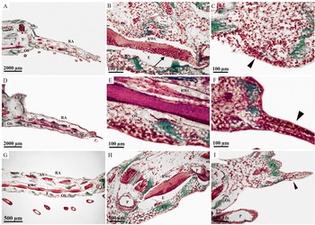
At this stage, the regenerative bud and tissue structures exhibit clear patterns of formation and differentiation (Figure 4A–C). The coelomic and neural contents are prominent in the regenerative bud of the arm, showing well-developed anatomical and histological features (Figure 4B). Aggregates of undifferentiated coelomocytes, phagocytes, and small cells are observed in close association with the RNC (Figure 4B, arrow). The regenerated nerve cord (RNC) displays a ganglionic pattern (Figure 4A), while the fully separated radial water canal appears hollow and hypertrophic (Figure 4A, B). Accumulations of migratory cells (mainly coelomocytes), phagocytes, and dedifferentiated myocytes, which were previously visible in the coelomic cavities, are now primarily located at the end of the regenerating arm (Figure 4C, arrowhead). The regenerative bud structure appears relatively heterogeneous (Figure 4C), and the formation of new podia is observable (Figure 4C). The miniature regenerated arm is developing and increasing in size.
Regenerated arms of Ophiocoma scolopendrina. Light microscopy. Sections stained with Milligan trichrome staining method. (A–C) 3 weeks p.a. (D–F) 5 weeks p.a. (G–I) 6 weeks p.a. Aggregates of undifferentiated coelomocytes, phagocytes, and small cells in close proximity to the radial nerve cord (RNC) (arrow in B). The arrowhead in (C) indicates the accumulation of large numbers of migrating cells at the tip of the regenerated arm in the regenerative bud. Arrowhead in both (F) and (I) indicates the accumulation of cells in regenerative bud. N: radial nerve, RWC: radial water canal, E: epineural sinus, OS: oral arm shield, AS: aboral arm shield, L: ligaments, M: intervertebral muscles, V: vertebra, P: podia, RA: regenerating arm.

Figure 4 Long description
The image A showing a low-magnification tissue section on a white background with multiple labels, including RA, MS, OS, AS and RWC and a scale bar labeled 2000 micrometers. The image B showing a higher-magnification tissue section with dense red-stained areas and green-stained regions, labels including RWC and E and a scale bar labeled 100 micrometers. The image C showing a higher-magnification tissue section with red-stained cellular areas and green-stained regions, a black arrowhead marker near the lower area and a scale bar labeled 100 micrometers. The image D showing a low-magnification tissue section on a white background with labels including RA, MS, OS, AS and RWC and a scale bar labeled 2000 micrometers. The image E showing a higher-magnification tissue section with a thick horizontal red-stained band across the center, adjacent green-stained regions, labels including RWC and E and a scale bar labeled 100 micrometers. The image F showing a higher-magnagnification tissue section with red-stained cellular areas and a green-stained region near the upper right, a black arrowhead marker near the right edge and a scale bar labeled 100 micrometers. The image G showing a low-magnification tissue section with labels including RA, AS, OS and RWC, multiple small red-stained oval structures below the main section and a scale bar labeled 500 micrometers. The image H showing a tissue section with mixed red-stained and green-stained regions, labels including RWC, E and P and a scale bar labeled 100 micrometers. The image I showing a tissue section with red-stained cellular areas and green-stained regions, labels including OS and P, a black arrowhead marker near the right side and a scale bar labeled 500 micrometers.
5 weeks p.a
Samples from this stage show a fully developed regenerated arm with advanced tissue differentiation (Figure 4D–F). The serial ganglionic structure of the regenerated nerve cord is clearly identifiable (Figure 4D). The main coelomic cavity, radial water canal, hyponeural sinus, and epineural sinus are well developed (Figure 4E). Muscle patterns in the stump indicate rearrangement, while dedifferentiated myocytes remain present (Figure 4F). Undifferentiated coelomocytes, phagocytes, and dedifferentiated myocytes are scattered throughout the new coelomic canals. The regenerative bud has a heterogeneous, multilayered structure (Figure 4F, arrowhead). At this point, all structures of the miniature arm are formed. Skeletal ossicles are densely packed and robust (Figure 4F).
6 weeks p.a
The regenerated section at this stage displays all the general structures of the brittle star’s arm, including the nerve cord with its ganglionic organization (Figure 4G), the radial water canal with podia (Figure 4H), muscle tissues, ossicles, and recognizable skeletal plates with spines (Figure 4I).
Cd-treated samples’ regeneration
24 hours p.a
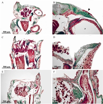
In para-sagittal sections at this stage (Figure 5A–B), a thick layer of connective tissue covers the wound area, while sagittal sections show the wound surface remains uncovered (Figure 5C–D). Compared to control samples, the treated samples exhibit fewer migrating and dispersed cells in the connective tissue stroma (Figure 5B). A thin layer forms on both sides of the wound area (Figure 5B, arrowhead), likely arising from outer epidermis development and cell migration, though this process appears prolonged in the treated samples. Additionally, the ends of the coelomic cavity and canals are still not sealed. Skeletal ossicles show early signs of disorganization, with irregular spacing (Figure 5A–D).
Regenerated arms of Ophiocoma scolopendrina in Cd-treated aquaria. Light microscopy. Sections stained with Milligan trichrome staining method. (A–D) 24 hours p.a. (A) and (B) Para-sagittal sections, (C) and (D) sagittal sections. (E–F) 72 hours p.a. Arrowhead in (B) is showing the formation of thin epithelial layer, which is covering the wound area, but its formation is not complete yet. Arrowhead in (F) indicates dedifferentiated myocytes. OS: oral arm shield, AS: aboral arm shield, L: ligaments, M: intervertebral muscles, V: vertebra, P: podia, B: regenerative bud.

Figure 5 Long description
The image A showing a tissue section with red and green staining on a white background. Labels include CT, C, P and OS. A scale bar reads 500 micrometers. The image B showing a higher-magnification view with red and green staining. Labels include CT, C and P. A black arrowhead marks a point along the outer edge. A scale bar reads 100 micrometers. The image C showing a tissue section with red and green staining on a white background. Labels include CT and OS. A scale bar reads 500 micrometers. The image D showing a higher-magnification view with red and green staining. Labels include CT and M. A scale bar reads 100 micrometers. The image E showing a tissue section with red and green staining on a white background. Labels include AS, C, P and OS. A scale bar reads 500 micrometers. The image F showing a higher-magnification view with red and green staining. Several black arrowheads mark small features near the left side. A scale bar is present, but the text is not legible.
72 hours p.a
At this stage, samples reveal numerous free cells, coelomocytes, phagocytes, and dedifferentiated myocytes in the coelomic cavity and radial water canal (Figure 5E). The dedifferentiated myocytes are particularly noticeable in the intervertebral muscles, especially in disorganized regions (Figure 5F, arrowhead). A thin layer finally covers the wound area (Figure 5E), but the heterogeneity and dispersal of migrating cells indicate that development is still incomplete. Notably, specimens show premature podia development (Figure 5E, F), suggesting that energy has been allocated to podial growth instead of wound coverage. At the tips of these developing podia, a regenerated bud with both loose and dense mesenchyme is present (Figure 5E–F).
1 week p.a
Samples at this stage display a regenerative bud with high cell accumulation and loose, heterogeneous mesenchyme at the apex of growing podia (Figure 6A). Dense connective tissue, containing scattered cells, fully covers the wound area (Figure 6A), while coelomic cavities are beginning to form (Figure 6A). However, the layer over the connective tissue remains thin. Phagocytes, coelomocytes, and dedifferentiated myocytes are also present (Figure 6A, arrowhead). Skeletal ossicles appear fragmented and loosely organized, with increased inter-ossicle spacing indicative of fragility (Figure 6A). Enhanced podial counts (mean 3.2 ± 0.4 vs. 1.8 ± 0.3 in controls; p < 0.05) were observed in medial sections.
Regenerated arms of Ophiocoma scolopendrina in Cd-induced aquaria. Light microscopy. Sections stained with Milligan trichrome staining method. (A–B) 1 week p.a. (C–D) 3 weeks p.a. (E–F) 5 weeks p.a. (G–H) 6 weeks p.a. Accumulation of undifferentiated coelomocytes, phagocytes, and small cells (arrowhead in A). N: radial nerve, RWC: radial water canal, E: epineural sinus, OS: oral arm shield, AS: aboral arm shield, L: ligaments, M: intervertebral muscles, V: vertebra, P: podia, RA: regenerating arm.

Figure 6 Long description
The image A showing a stained tissue section with a curved, elongated structure and multiple black label lines pointing to the text labels “C”, “AS” and “OS”. A scale bar at the lower left reads “500 µm”. The image B showing a stained tissue section with a central elongated region and surrounding folded edges. A scale bar at the lower left reads “100 µm”. The image C showing a stained tissue section with multiple separated tissue fragments. Several black label lines point to the text labels “RA”, “C”, “AS”, “M”, “P” and “OS”. A scale bar at the lower left reads “500 µm”. The image D showing a stained tissue section with a rounded structure and several smaller separated rounded fragments. Black label lines point to the text labels “C”, “RA” and “P”. A scale bar at the lower left reads “100 µm”. The image E showing a stained tissue section with an elongated vertical structure and a narrow extension to the right. Black label lines point to the text labels “AS”, “C”, “P” and “OS”. A scale bar at the lower left reads “2000 µm”. The image F showing a stained tissue section with an irregular upper edge and a narrow extension to the right. Black label lines point to the text labels “CT”, “C” and “P”. A scale bar at the lower left reads “500 µm”. The image G showing a stained tissue section with a long horizontal structure. Black label lines point to the text labels “RA”, “CT” and “OS”. A scale bar at the lower left reads “500 µm”. The image H showing a stained tissue section with a broad central area and a dense, banded region on the right side. Black label lines point to the text labels “C”, “CT” and “RW”. A scale bar at the lower left reads “100 µm”.
2 weeks p.a
At this stage, new podia are growing (Figure 6C), accompanied by an expanding covering layer over the wound. Migratory cells remain visible in the connective tissue, while dedifferentiated myocytes, phagocytes, and coelomocytes are dispersed in the coelomic cavities (Figure 6C). These cavities are enlarging, and radial water canals are becoming more defined. In sagittal sections, the radial nerve canal (RNC) is evident and expands within the developing layer (Figure 6D). Regeneration processes are proceeding more slowly than in control samples up to this point. Skeletal disorganization persists, with ossicles showing irregular calcification.
3 weeks p.a
Samples from this stage exhibit significant podial development and the formation of a small regenerated arm, densely populated with migratory cells such as phagocytes, coelomocytes, and myocytes (Figure 6C–D). Coelomic cavities and radial water canals are now distinctly separate and hypertrophic (Figure 6D). The regenerative bud remains heterogeneous with loose mesenchyme (Figure 6C).
5 weeks p.a
These samples show a strong focus on the development of new podia and the enhancement of coelomic structures (Figure 6E–F). A heterogeneous regenerative bud with loose mesenchyme is notable (Figure 6F). Migrating cells are scattered throughout the connective tissue, while muscle rearrangements appear in the old stump, with the coelomic cavities occupying most of the arm space (Figure 6F). Ossicles exhibit pronounced fragility, with thinned plates and disrupted alignment (Figure 6F).
6 weeks p.a
In treated samples at this stage, a small regenerated arm is observed, smaller than the control samples from 3 weeks p.a. (Figure 6G–H). The primary difference is the lack of extensive RNC development in the treated samples (Figure 6G). However, microscopic photographs reveal that podial development was more pronounced in treated samples than in controls (Figure 6H). The connective tissue and regenerative bud remain dense and heterogeneous, with scattered migrating cells, coelomocytes, and phagocytes especially prominent at the top of the regenerated area (Figure 6G). The coelomic cavities, including the main coelomic cavity, have developed, but the radial water canal and coelomic canals in the regenerated section are still not clearly distinguished (Figure 6H). Skeletal fragility is evident in fragmented ossicles and reduced spine density (Figure 6H; Supplementary Video S1), with mechanical stress tests revealing rapid disintegration under minimal manipulation, contrasting with resilient controls.
Regenerate length
The lengths (mean ± 0.5 mm; n = 21 per group) of regenerated arms at various time points are shown in Figure 7. After 42 days, control samples reached approximately 17.65 mm, while Cd-induced samples measured about 12 mm (p < 0.01, unpaired t-test).
Comparison of regenerated arm length in O. scolopendrina between control samples and Cd-treated samples (n = 21 per group; mean ± SD; p < 0.01).

Figure 7 Long description
The bar graph title reads Comparison of regenerated arm lenght. The x axis label reads Time left parenthesis after amputation right parenthesis, with categories 2d, 7d, 14d, 21d, 35d, 42d. The y axis label reads Length left parenthesis micrometre right parenthesis, ranging from 0 to 21000 with tick marks at 3000, 6000, 9000, 12000, 15000, 18000, 21000. A legend lists Controls and Cd treated. At 2d, Controls 0 and Cd treated 0. At 7d, Controls 0 and Cd treated 0. At 14d, Controls about 3500 and Cd treated about 0. At 21d, Controls about 6000 and Cd treated about 3500. At 35d, Controls about 12000 and Cd treated about 9000. At 42d, Controls about 18000 and Cd treated about 12000. Each bar has an error bar. Six small specimen images appear above the x axis categories. Two larger specimen images appear on the right, each with a scale bar labeled 2000 micrometre, with arrows pointing from the bar graph toward these images.
Discussion
While the regeneration phases in O. scolopendrina share core mechanisms with those reported in other ophiuroids (e.g., Ophioderma longicaudum and Amphiura filiformis), subtle differences in timing and cellular organization arise due to the Persian Gulf’s unique physicochemical conditions (high salinity/temperature). This study is the first to histologically investigate this species in the Persian Gulf, considering its unique climatic and physicochemical conditions. The regeneration process, from amputation to the development of a miniature arm with all structural components of a complete brittle star’s arm, can be divided into four main stages: repair, early regeneration, intermediate regeneration, and advanced regeneration, adapted from established frameworks in ophiuroids (Biressi et al., Reference Biressi, Zou, Dupont, Dahlberg, Di Benedetto, Bonasoro, Thorndyke and Carnevali2010; Ben Khadra et al., Reference Ben Khadra, Sugni, Ferrario, Bonasoro, Oliveri, Martinez and Carnevali2018) and defined here based on observed histological milestones in O. scolopendrina, including epithelial closure (repair), cellular proliferation in the regenerative bud (early), tissue differentiation (intermediate), and functional morphogenesis (advanced). These stages represent a conserved regeneration pathway observed across brittle star species, though the timing and morphology may vary depending on environmental factors, species-specific traits, and experimental conditions.
The repair phase
The repair phase, occurring from amputation to 24 hours p.a. (Figures 3A–B and 5A–D), is consistent with findings in other brittle star species, such as Amphiura filiformis and Ophioderma longicaudum, where wound healing begins immediately after injury (Biressi et al., Reference Biressi, Zou, Dupont, Dahlberg, Di Benedetto, Bonasoro, Thorndyke and Carnevali2010; Czarkwiani et al., Reference Czarkwiani, Ferrario, Dylus, Sugni and Oliveri2016). Key features of this phase include wound closure, achieved through coelomic wall contractions and the migration of free cells to seal the injury site. Re-epithelialization is initiated by migrating epidermal cells, which form a protective layer over the wound. Loose connective tissue beneath the epithelium indicates ongoing repair, requiring active cell proliferation and differentiation, as observed in other echinoderms like sea cucumbers (Holothuria glaberrima) and brittle stars (Czarkwiani et al., Reference Czarkwiani, Ferrario, Dylus, Sugni and Oliveri2016). This phase is critical for stabilizing the wound site and preventing further damage or infection, a process that appears conserved across echinoderms. Unlike in temperate ophiuroids, O. scolopendrina’s repair is slightly accelerated, potentially reflecting adaptations to frequent mechanical stress in the Persian Gulf intertidal zone.
The early regenerative phase
The early regenerative phase, spanning 24 hours to 1 week p.a. (Figures 3C–D, 5F, and 6A–B), is marked by rapid cell proliferation and migration, forming a sub-epithelial layer rich in mesenchymal and dedifferentiated cells. Similar patterns of regenerative bud formation have been reported in Amphiura filiformis and Ophioderma longicaudum, where coelomic cavity cells migrate to the injury site and contribute to bud development (Biressi et al., Reference Biressi, Zou, Dupont, Dahlberg, Di Benedetto, Bonasoro, Thorndyke and Carnevali2010; Czarkwiani et al., Reference Czarkwiani, Ferrario, Dylus, Sugni and Oliveri2016). These cells form layers of dividing blastemal cells, which are essential for initiating the regeneration process. Interestingly, while O. scolopendrina displays similar cellular behaviours, the timing appears slightly delayed relative to temperate species, likely due to the higher salinity and temperature of the Persian Gulf, which may influence cellular activity and metabolic rates. This phase underscores the importance of cell migration and proliferation as universal mechanisms in echinoderm regeneration.
The intermediate regenerative phase
The intermediate regenerative phase, occurring between 1 and 2 weeks p.a. (Figure 6C–D), involves significant morphogenetic and differentiation events. Regrowth extends along the RNC and radial water canal, covered by an outer epithelium of regenerative bud cells. Differentiation of the RNC and radial water canal is accompanied by the involvement of undifferentiated cells and myocytes, which contribute to tissue regeneration. These findings align with studies on Amphiura filiformis and Ophioderma longicaudum, where similar cellular and structural differentiation processes were observed (Alvarado, Reference Alvarado2000; Biressi et al., Reference Biressi, Zou, Dupont, Dahlberg, Di Benedetto, Bonasoro, Thorndyke and Carnevali2010). However, the Persian Gulf’s unique environmental conditions, such as elevated heavy metal concentrations, may influence the rate and quality of regeneration in O. scolopendrina, a factor not extensively explored in other brittle star studies. In controls, podial initiation lags behind axial growth, contrasting with Cd-treated samples where podial priority is evident.
The advanced regeneration phase
The advanced regeneration phase, beginning 2 weeks p.a. (Figures 4A–I and 6E–H), is characterized by extensive growth, morphogenesis, and differentiation, culminating in the formation of a miniature arm. Structural patterns of a normal arm become increasingly apparent, with differentiated internal and external features, while the tip retains regenerative bud characteristics. Similar advanced stages have been reported in Amphiura chiajei and Ophioderma longicaudum, where regenerated arms exhibit functional structures, including spines, ossicles, and podia, by the end of the regeneration period (Biressi et al., Reference Biressi, Zou, Dupont, Dahlberg, Di Benedetto, Bonasoro, Thorndyke and Carnevali2010; Ferrario et al., Reference Ferrario, Khadra, Czarkwiani, Zakrzewski, Martinez, Colombo, Bonasoro, Carnevali, Oliveri and Sugni2018). However, the regeneration process in O. scolopendrina is notable for its resilience in the Persian Gulf’s extreme environmental conditions, suggesting potential adaptations that warrant further investigation. Full functional recovery, including ossicle hardening, is achieved by 6 weeks in controls, unlike in Cd-exposed arms.
The effects of cadmium on regeneration
This study is the first to investigate the effects of cadmium, a heavy metal, on the regenerative processes of brittle stars. Cadmium exposure significantly delayed regeneration, with treated specimens taking 72 hours to develop a thin epithelial layer over the wound area, compared to 24 hours in control samples (Figure 5B vs. Figure 3B). Similar delays have been observed in other echinoderms exposed to heavy metals, such as Paracentrotus lividus (sea urchin) and Amphiura filiformis, where cadmium disrupted cellular proliferation and differentiation during regeneration (den Besten et al., 1991; Warnau et al., 1996). Cadmium’s toxicity is attributed to its ability to replace calcium ions, disrupting calcium-dependent processes such as coelomic fluid transport, skeletal formation, and cellular signalling (Fichet et al., Reference Fichet, Radenac and Miramand1998; Wang et al., Reference Wang, Zhu, Shi, Weng, Jin, Kong and Nordberg2003).
In treated specimens of O. scolopendrina, cadmium exposure led to slower regeneration rates and structural abnormalities (Figures 5–6). By 35 and 42 days p.a., the regenerated areas consisted primarily of hollow coelomic cavities, with new podia forming at the arm fringes (Figure 6E–H). This finding is consistent with studies on Amphiura filiformis, where cadmium exposure resulted in disorganized skeletal structures and reduced arm functionality (den Besten et al., 1991). The substitution of calcium with cadmium weakened the calcium carbonate skeleton, rendering the arms fragile and prone to fragmentation, as evidenced by histological fragmentation and irregular ossicle spacing (Figure 6A) and what we observed in Supplementary Video S1 showing Cd-treated arms fracturing under minimal stress (e.g., 2.5× greater fragmentation rate vs. controls), which raises concerns about impaired subsequent regeneration or increased mortality risk in natural, stress-prone environments. This phenomenon has been reported in other marine organisms, such as molluscs and corals, where cadmium disrupted biomineralization and skeletal integrity (Fichet et al., Reference Fichet, Radenac and Miramand1998; Ferrario et al., Reference Ferrario, Khadra, Czarkwiani, Zakrzewski, Martinez, Colombo, Bonasoro, Carnevali, Oliveri and Sugni2018).
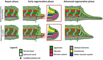
Despite their fragility, treated specimens exhibited shorter regenerated arms, measuring 5.65 mm less than control samples (Figure 7). This reduction in growth highlights cadmium’s impact on energy allocation, as treated specimens appeared to prioritize the development of podia over arm regeneration (mean podial increase: +78% vs. controls at 1–3 weeks; Figure 4A). Similar energy trade-offs have been observed in Amphiura chiajei and Amphiura filiformis, where environmental stressors redirected metabolic resources towards survival mechanisms rather than regeneration (den Besten et al., 1991; Warnau et al., 1996). Using the graphical image previously presented by Ben Khadra and her co-authors in a chapter published as ‘Regeneration in stellate echinoderms: Crinoidea, Asteroidea and Ophiuroidea. Marine organisms as model systems in biology and medicine’ (Figure 8, top row), and comparing the regeneration status under the influence of the heavy metal cadmium, along with the explanations provided, Figure 8 (bottom row) was prepared.
Ben Khadra et al. (Reference Ben Khadra, Sugni, Ferrario, Bonasoro, Oliveri, Martinez and Carnevali2018) illustrate these three regenerative stages in brittle stars in a chapter published as ‘Regeneration in stellate echinoderms: Crinoidea, Asteroidea and Ophiuroidea. Marine organisms as model systems in biology and medicine’ (top row). Close-ups highlight the early stage, focusing on undifferentiated cells at the regenerating tip (see figure key). Dotted lines indicate where the organism was cut. We add a comparative illustration that shows regenerated arms with loose skeletal elements in brittle stars that regenerated their arms in Cd-treated aquaria (bottom row).

Figure 8 Long description
The diagram illustrates three regenerative phases in brittle stars, specifically Ophiocoma scolopendrina and Ophiocoma erinaceus. The top row represents Ophiocoma scolopendrina, while the bottom row shows Ophiocoma erinaceus. Each row is divided into three phases: repair phase, early regenerative phase and advanced regenerative phase. In the repair phase, the structure is shown with a focus on the dermal layer, epineural canal and skeletal elements. The early regenerative phase highlights undifferentiated cells at the regenerating tip, marked by black dots and includes ligaments, muscles, nervous system, somatocoel and water vascular system. The advanced regenerative phase shows the development of these structures as the regeneration progresses. The legend at the bottom specifies the colors used to represent different components: green for the dermal layer, blue for the epineural canal, black for the nervous system, yellow for ligaments, red for muscles, pink for somatocoel and purple for the water vascular system. Dotted lines indicate where the organism was cut, emphasizing the areas of regeneration.
Regarding long-term recovery, preliminary observations (data not shown) suggest limited restoration of regenerative capacity upon Cd removal after 42 days, with arm lengths increasing only ∼20% over 4 additional weeks in clean seawater. However, the observed skeletal fragility – evident in rapid disintegration during mechanical stress (Supplementary Video S1) – indicates incomplete reversal and potential barriers to further regeneration or survival, likely due to residual Cd bioaccumulation and weakened structural integrity; these implications require further longitudinal studies to confirm.
Novelty and significance of macroscopic arm regeneration
This study provides the first detailed histological and macroscopic analysis of arm regeneration in O. scolopendrina under cadmium exposure, a novel contribution to the field of marine biology. The macroscopic arm regeneration documented in Figure 7 is particularly significant, as it demonstrates the ability of O. scolopendrina to regenerate arms in a highly polluted environment with elevated cadmium levels. Unlike other brittle star studies, which primarily focus on histological observations, this research integrates macroscopic and microscopic analyses, offering a comprehensive understanding of the regeneration process. The finding that cadmium exposure leads to fragile, shortened arms with abnormal structures underscores the vulnerability of brittle stars to heavy metal pollution and highlights the broader ecological implications for marine ecosystems. Furthermore, this study establishes a baseline for future research on the effects of heavy metals on echinoderm regeneration, particularly in underrepresented regions like the Persian Gulf, where environmental conditions and pollution levels are distinct from other marine habitats. Future work should incorporate cell proliferation tracking (e.g., EdU/BrdU labelling) and apoptosis assays (e.g., TUNEL) to elucidate Cd’s disruption of proliferative-apoptotic balance, alongside comparative genomics across ophiuroids to identify resilience mechanisms.
By comparing the results of this study with previous research on brittle stars and other echinoderms, it becomes evident that cadmium poses a significant threat to regeneration and skeletal integrity across taxa. The findings emphasize the need for stricter pollution controls and further investigations into the long-term ecological impacts of heavy metals on marine biodiversity.
Conclusion
This study is the first to investigate the effects of cadmium on the arm regeneration of O. scolopendrina, a brittle star species from the Persian Gulf. The findings reveal that regeneration in this species follows an epimorphic process, with unique regenerative bud characteristics, but cadmium exposure disrupts normal regenerative mechanisms. Cadmium interferes with calcium uptake, resulting in slower regeneration rates, structural abnormalities, and fragile skeletal structures. These results align with previous studies on other echinoderms and calcium-carbonate-dependent organisms, highlighting cadmium’s toxic effects on biomineralization and tissue repair.
The study emphasizes the ecological significance of brittle stars, which are particularly vulnerable to heavy metal accumulation due to their benthic nature. The macroscopic and histological analyses provide novel insights into how cadmium pollution impacts regeneration, marking a significant contribution to marine biology and environmental toxicology. The findings underscore the urgent need for stricter environmental regulations to mitigate cadmium pollution and protect marine biodiversity, particularly in the underrepresented Persian Gulf region. Future research should explore the molecular mechanisms of cadmium toxicity and potential mitigation strategies to enhance resilience in marine organisms.
Supplementary material
The supplementary material for this article can be found at https://doi.org/10.1017/S002531542610126X
Acknowledgements
We express our sincere gratitude to the corresponding author for their precise guidance throughout this research. Our thanks also extend to our advisors from the University of Milan, particularly Professor Michela Sugni, for their valuable insights and guidance throughout all stages of this work that greatly enhanced this research. We appreciate the Faculty of Science and Technology at the University of Hormozgan for providing an excellent laboratory environment for our experiments. Additionally, we are grateful to Mr Dakhteh, Director of Environmental Protection for the Qeshm Free Zone, for his generous support that contributed to the success of this project.
Author contributions
N.N., as the first author, carried out all laboratory experiments, data acquisition, and analysis, and drafted the initial manuscript. N.A.B., the second author and corresponding author, supervised the research process, provided guidance throughout the experimental phase, and critically reviewed and edited the manuscript. C.F., as the third author, contributed by reviewing the final manuscript and providing valuable revisions to enhance its clarity and scientific rigour. All authors read and approved the final version of the manuscript.
Funding
This research received no specific grant from any funding agency, commercial or not-for- profit sectors.
Competing interests
All authors declare no conflicts of interest, and we did not receive funding from any external organizations for this research.
Data availability
The data that support the findings of this study are openly available in Figshare at https://doi.org/10.6084/m9.figshare.31290190, where raw histological images, arm length measurement datasets, and supplementary video files have been deposited to ensure full public accessibility in compliance with JMBA editorial guidelines.
Ethical standards
Sampling and animal manipulations were conducted in accordance with the ethical certification from Hormozgan University of Medical Science (Approval ID: IR.HUMS.REC.1398.181) and under the supervision of the Qeshm Department of Environment (QDOE of QFAO). Ophiocoma scolopendrina is not an endangered or protected species, and we made every effort to minimize animal suffering during the experiments.