Most gastrointestinal disease-related hospital admissions are due to pancreatitis, which has a significant risk of morbidity, death and socio-economic hardship(Reference Mayerle, Sendler and Hegyi1). Acute pancreatitis (AP) is an unpredictable and potentially lethal disease. The critical determinants of prognosis are the development of organ failure and subsequent infection of pancreatic or peripancreatic necrosis(Reference Boxhoorn, Voermans and Bouwense2). The incidence and mortality rates for AP were estimated to be 33·74 cases (95 % CI 23·33, 48·81) per 100 000 person-years and 1·60 fatalities (95 % CI 0·85, 1·58) per 100 000 person-years globally(Reference Xiao, Tan and Wu3). The last 10 years have seen a shift in the management of AP towards a multidisciplinary, individualised and minimally invasive strategy. Despite advances in therapy and critical care, severe AP still has a significant death rate.
In up to 20 % of patients, severe AP may develop(Reference Besselink, van Santvoort and Boermeester4,Reference Banks, Bollen and Dervenis5) . In the early phase of severe AP, a pro-inflammatory response develops that is characterised by the release of large quantities of cytokines. This cytokine storm may trigger a systemic inflammatory response syndrome) and often leads to organ failure. Organ failure is associated with a mortality of up to 35 % in AP(Reference van Santvoort, Besselink and Bakker6,Reference Rhee, Dantes and Epstein7) .
n-3 fatty acids (FA) are found in fish oil and contain EPA and DHA(Reference Calder8). Multiple clinical studies have found that n-3 fatty acids may improve the prognosis of AP and reduce in-hospital deaths and ICU admissions(Reference Lasztity, Hamvas and Biró9–Reference Smith and Ebrahim16). However, the specific role of n-3 fatty acids in AP is not well understood. The pieces of evidence for these studies are derived from clinical observational studies. Unmeasured confounding factors or reverse causality are continuously interfered with conventional observational findings. Considering the vulnerability of observational studies to confounding and reverse causality, it is still uncertain the causal impact of circulating n-3 fatty acid levels on the elevated risk of AP.
Genome-wide association studies (GWAS) involve testing genetic variants across the genomes of many individuals to identify genotype–phenotype associations. GWAS have revolutionised the field of complex disease genetics over the past decade, providing numerous compelling associations for complex human traits and diseases. Genetic tools are used as instrumental variables (IV) in the Mendelian randomisation (MR) design to distinguish between correlation and causality in observed data. It accomplishes causality exploration by decreasing the possibility of reverse causality and minimising residual confusion. Individual qualities are often unrelated because genetic variation is randomly distributed after conception. This procedure is comparable to a randomised controlled trial (RCT), randomly assigning individuals to experimental and control groups. It ensures that those with genetic variations linked to higher risk factors are distributed equally among the groups, minimising the possibility that these risk factors would have confusing effects. Since alleles are fixed and unaffected by the beginning or progression of the disease, MR analysis also helps to avoid the problem of reverse causality(Reference Smith and Ebrahim16). Even though MR has been used in pancreatitis(Reference Mao, Mao and Sun17–Reference Ling, Liang and Mo19), the exposure factors currently being studied are only traditional risk factors. It is still very encouraging to use MR to further investigate the function and value of metabolomics in pancreatitis at the gene level.
Therefore, in order to further explore the role of n-3 fatty acids in AP and whether there is a causal effect on the pathogenesis of AP, we conducted a systematic review and meta-analysis of RCT of n-3 fatty acids in the treatment of AP. Furthermore, we investigated the causal effect of n-3 fat on AP based on MR analysis.
Materials and methods
Study design
This work developed primary analyses to research the effects of n-3 fatty acids complementary therapy on AP based on system review and meta-analysis. We performed a meta-analysis of RCT studies involving n-3 fatty acids in the treatment of AP. The evaluation indicators included mortality, infectious complications, length of hospital stay and length of ICU stay. Furthermore, to further investigate the causal effect of n-3 fatty acids on the onset of AP, we performed a MR analysis. The IV of n-3 fatty acids came from one metabolomics quantitative trait loci study on circulating metabolites. The studies used the 249 circulating metabolites linked to human genetic variations through genome-wide association scans and high-throughput metabolic profiling. We achieved these circulating metabolites from the UK Biobank (unpublished, accessible via the MRC IEU OpenGWAS database). To strengthen the validity of our conclusions, we developed two-sample MR analyses. We used publicly available summary statistics. Therefore, it is unnecessary to obtain new ethical approval.
The search strategy of meta-analysis
Keywords, including ‘n-3 fatty acids’, ‘n-3 FA’ and ‘acute pancreatitis’, were searched in the PubMed, Embase and Cochrane Library databases up to 31 January 2024. We screened studies according to the following inclusion and exclusion criteria (see below). This study followed the guidelines of the Cochrane Collaboration Handbook, observational studies in epidemiology statement(Reference Tu and Greenwood20), Meta-Analysis and Systemic Reviews of Observational Studies (MOOSE) and Preferred Reporting Items for Systematic Review and Meta-Analysis (PRISMA)(Reference Moher, Liberati and Tetzlaff21). This systematic review and meta-analysis protocol was registered in the International Prospective Register of Systematic Reviews (PROSPERO Number: CRD42022353127).
Inclusion and exclusion criteria
The inclusion criteria are as follows: (1) reported the definition of n-3 fatty acids intervention duration, (2) available outcome events (mortality, infectious complications, length of hospital stay and length of ICU stay) or data for meta-analysis and (3) RCT. The exclusion criteria are as follows: (1) if the report is duplicated, only the most recent report is included and (2) the whole research process is not reported in detail (conference paper without full text).
Study selection, data extraction and quality assessment
Studies were independently selected and assessed by three reviewers (TBB, GAJ and YG). Duplicated articles were excluded by reviewing the title and abstract. The three reviewers independently assessed these studies for eligibility. Then, data including author, study type, region, study size (patients/controls), duration of intervention, mean age, dose, route of nutrition, severity criteria of use and the interest outcomes (e.g. mortality, infectious complications, length of hospital stay and length of ICU stay) in each study and baseline characteristics in each study were extracted by the reviewers. The Cochrane risk-of-bias tool was used to evaluate the quality of the included RCT. This tool assessed bias across the following seven domains: (i) random-sequence generation, (ii) allocation concealment, (iii) blinding of participants and personnel, (iv) blinding of outcome assessment, (v) incomplete outcome data, (vi) selective reporting and (vii) other bias. Each domain was determined as low risk, unclear risk or high risk.
Data sources
Metabolic profile for analyses
Summary-level datasets on 249 circulating metabolites used in primary analysis were obtained from Nightingale Health Metabolic Biomarkers Phase 1 release study in UK Biobank (June 2019–April 2020). This study included 115 078 randomly selected participants. Metabolic biomarkers were measured with non-fasting baseline EDTA plasma samples by high-throughput NMR (https://biobank.ndph.ox.ac.UK/ukb/label.cgi?id=220). The biomarkers include 168 absolute metabolites (unit, mmol/l) and eighty-one metabolite ratios spanning multiple metabolic pathways such as lipoproteins, fatty acids, amino acids and ketone bodies. The details of sample collection and NMR profiling have been depicted in previous publications(Reference Mi, Jiang and Liu22–Reference Cohen, Chalumeau and Cohen24).
Instrumental variable selection
We screened SNP relevant to metabolite biomarkers by a traditional genome-wide association significance criterion (P < 5 × 10⁻⁸). We excluded SNP that were in linkage disequilibrium (R2 > 0·001 or within ±10 000 kb of the 1000 Genomes European-ancestry Reference Panel). The F-statistics were applied to measure the strength of IV. We computed mean F-statistics to test for weak instruments. Calculation of the F-statistic (β 2/SE2) to assess weak IV bias. An F-value below 10 indicates a weak IV bias, which might lead to an underestimation of statistical power.
Genome-wide association studies summary data for acute pancreatitis
GWAS refer to multi-centre, large sample and repeatedly validated studies on the association between genes and diseases at the whole genome level. It is a research method that uses high-density genetic markers (such as SNP) to genotype large-scale population DNA samples in order to identify genetic factors associated with complex diseases and comprehensively reveal genetic genes related to disease occurrence, development and treatment. We searched GWAS summary data for AP from the GWAS catalog accession. The GWASID is finn-b-K11_ACUTPANC (https://gwas.mrcieu.ac.UK/datasets/finn-b-K11_ACUTPANC/). In particular, the GWAS data with 16 380 428 SNP were obtained from the Finland Biobank-related AP, with a total sample size of 198 166 Europeans, 3022 cases and 195 144 controls.
Mendelian randomisation
This study used ten MR analytic tools. They are MR-Egger and MR-Egger (bootstrap), the Wald ratio, inverse-variance weighted (IVW) (fixed-effect and random-effect), simple mode, simple median, weighted mode and penalised weighted median.
The IVW method was utilised as the primary method for causal estimation in that it provides a valid causal estimate despite heterogeneity. Wald ratios of the impacts of each SNP on the outcome were coupled with either a fixed-effect IVW for IV ≤ 3 or a random-effect IVW for IV > 3. Cochran’s Q values, I2 and the H-statistics were used to estimate the heterogeneities of the IVW studies(Reference Hartwig, Davey Smith and Bowden25). P < 0·05 from the Cochran Q test was seen as a sign of outcome heterogeneity(Reference Hartwig, Davey Smith and Bowden25). Further sensitivity studies used weighted mode, weighted median and MR-Egger(Reference Bowden, Davey Smith and Burgess26,Reference Hemani, Zheng and Elsworth27) . Due to its ability to identify and correct horizontal pleiotropy, the MR-Egger technique may provide reliable causal estimates even when pleiotropy is present (P for intercept < 0·05)(Reference Hemani, Zheng and Elsworth27). When up to 50 % of the weight in the MR analyses originated from unreliable instrument variables, the weighted median is a strategy that can be utilised to reinforce the causal estimations(Reference Bowden, Davey Smith and Burgess26). The effect estimate supported by the most significant number of genetic instruments is reported using the weighted mode technique(Reference Hemani, Zheng and Elsworth27). In R version 3.4.1 (R Foundation for Statistical Computing), all statistical analyses were performed using the ‘TwoSampleMR’ and ‘Mendelian randomisation’(Reference Yavorska and Burgess28) packages. Statistical significance was determined to have a two-tailed P-value of less than 0·05.
Results
Inclusion of studies
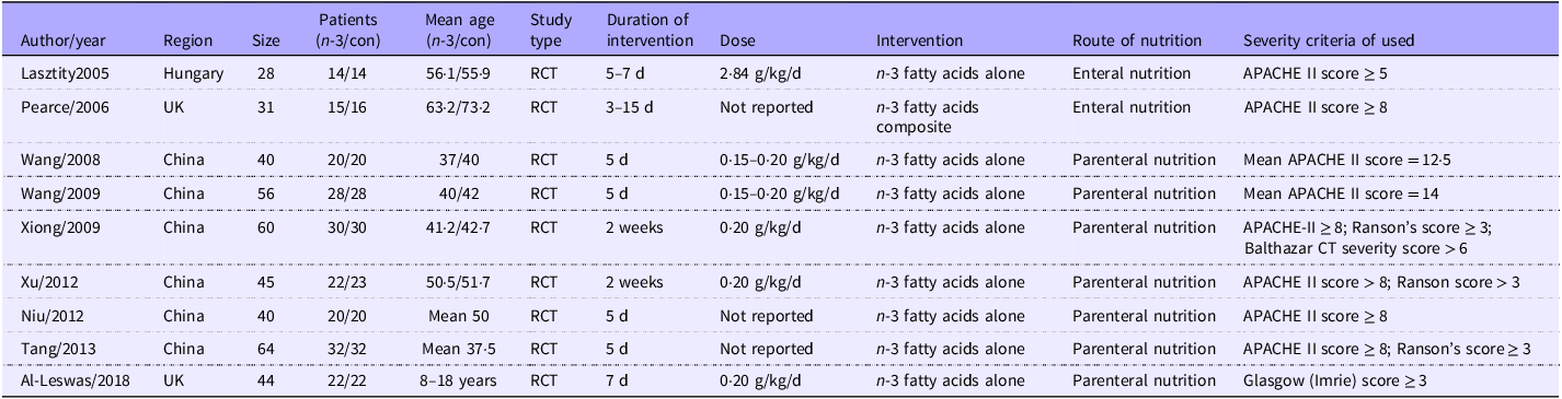
Based on the search strategy, 235 studies were screened out. After removing unrelated ones, nine RCT were finally included in our study. The screening strategy is shown in Fig. 1. The general information of these studies is shown in Table 1. The risk bias score for the study is presented in online Supplementary Fig. 1. Among the included 408 individuals, 305 (74·75 %) individuals have come from China. Two hundred and three patients were randomised to n-3 fatty acids treatment, and 205 patients were placed in the control group. Seven studies administered parenteral nutrition, while two studies used enteral nutrition. The subjects were middle-aged and elderly patients. The duration of the intervention was over 5 d. The intervention dose was a standard dose of 0·15–0·20 g/kg/d.
Flow chart illustrating the study selection process.

Characteristics of included studies

RCT, randomised controlled trial; APACHE, Acute Physiology and Chronic Health Evaluation.
Search findings
Of these studies, eight studies reported the mortality, and the results of the meta-analysis showed that complementary therapy of n-3 fatty acids significantly reduced mortality (RR: 0·30; 95 % CI 0·14, 0·65, P < 0·05; Fig. 2). Seven studies reported the infectious complications, and the results of meta-analysis showed that complementary therapy of n-3 fatty acids significantly reduced infectious complications in AP (RR: 0·45; 95 % CI 0·27, 0·77, P < 0·05; Fig. 3). Six studies reported the length of hospital stay, and the results of meta-analysis showed that complementary therapy of n-3 fatty acids significantly reduced length of hospital stay in AP (MD: –1·02; 95 % CI –1·85, –0·20, P < 0·05; Fig. 4). Four studies reported the length of ICU stay, and the results of meta-analysis showed that complementary therapy of n-3 fatty acids also reduced length of ICU stay in AP (MD: –0·49; 95 % CI –1·29, –0·31, P < 0·05; Fig. 5) but without significant difference.
Forest plot of the effect of n-3 fatty acids on mortality in acute pancreatitis. IV, inverse variance (statistical method).

Forest plot of pooled estimates of n-3 fatty acid supplementation on infectious complications in acute pancreatitis. IV, inverse variance (statistical method).

Forest plot of pooled estimates of n-3 fatty acid supplementation on length of hospital stay in acute pancreatitis. IV, inverse variance (statistical method).

Forest plot of pooled estimates of n-3 fatty acid supplementation on length of ICU stay in acute pancreatitis. IV, inverse variance (statistical method).

Mendelian randomisation study
Based on previous studies, we comprehensively reviewed the risk factors for AP and found that seventy-five genome-wide SNP were significantly associated with n-3 fatty acids. Checking these seventy-five SNP in the PhenoScanner V2 web(Reference Kamat, Blackshaw and Young29), we discovered that one SNP was associated with the risk of AP, and it was removed from the raw data. Ultimately, seventy-five SNP were included in the two-sample MR analysis. The characteristics of these seventy-five SNP are listed in online Supplementary Table 1. The results derived from nine MR analysis methods are shown in Fig. 6(a). Nine MR analysis approaches include fixed-effect, random-effect IVM, simple mode, weighted mode, simple median, weighted median, penalised weighted median and MR-Egger (bootstrap). The fixed-effect and random-effect IVW models (OR, 0·887; 95 % CI 0·797, 0·986, P = 0·027; OR, 0·887; 95 % CI 0·792, 0·993, P = 0·037, respectively, Fig. 5) all showed that n-3 fatty acids has a significant causal effect on the AP risk. Other MR methods such as simple mode (OR, 0·821; 95 % CI 0·575, 1·172, P = 0·280), weighted method (OR, 0·806; 95 % CI 0·694, 0·937, P = 0·006), simple median (OR, 0·853; 95 % CI 0·686, 1·060, P = 0·151), weighted median (OR, 0·803; 95 % CI 0·683, 0·944, P = 0·008), penalised weighted median (OR, 0·799; 95 % CI 0·676, 0·944, P = 0·008), MR-Egger (OR, 0·901; 95 % CI 0·760, 1·068, P = 0·233) and MR-Egger (bootstrap) (OR, 0·833; 95 % CI 0·721, 0·964, P = 0·003) method were also showed a causal relationship between n-3 fatty acids and risk of developing AP (online Supplementary Table 2). There was no heterogeneity in the MR-Egger analysis (Q = 82·184, P = 0·193) and the IVW analysis (Q = 82·256, P = 0·215), as shown in online Supplementary Table 3. Moreover, MR-Egger regression analysis demonstrated no directional pleiotropic effect across the genetic variants (intercept, –0·002; P = 0·803), as shown in online Supplementary Table 4. The leave-one-out sensitivity analysis indicated that no single SNP significantly contributed to the association between n-3 fatty acids and the high risk of developing AP (Fig. 6(c)).
(a) Scatter plot to visualise causal effect of n-3 fatty acid on acute pancreatitis. The slope of the straight line indicates the magnitude of the causal association; (b) fixed-effect IVW analysis of the causal association of n-3 fatty acid with acute pancreatitis. The black dots and bars indicated the causal estimate and 95 % CI using each SNP. The red dot and bar indicated the overall estimate and 95 % CI meta-analysed by MR-Egger and fixed-effect inverse-variance weighted method. (c) MR leave-one-out sensitivity analysis for n-3 fatty acid on acute pancreatitis. Circles indicate MR estimates for n-3 fatty acid on acute pancreatitis using inverse-variance weighted fixed-effect method if each SNP was omitted in turn. (d) MR funnel plot for n-3 fatty acid on acute pancreatitis. IVW, inverse-variance weighted; MR, Mendelian randomization.

Discussion
To the best of our knowledge, this is the first research integrated into a meta-analysis of clinical observational RCT and MR analysis of genetic evidence to investigate the role of n-3 fatty acids in AP. The results of our study found that n-3 fatty acids complementary therapy can significantly reduce in-hospital deaths and improve infectious complications of AP. Furthermore, genetically predicted serum levels of n-3 fatty acids significantly reduced the risk of AP.
n-3 fatty acids were classified as essential because the organism cannot synthesise them; hence, the consumption of food rich in n-3, such as fish from cold waters, nuts and seed oils, is mandatory(Reference Simopoulos30). Previous studies have confirmed the critical role of n-3 fatty acids in autoimmune diseases, heart failure and lung function(Reference Hahn, Cook and Alexander31–Reference Patchen, Balte and Bartz33). Our study found that n-3 fatty acids were also an excellent complementary therapy for AP. Treatment with n-3 fatty acids for more than 5 d significantly reduced in-hospital mortality (70 % mortality) and infectious complications (55 % mortality) of AP and substantially reduced hospital stay (1·1 d). Several previous meta-analyses reported the effect of n-3 fatty acids on AP. Consistently published meta-analyses(Reference Jafari, Feizi and Askari34–Reference Lei, Wang and Xia36), the results of this study also supported that supplementary n-3 fatty acids significantly reduce infection complications, mortality and length of hospital stay. In addition to these outcomes, previous authors discovered a noticeable reduction in organ failure in patients with n-3 fatty acids(Reference Zhou, Xue and Liu35,Reference Wolbrink, Grundsell and Witteman37) . In a network meta-analysis comparing different immune nutrients in AP patients, n-3 polyunsaturated performed the best efficacy in decreasing mortality, length of hospital stay and intensive care unit stay, and C-reactive protein (CRP)(Reference Tao, Yang and Xu38).
The mechanism by which n-3 fatty acids improve hospitalisation outcomes in AP and reduce infectious complications may be related to the anti-inflammatory effects of n-3 fatty acids. Inflammation is an essential part of host defence, firstly by creating a hostile environment for microbes and later by initiating tissue repair, recovery and maintenance of homoeostasis. However, prolonged (unresolved) inflammation and continuous release of pro-inflammatory mediators can cause tissue damage, metabolic changes and loss of function(Reference Calder, Albers and Antoine39–Reference Barnig, Bezema and Calder41). In AP, a large number of inflammatory cytokines were released, leaving the whole body in a state of ‘hyperinflammation’. The increased serum levels of n-3 fatty acids are linked with decreased levels of other inflammatory markers, including various cytokines and chemokines, acute-phase proteins and adhesion molecules(Reference Calder42–Reference Djuricic, Mazic and Kotur-Stevuljevic45). Additionally, n-3 fatty acids decreased the production of arachidonic acid-derived eicosanoids. They do this partly by competing with arachidonic acid for incorporation into cell membrane phospholipids, partly by reducing the release of arachidonic acid from membranes, partly by inhibiting the action of the enzymes Cyclooxygenase (COX)-2 and 5-Lipoxygenase (LOX) on arachidonic acid and partly by competing with arachidonic acid for metabolism by COX and LOX enzymes(Reference Djuricic and Calder46). Innate antioxidant defence mechanisms, production of anti-inflammatory mediators (e.g. IL-10) and intracellular signalling pathways are promoted by n-3 fatty acids. The anti-inflammatory effects of n-3 fatty acids were often reported to involve decreased activation of the pro-inflammatory transcription factor NF-κB in response to inflammatory stimuli as a result of the inhibition of phosphorylation of the inhibitory subunit of NF-κB, IκB(Reference Weatherill, Lee and Zhao47,Reference Lee, Sohn and Rhee48) . Therefore, early intervention with n-3 fatty acids is an appropriate treatment strategy for severe AP.
n-3 in the diet experiences enzymatic changes to generate EPA and DHA molecules, which then become components of the phospholipid structure of cell membranes and affect inflammation(Reference Ariturk, Cilingir and Kolgazi49). The EPA and DHA have been intensively researched in terms of their capacity to regulate inflammation. They are able to lessen the generation of reactive oxygen species and pro-inflammatory cytokines, which helps to improve oxidative damage and inflammation in a variety of tissues, including the lungs. The underlying mechanisms entail the activation of critical signalling pathways, including the nuclear factor erythroid 2-related factor 2 pathway, which is involved in the expression of pro-inflammatory genes and plays an essential role in the cellular antioxidant response. This effect reduces the production of inflammatory cytokines, including TNF-x, IL-1β and IL-6(Reference Calder50,Reference Lee and Johnson51) . The impact of EPA and DHA on inflammation has been applied to several inflammatory diseases, such as lung disease and inflammatory bowel disease. Numerous studies have shown the positive effects of n-3 fatty acids in the setting of lung disorders. In models of acute respiratory distress syndrome, asthma and chronic obstructive pulmonary disease, for example, dietary fish oil has been demonstrated to reduce symptoms and enhance lung function. The above effect of n-3 fatty acids on lessening the respiratory system’s oxidative stress and inflammation is attributed to EPA and DHA(Reference Brustad, Bønnelykke and Chawes52–Reference Fekete, Szarvas and Fazekas-Pongor54). Previous studies demonstrated the association between EPA and DHA and inflammatory bowel diseases, as shown by the National Health and Nutrition Examination Survey. They recommended that the consumption of EPA (0·0045–0·01 g) and DHA (>0·073 g) every day effectively prevent the development of inflammatory bowel disease(Reference Wang, Dou and Pan55). In addition, researchers discovered that ulcerative colitis patients taking EPA (3·2 g) and DHA (2·16 g) every day had notablely lower colonoscopic scores(Reference Dichi, Frenhane and Dichi56). A meta-analysis concluded that DHA and EPA can relieve body burden due to inflammatory factors(Reference Yue, Zeng and Wang57).
In the present study, we also confirmed that n-3 fatty acids have a causal effect on reduced risk of AP. Globally, the distribution of aetiologies of AP differs by region, with gallstones and alcohol as the leading causes in the USA and a predominance of gallstones in southern Europe. The increasing trend of hypertriacylglycerolaemia in China has been postulated to be related to the changing lifestyle and behaviours of the Chinese population, as well as the increasing prevalence of metabolic syndrome and increasing energetic intake(Reference Yang and McNabb-Baltar58). However, an aetiology is not established in approximately 10 – 30 % of AP cases, which can classified as idiopathic AP(Reference Mohan59). One recent study indicated that combined intake of n-3 fatty acids near linearly lowers TAG and non-HDL-cholesterol(Reference Wang, Zhang and Zhou60). Lowering TAG and non-HDL-cholesterol may be an important mechanism by which n-3 fatty acids reduce the risk of AP. Furthermore, n-3 fatty acids have strong antioxidant stress and anti-inflammatory effects(Reference Nadeem, Imran and Taj61). Dietary supplementation of n-3 fatty acids can down-regulate serum IL-1β, IL-6, IL-8, TNF-α and caspase-1(Reference Ibrahim, Arisha and Khater62). Oxidative stress and inflammatory response are the key pathological bases of AP. Therefore, early intervention of n-3 fatty acids can significantly improve the hospitalisation prognosis of patients with AP, and daily supplementation of appropriate n-3 fatty acids dose may effectively prevent the occurrence of AP.
A strength of our study was that it integrated meta-analysis and MR design. Meta-analysis based on RCT studies can provide evidence with sufficient confidence. However, in order to overcome the interference of confounding factors as much as possible, we can use MR to provide genetic evidence. MR studies that leverage genetic variants in genomic regions (e.g. n-3 fatty acids) with a biological link to the metabolism of the exposure of interest (e.g. n-3 fatty acids) are less likely to be affected by confounding compared with traditional observational studies. The observed consistency in a meta-analysis of RCT and genetic evidence of MR studies strengthens the proof of causality. This research methodology is groundbreaking and can provide a reference for the subsequent research. A potential limitation of our work is that the results of genetic evidence might only be generalisable to European populations. In addition, with only two genetic instruments and summary-level data, we could not use statistical methods to investigate possible pleiotropy biasing the MR estimates or non-linear relationships between plasma n-3 fatty acids and AP risk. We, therefore, cannot rule out a threshold effect, with a protective effect only seen, for example, with very high plasma levels of n-3 fatty acids. This study provided evidence to support the protective effect of n-3 fatty acids on AP by meta-analysis and MR analysis. This suggestion supplies a potential adjuvant intervention for clinicians to prevent and treat AP. Patients also attach importance to the dietary intake of n-3 fatty acids.
Conclusions
n-3 fatty acids complementary therapy may improve the prognosis of AP. Specifically, early intervention with n-3 fatty acids can significantly reduce in-hospital mortality and infectious complications in AP patients. Furthermore, genetically predicted serum levels of n-3 fatty acids can substantially reduce the risk of AP. Therefore, early intervention of n-3 fatty acids is an essential strategy for the treatment of AP.
Limitation
This study reported a genetic association between n-3 fatty acids and AP in MR analysis. However, several host factors may influence serum levels of n-3 fatty acids, such as metabolites, exercise, lifestyle and diet. MR analysis cannot establish causality for these factors. Further investigations are required to explore more factors influencing the association between n-3 fatty acids and AP.
Supplementary material
For supplementary material/s referred to in this article, please visit https://doi.org/10.1017/S0007114524002812
Acknowledgements
The authors thank all participants for their support for UK Biobank.
No financial support provided.
B. T. developed the main research plan. Y. M., A. G. and B. T. analysed the data and generated charts, and Y. M. wrote the manuscript. G. Y. collected the data and assemble references. All authors contributed to the article and approved the submitted version.
The authors declare that the research was conducted in the absence of any commercial or financial relationships that could be construed as a potential conflict of interest.