

Protein digestion is a complex process resulting in the concerted action of digestive enzymes on dietary proteins and depending on many factors such as the type of dietary proteins, gastric and intestinal pH, peptic activity, endogenous secretions, and motility.
Major differences in the composition of breast milk and infant formula (protein content, lipids, lactose, growth factors, immunoglobulins, enzymes, etc.) can make the way proteins are digested by the newborn different and can therefore modulate the physiology of the gut( Reference Le Huërou-Luron, Blat and Boudry 1 ). Although studies have been performed on different aspects of digestion (metabolism, gastric emptying, protein flow, etc.), there is very limited information in the literature on protein digestion in the infant.
During the neonatal period, the gastric hydrolysis of milk proteins by pepsin is limited due to the buffering capacity of milk that increases the pH, thus limiting the activity of this acid protease( Reference Mason 2 ). Gastric emptying was shown to be faster with human milk than with casein-based formula- and cows' milk-fed children( Reference Driessche, Peeters and Marien 3 , Reference Billeaud, Guillet and Sandler 4 ). However, identical gastric emptying was found in preterm infants fed either a soluble milk protein (SMP)-predominant (caseins:SMP ratio 40:60) or a casein-predominant (caseins:SMP ratio 82:18) formula, the amount of all other nutrients and osmolality being similar( Reference Thorkelsson, Mimouni and Namgung 5 ).
Several studies have been conducted in vitro on the digestion of milk proteins using various types of milk matrices including infant formula( Reference Dupont, Mandalari and Molle 6 – Reference Sakai, Yoshino and Satter 8 ). In human adults, it has been shown that intact protein can escape gastric digestion and reach the small intestine( Reference Mahé, Roos and Benamouzig 9 ). The major SMP, i.e. β-lactoglobulin (β-lg), when ingested alone, was found completely intact in the jejunum of human adults whereas only 64 % of β-lg remained intact when adults were fed skimmed milk( Reference Mahé, Messing and Thuillier 10 ). In contrast, caseins were extensively hydrolysed and no intact protein was found in the small intestine. In calves aged 1 month, 60 % of β-lg remained intact in the stomach after 7 h raw skimmed milk ingestion( Reference Scanff, Yvon and Pelissier 11 ). Despite these numerous studies, an exhaustive characterisation of the extent of milk proteolysis in vivo in neonates has never been done for ethical reasons.
A myriad of bioactive peptides is contained in milk proteins and can be released during protein digestion. Numerous bioactivities, i.e. immunomodulating, antimicrobial, antioxidative, opiate, antihypertensive, osteoprotective and antilipaemic, have been described after in vitro and in vivo digestion of purified proteins, milk and dairy products. These bioactive peptides may exert their actions both locally on the intestinal barrier and the gut-associated lymphoid tissues and outside the gastrointestinal tract after passage in the blood, but in both cases need to survive the action of digestive enzymes. However, kinetics of appearance and nature of the peptides released in the gut after ingestion of a complex matrix-like infant formula have never been exhaustively studied in vivo.
The present study was performed to assess the hydrolysis of infant formula proteins in the different compartments (stomach, proximal jejunum, median jejunum and ileum) of the gut at different postprandial times using the piglet as a model for the human infant. This model has previously been shown to be relevant for studying different aspects (gastric emptying, evolution of stomach pH, etc.) of protein digestion in human infants( Reference Darragh and Moughan 12 , Reference Moughan, Birtles and Cranwell 13 ). Residual proteins were quantified in different compartments of the gut at three times after the meal (30, 90 and 210 min) by using an inhibition ELISA; peptides were identified by MS.
Materials and methods
Diets
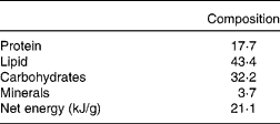
A formula adapted to the energy and protein requirements of piglets was manufactured by Lactalis (Retiers). Compared with a standard infant formula, it contained higher amounts of proteins and lipids (and a lower amount of lactose) but the protein:lipid ratio was kept constant. Its chemical composition is given in Table 1. The formula contained a mixture of skimmed milk powder and SMP in order to reach a caseins:SMP ratio of 40:60. The acronym SMP was used rather than whey proteins because the proteins used in the present study were obtained from the soluble phase of microfiltred and ultrafiltred milk and were native, whereas whey proteins are usually obtained after cheese manufacture and have been pasteurised. Lipids consisted of a mixture of vegetable oils. The formula was rehydrated at 20 % in water (w/v) before distribution to piglets.
Composition (% DM) of infant formula adapted to piglets
Animals and feeding
The experiment was conducted in compliance with the guidelines of the French Ministry of Agriculture for Use of Animals in Research (certificate of authorisation to experiment on living animals, no. 7676). Crossbred (Pietrain × (Large White × Landrace)) piglets from the experimental herd of INRA (Saint-Gilles, France) were used, regardless of sex. A total of eighteen piglets were separated from their mothers after 2 d and fed the formula with an automatic milk feeder as described previously( Reference Morise, Sève and Macé 14 ) for 26 d. The experimental design was a complete block design with a 1 × 3 factorial arrangement of one diet and three slaughter times after the last meal (30, 90 and 210 min). The daily net energy ration of 1450 kJ/body weight0·75 was partitioned into ten meals automatically distributed during the day. Body weights were recorded weekly, and feeding schedules were adjusted accordingly.
Collection of digested samples
At the age of 28 d, piglets were allocated to three groups according to their slaughter times after the last meal: 30, 90 and 210 min. They were slaughtered by electronarcosis immediately followed by exsanguination. Immediately after, the digestive tract was removed, dissected and digested samples were collected from four segments: stomach, proximal jejunum (2·5 m from the beginning of the small intestine), median jejunum (4–6 m of the small intestine between the proximal jejunum and ileum) and ileum (1·5 m of the extremity of the small intestine).
The total content of each segment was collected, dispersed by Ultra-Turrax (IKA Ultra Turrax T18 basic; IKA® Werke GmbH & Co.) for 1 min at 15 600 rpm, and pH was measured. Sodium benzoate and phenylmethylsulfonyl fluoride (10 and 0·37 g/kg content, respectively) were added to each digested sample in order to avoid further protein breakdown. All effluents were stored at − 20°C until further analyses.
Chemical and biochemical analyses
The digested samples collected from each piglet were analysed in duplicate for DM and total N. The DM of effluents was determined after drying samples in an oven at 105°C for 7 h. Total N was determined in both formula and digested samples by the Dumas method( 15 ).
Antibodies
Mouse monoclonal antibodies specific for α-lactalbumin (α-la)( Reference Jeanson, Dupont and Grattard 16 ) and β-lg( Reference Johansson, Lugand and Rolet-Repecaud 17 ) were obtained as previously described. Casein-specific monoclonal antibodies taken from INRA's collection were directed against the following fragments: β-casein (f133–150) (f39–19); αs1-casein (f5–13) (f133–150); αs2-casein (f190–207) (f16–35); κ-casein (98–115) (150–169). Rabbit polyclonal antibodies specific for proteins were of commercial origin, i.e. β-lg, α-la (Tebu-Bio) and total caseins (Gene Tex, Inc.).
SDS-PAGE
SDS-PAGE was performed using 10 % polyacrylamide NuPAGE® Novex® Bis-Tris precast gels (Invitrogen) according to the manufacturer's instructions. All samples were centrifuged for 10 min at 12 000 g , defatted and supernatant fractions were diluted 4-fold with distilled water. Diluted samples (32·5 μl) were treated with 5 μl of 0·5 m-dl-dithiothreitol and 12·5 μl of NuPAGE® LDS sample buffer before analysis.
Gels were fixed in 50 % (v/v) ethanol, 10 % (v/v) acetic acid and were rinsed for 15 min in deionised water before staining with Coomassie Blue. Molecular-weight markers used were the pure proteins β-casein, β-lg and α-la. α-La( Reference Caussin, Famelart and Maubois 18 ), β-lg( Reference Leonil, Molle and Fauquant 19 ) and β-casein( Reference Senocq, Dupont and Rolet-Repecaud 20 ) were purified as described previously. Image analysis of SDS-PAGE gels was carried out using Image Scanner II (Amersham Biosciences).
Immunoblotting
Digested samples and undigested infant formula were submitted to SDS-PAGE electrophoresis as described above. Immediately after separation, proteins and peptides were transferred onto a 0·2μm pore size polyvinylidene difluoride (PVDF) membrane (Bio-Rad) at 30 V and 250 mA for 90 min using a semi-dry transfer cell (Trans-Blot SD; Bio-Rad) using a 39 mm-glycine, 48 mm-2-amino-2-hydroxymethyl-propane-1,3-diol (Tris), 0·0375 % (w/v) SDS and 20 % (v/v) methanol transfer buffer. Immunodetection was conducted according to the following procedure. The membrane was incubated at room temperature for 1 h periods in PBS with 0·3 % (v/v) Tween-20 (PBS-T) with, successively, 5 % (w/v) decomplemented horse serum (Invitrogen), β-lg-, α-la- or caseins-specific mouse monoclonal antibodies (1 ml of each monoclonal antibodies at 0·5 μg/ml) and goat anti-mouse Ig alkaline phosphatase conjugate at 1:500 (v/v). The membrane was washed between each step by soaking for 10 min in three changes of the same buffer (PBS-T). After the last washing, the membrane was stained with BCIP/NBT (5-bromo-4-chloro-3-indolyl phosphate/nitro blue tetrazolium tablets) (Sigma) at 2·5 mg/ml in water. A negative control was made using a blot without monoclonal antibodies in order to detect non-specific bands.
Inhibition ELISA
Inhibition ELISA was performed as previously described( Reference Dupont, Mandalari and Molle 6 ) using β-lg-, α-la- and caseins-specific polyclonal antibodies to determine the residual immunoreactivity of each protein present in the four digestive compartments during the digestion process.
Nano-liquid chromatography–MS/MS
Jejunums (proximal and median jejunum pooled) and ileum contents of each piglet were analysed by liquid chromatography (LC)–MS/MS in order to identify the peptides remaining after digestion. After centrifugation for 10 min at 12 000 g , supernatant fractions were diluted in water (1:5, v/v) and loaded onto a C18 cartridge (UPTI-CLEAN REC18; Interchim) and 200 μl of 35 % acetonitrile were added three times in order to recover peptides that were then dried in a Speed Vac Concentrator (SVC 100H; Savant Instruments Inc.) to eliminate all traces of acetonitrile. Dried samples were then diluted in 0·1 % trifluoroacetic acid (Pierce), and 10 μl were injected and trapped onto a micro-pre-column cartridge (C18 PepMap 100, 300 μm internal diameter × 5 mm; Dionex) before separation of peptides onto a column (C18 PepMap 100, 75 μm internal diameter × 150 mm; Dionex). The separation started with 5 % solvent B (95 % acetonitrile, 0·08 % formic acid and 0·01 % trifluoroacetic acid in LC-grade water) and 95 % solvent A (2 % acetonitrile, 0·08 % formic acid and 0·01 % trifluoroacetic acid in LC-grade water) for 5 min and a linear gradient from 5 to 70 % of solvent B and from 95 to 30 % of solvent A for 65 min was performed at a flow rate of 200 nl/min.
The online separated peptides were analysed by electrospray ionisation quadrupole-time of flight MS in positive ion mode. An optimised voltage of 3·2 kV was applied to the nanoelectrospray ion source (Proxeon Biosystems A/S). MS and MS/MS data were acquired in continuum mode. Data-direct analysis was employed to perform MS/MS analysis on 1+ to 4+ charged precursor ions. Precursor selection was based upon ion intensity and charge state; if the precursors had been previously selected for fragmentation they were excluded from the rest of the analysis. Spectra were collected in the selected mass range of 400–2000 m/z for MS spectra and 60–2000 m/z for MS/MS. The mass spectrometer was operated in data-dependent mode automatically switching between MS and MS/MS acquisition using Analyst QS 1·1 software (Applied Biosystems) when the intensity of the ions was above 10 counts per s. To identify peptides, all data (MS and MS/MS) were submitted to MASCOT (v.2·2; Matrix Science). The search was performed against a homemade database dealing with major milk proteins which represents a proportion of the Swissprot database (http://www.expasy.org). No specific enzyme cleavage was used and the peptide mass tolerance was set to 0·3 Da for MS and MS/MS. For each peptide identified, a minimum MASCOT score corresponding to a P value < 0·05 was considered as a prerequisite for peptide validation.
Statistical analysis
The data were analysed using the GLM procedure of SAS (version 8·1; SAS Institute, Inc.) using two separate ANOVA. Effect of time after the last meal and effect of proteins at each digestive site were tested. The values presented are least square mean values with their standard errors and effects were considered significant at P ≤ 0 ·05. Tendencies were reported when 0·05 ≤ P ≤ 0·10.
Results
All piglets were in good health throughout the experiment. Food intake and body-weight gain during the experimental period were similar in the three groups (30, 90 and 210 min after the last meal) of 28-d-old piglets. Formula intake during the last meal was 149 (sem 14·4) g.
Table 2 shows the content, DM and N contents and residual immunoreactive proteins in the stomach, proximal jejunum, median jejunum and ileum at 30, 90 and 210 min after the last meal. The drop in gastric pH with time was not significant (4·49, 4·46 and 3·1, respectively). The mean pH of small intestine contents was 5·7, 6·6 and 7·2 in the proximal and median jejunum and ileum, respectively, whatever the time after the last meal was. N significantly decreased with time in the stomach and proximal jejunum (P = 0·0034 and P = 0·02, respectively; Table 2). At time 30 min, 79 (sem 7·2) % of ingested N was still present in the stomach of the piglet but only 39 (sem 5·1) and 10 (sem 3·2) % were found in the stomach at 90 and 210 min, respectively. In the proximal jejunum, ingested N was significantly higher at 30 min than at 90 and 210 min after the last meal (4·47 (sem 0·44), 3·17 (sem 0·55) and 1·89 (sem 0·33) %, respectively).
Content, DM and nitrogen content and residual immunoreactive proteins in the stomach, proximal jejunum, median jejunum and ileum at 30, 90 and 210 min after the last meal (Mean values with their standard errors)

β-Lg, β-lactoglobulin, α-La, α-lactalbumin.
a,b,c Mean values within a row with unlike superscript letters were significantly different (P < 0·05).
SDS-PAGE and immunoblotting
SDS-PAGE gels showed different patterns between the three groups (Fig. 1). Only a few bands were detected in gastric contents after 210 min compared with those detected after 30 and 90 min. SDS-PAGE showed an intense band corresponding to β-lg in the stomach at 30 and 90 min that totally disappeared 210 min after the last meal. Bands corresponding to α-la also appeared in the stomach at 30 and 90 min.
SDS-PAGE analysis of undigested infant formula (IF) and contents of the stomach (S), proximal jejunum (P), median jejunum (M) and ileum (I) at 30, 90 and 210 min after the last meal. Mr, molecular weight.

Western blotting with monoclonal antibodies that allow specific detection of β-lg, α-la and caseins were performed on the same effluents and on the undigested infant formula (Fig. 2(A), (B) and (C)). For β-lg, a major band at about 18 kDa was revealed in the undigested infant formula and most, if not all, of the effluents analysed showed the presence of undigested β-lg in these samples (Fig. 2(A)). Bands at 25 and 36 kDa were detected in the undigested infant formula and also in the stomach and proximal jejunum effluents, the latter probably corresponding to dimers of β-lg. Most of the high-molecular-weight proteins revealed by immunoblot were the result of non-specific cross-reactions between the secondary antibody and the transferred proteins (Fig. 2(D)). For α-la, a major band at about 14 kDa was detected in the undigested infant formula and the stomach and proximal jejunum effluents (Fig. 2(B)). Dimers of α-la were detected with the purified protein, the undigested infant formula and the stomach content collected 30 min after meal ingestion. Finally, caseins were revealed only in the purified protein control and undigested infant formula (Fig. 2(C)). Fragments of lower molecular weight (between 15 and 20 kDa were visible in the median jejunum and ileum and might correspond to casein proteolysis products. These data clearly show that both β-lg and α-la partly resist digestion and can be found as intact proteins in the effluents in contrast with caseins that are hydrolysed.
Western blotting analysis of undigested infant formula (IF) and contents of the stomach (S), proximal jejunum (P), median jejunum (M) and ileum (I) at 30, 90 and 210 min after the last meal using β-lactoglobulin (β-lg) (A), α-lactalbumin (α-la) (B) and β-casein (β-Cn) (C) monoclonal antibodies. (D) Negative control without monoclonal antibodies.

Evolution of protein immunoreactivity during digestion
A significant decrease in protein immunoreactivity with time was observed in the stomach (Fig. 3). At 30 min after the last meal, casein residual immunoreactivity was low in the stomach (23·7 (sem 5·6) %) and disappeared completely at 210 min. In contrast, β-lg partially resisted gastric digestion after 30 min (45·6 (sem 12·3) %) and completely disappeared at 210 min (1·37 (sem 0·42) %). α-La showed a similar behaviour to that of β-lg (42·6 (sem 8·4) % of α-la remained immunoreactive in the stomach at 30 min).
β-Lactoglobulin (
![]() ), α-lactalbumin (
), α-lactalbumin (
![]() ) and casein (
) and casein (
![]() ) residual immunoreactivity (% amount of respective ingested proteins) in the stomach (A), proximal jejunum (B), median jejunum (C) and ileum (D) determined by inhibition ELISA. Values are means, with standard errors of the mean represented by vertical bars. a,b Mean values at a postprandial time with unlike letters were significantly different (P < 0·05).
) residual immunoreactivity (% amount of respective ingested proteins) in the stomach (A), proximal jejunum (B), median jejunum (C) and ileum (D) determined by inhibition ELISA. Values are means, with standard errors of the mean represented by vertical bars. a,b Mean values at a postprandial time with unlike letters were significantly different (P < 0·05).

Surprisingly, 1 % of casein residual immunoreactivity was still detected in the ileum at the three times after the last meal, whereas a low amount of immunoreactive β-lg and α-la was detected in the ileum.
Identification of peptides in jejunums and ileum by liquid chromatography–MS/MS
Jejunum (proximal and median jejunum pooled) and ileum contents were subjected to LC–MS/MS to allow the identification of peptides with a minimum molecular mass of 400 Da. Only the peptides identified unambiguously (P < 0·05) as derived from β-lg, α-la and caseins are shown in Fig. 4(A) and (B). Figure 4. gives only the occurrence of peptides and their size distribution since the LC–MS/MS analysis, as it was performed in the present study, is rather qualitative than quantitative.
Peptide identification in the jejunum (A) and ileum (B) of piglets at 30, 90 and 210 min after the last meal using liquid chromatography–MS/MS. Cn, casein; lg, lactoglobulin.

The mean number of peptides identified in the jejunum at each time was 13 (sem 3), 12 (sem 4) and 0·9 (sem 0·3) peptides at 30, 90 and 210 min, respectively (Fig. 4(A)). In the ileum, 1·3 (sem 1·2), 1·0 (sem 0·6) and 1·1 (sem 0·9) peptides were detected at 30, 90 and 210 min after the last meal, respectively (Fig. 4(B)).
Most of the peptides detected in the jejunum at the three times after the last meal were derived from β-casein. Their length varied between five and twenty-two amino acids (653 and 2510 Da) at 30 min, four and eighteen amino acids (552 Da and 2085 Da) at 90 min and only four and six amino acids (509 and 788 Da) at 210 min. In the ileum, only three peptides of β-casein were detected, with length ranging from five to eight amino acids (607 and 966 Da) at the three times. In contrast, αs1-casein generated only a very few peptides that were mainly identified in the jejunum at 30 and 90 min. Finally, only two peptides of κ-casein, i.e. κ-casein (f155–161) and κ-casein (f155–160), were detected in the jejunum at 30 min and two peptides from the same protein, i.e. κ-casein (f155–161) and κ-casein (f115–121), in the jejunum at 90 min.
Peptides of β-lg were detected in both jejunums and the ileum. Small peptides (seven to fifteen amino acids) of β-lg were detected in the jejunum at 30 and 90 min but none was detected in the jejunum after 210 min. Two large peptides (forty and twenty-three amino acids) of β-lg, i.e. β-lg (f1–40) and β-lg (f102–124), with molecular weights of 4276 and 2674 Da, respectively, were identified in the ileum at 30, 90 and 210 min for the smaller one, and 30 and 210 min for the biggest.
Discussion
The aim of the present study was to evaluate the kinetics of milk protein hydrolysis and peptide release in the gut using the piglet as a model of the human infant. The results obtained clearly show that caseins and SMP behave differently in the gastrointestinal tract of infant formula-fed piglets. β-Lg and α-la appeared as the most resistant proteins towards digestion and were able to remain intact in the piglet stomach and proximal jejunum for more than 1 h after the meal ingestion. Although the hydrolysis of caseins in the upper gastrointestinal tract was very rapid, immunoreactive fragments and peptides were surprisingly detected in both the jejunum and ileum. Two large peptides of β-lg were also identified in the ileum. The rapid hydrolysis of caseins as compared with SMP has already been observed in vivo in 4-week-old rhesus monkeys( Reference Lindberg, Engberg and Jakobsson 21 ), human adults( Reference Mahé, Messing and Thuillier 10 ), preruminant calves( Reference Scanff, Yvon and Pelissier 11 ) and also demonstrated in in vitro experiments( Reference Dupont, Mandalari and Mollé 22 – Reference Eriksen, Holm and Jensen 24 ). We showed that about 45 % of β-lg and 43 % of α-la remained detectable by ELISA in the stomach of piglets 30 min after the last meal while only 24 % of caseins were detected. In human adults with a jejunostomy, Mahé et al. ( Reference Mahé, Messing and Thuillier 10 ) observed that 30 min after skimmed milk ingestion, 64 and 44 % of β-lg and α-la, respectively, remained intact, whereas caseins were entirely degraded.
β-Lg has been shown to be resistant to digestion in many in vivo and in vitro studies but α-la is a protein known to be prone to pepsin digestion in vitro. However, in the present study, β-lg and α-la have the same patterns toward digestion. Moreno et al. ( Reference Moreno, Mackie and Mills 25 ) have found that the breakdown of α-la during gastric digestion was slowed in the presence of phosphatidylcholine, with little effect being observed during subsequent duodenal digestion. At low pH, α-la has a partially unfolded form that allows it to penetrate into the phosphatidylcholine vesicles. These interactions are probably responsible for the slowing of gastric digestion by reducing the accessibility of the protein to pepsin. Mandalari et al. ( Reference Mandalari, Mackie and Rigby 26 ) showed the same results with β-lg.
N determination in the stomach showed that 30 min after ingestion, 79 (sem 7·2) % of N was still present in the stomach of the piglet whereas 39 (sem 5·1) and 10 (sem 3·2) % were found in the stomach after 90 and 210 min, respectively. Only 24 % of caseins and 45 % of SPM were detected by ELISA 30 min after ingestion when 79 % of the N (dietary + endogenous proteins) is still present in the stomach. The difference observed between the 79 % residual N (dietary + endogenous) and the 45 % residual SPM and 24 % residual caseins can have different explanations: it can be either due to significant endogenous secretions or to an extensive hydrolysis and/or emptying of milk proteins making them not detectable by ELISA. The pH of stomach contents did not drop below 4 until 210 min after meal ingestion. The gastric pH levels are comparable with those reported by other workers for piglets at approximately the same age( Reference Moughan, Cranwell and Smith 27 , Reference Wilson and Leibholz 28 ) and for breast-fed neonates( Reference Mason 2 ). These relatively high pH can prevent casein coagulation and induce a fast gastric emptying of these proteins.
Despite the physiological differences between adults and infants (lower acid and enzyme secretions in infants), the present study demonstrated that the behaviour of milk proteins towards digestion is similar between infant and adult models, with differences in the extent of protein hydrolysis. This is in agreement with our recent in vitro data( Reference Dupont, Mandalari and Mollé 22 ). In vitro digestion of β-lg was similar between the infant and adult model during the gastric phase but in the duodenal phase 56 and 72 % of β-lg remained intact in the infant and adult model, respectively. This higher resistance of β-lg towards digestion was attributed to an interaction between the protein and gastric phosphatidylcholine vesicles that were present at higher concentration in the adult. The high resistance of β-lg and α-la to digestion can also be explained by the highly compact conformation assumed by β-lg and α-la for the presence of two and four intramolecular disulfide bridges, respectively( Reference Picariello, Ferranti and Fierro 29 ).
The residual immunoreactivity of caseins, expressed as the percentage amount of ingested casein detected in the ileum at all the times tested, was higher than the residual activity of caseins in the proximal and median jejunum. It is hard to conclude whether the 1·52 % of immunoreactive caseins detected in the ileum 30 min after meal ingestion come from the test meal or a previous one. Our current hypothesis is that caseins have domains that are resistant to proteolysis in the gut( Reference Dupont, Mandalari and Molle 6 ) and that these undigested fragments might be stored in the ileum due to the ileocaecal valve which slows undigested materials before passing into the large intestine. Similarly, about 1 % immunoreactive fragments derived from caseins was found in the ileum of piglets at 90 and 210 min. Are these fragments identical to the ones already described as being resistant to digestion? The intact fragments of caseins detected in the jejunum and ileum were mainly peptides from β-casein that were detected by LC–MS/MS in both the jejunum and ileum at 30, 90 and 210 min. We observed many peptides from the area 74–91 of β-casein. This fits perfectly with the casein domains resisting proteolysis during digestion that have been identified in vitro using an infant digestion model as being the most resistant area of β-casein in raw, pasteurised and sterilised milks and yogurt( Reference Dupont, Mandalari and Molle 6 ). Picariello et al. ( Reference Picariello, Ferranti and Fierro 29 ) found that the C-terminal region of β-casein gave rise to very intense peptide signals after in vitro digestion of caseins. However, in the present study only few peptides from the C-terminal region of β-casein were detected in the jejunum 90 min after the meal. β-Lg peptides of seven to fifteen amino acids were detected in the jejunum at 30 and 90 min and large peptides of β-lg of twenty-three and forty amino acids were observed in the ileum. These peptides persist in the ileum until 210 min after the meal.
Some of the β-casein peptides detected in the present study are known to promote bioactivities, such as peptide β-casein (f60–66) and peptide β-casein (f80–90) that carry immunomodulatory activity in vitro and anti-hypertensive activity in spontaneously hypertensive rats, respectively( Reference Abubakar 30 – Reference Kayser 32 ). Other peptides detected in both the jejunum and ileum can exert bioactivities. Peptides from β-lg and α-la released by the action of digestive enzymes were shown to have the potential to influence the specific immune response through the modulation of murine splenocyte proliferation and cytokine secretion( Reference Saint-Sauveur, Gauthier and Boutin 33 , Reference Jacquot, Gauthier and Drouin 34 ).
Peptide identification by MS is hard to achieve in complex samples such as intestinal contents. The present study is, to our knowledge, the first exhaustive characterisation of dietary peptides released during in vivo milk protein digestion.
In conclusion, the present study has established that β-lg and α-la were more resistant than caseins to digestion in the stomach of piglets receiving infant formula 30, 90 and 210 min after the last meal. However, peptides derived mainly from β-caseins were present in the jejunum and ileum of piglets and can promote bioactivity. Caseins appear to be rapidly hydrolysed into large peptides in the stomach, since only 24 % were detected 30 min after meal ingestion. However, it appears that this hydrolysis is incomplete and leads to the survival of domains that were detected in the ileum.
This finding provides support for studying the possibility that peptides found in the intestinal lumen modify intestinal functions and exert bioactivities.
Acknowledgements
The present study was presented at the 8th day of the French nutrition conference, 7–10 December 2010, Lille, France. The authors thank F. Kest for diet manufacturing, all of the staff involved in the animal care and feeding and in animal slaughtering, as well as G. Savary for the maintenance of incubators and automatic formula feeders. We also acknowledge O. Menard, Y. Le Gouar and M. Formal for their expert technical assistance. The authors are involved in the FA1005 COST Action INFOGEST on food digestion. The authors are solely responsible for the work described in this article. D. D. F. M. and I. L. were responsible for the project development. K. B. and I. L. conducted the experiments in piglets. K. B. D. D. and I. L. were responsible for analysis. K. B. and J. J. were responsible for LC–MS/MS analysis. I. C. was responsible for diet formulation. K. B. and I. L. were responsible for generating statistical analysis. K. B., I. L., C. B., J. J. and D. D. were responsible for drafting the manuscript.
There are no conflicts of interest.


