


Introduction
The taxonomy of the calcicolous, Mediterranean lichen formerly known as Caloplaca subochracea auct. (Teloschistaceae Zahlbr.) was clarified by Arup et al. (Reference Arup, Bertrand, Navarro-Rosinés, Nimis, Roux and Søchting2023) on the basis of morphological, chemical and molecular data. Their results showed that this lichen belongs to Gyalolechia A. Massal. as redefined by Arup et al. (Reference Arup, Søchting and Frödén2013), and that two closely related species can be recognized, formerly treated as varieties. In their phylogenetic tree, the two formed a fully supported clade but were separated on two distinct branches, indicating the existence of two saxicolous species: G. marmorata (Bagl.) Nimis & Arup (with a white thallus) and G. luteococcinea (Clauzade & Cl. Roux) Cl. Roux et al. (with a yellow thallus). Arup et al. (Reference Arup, Bertrand, Navarro-Rosinés, Nimis, Roux and Søchting2023) had also included in the molecular analysis a sample from the Canary Islands (Tenerife) which was morphologically very similar to G. luteococcinea, but was epiphytic on Euphorbia balsamifera Aiton. That sample differed from G. luteococcinea in 17 bases in the ITS gene, suggesting that a third species could be present in the complex. This result reminded one of the authors of the present paper (PLN) of an epiphytic lichen that he had collected on coastal shrubs in several localities in Central and Southern Italy, which was very similar to G. luteococcinea but remained unidentified due to the very different substratum (Nimis Reference Nimis1993: 184). However, despite several attempts, these samples proved to be too old for successful DNA extraction. Fresh epiphytic samples collected by JV in Rhodes (Greece) were molecularly analyzed, and proved to cluster together with the sample from Tenerife, prompting the search for fresh material of epiphytic samples from Italy; this was finally obtained from four localities in Latium and Sardinia in 2024. The molecular analysis of these samples confirmed the existence of a third, epiphytic species, which is formally described in this paper.
Materials and Methods
Anatomical measurements
Anatomical measurements were carried out in water, except those of paraphyses and excipular hyphae, which were observed and measured after treatment with 10% potassium hydroxide (K). Crystals were observed in polarised light (POL). Spot tests were carried out using K, commercial bleach (C) and paraphenylenediamine (P). Characters were measured according to the guidelines in Vondrák et al. (Reference Vondrák, Frolov, Arup and Khodosovtsev2013).
Secondary chemistry
Secondary metabolites were identified using HPLC according to Søchting (Reference Søchting1997). Thallus and apothecia were analyzed separately, but fragments of apothecium initials could not be avoided completely in the thallus sample. The relative composition of the secondary compounds was calculated based on absorbance at 270 nm, according to Søchting (Reference Søchting1997).
Sequence alignment
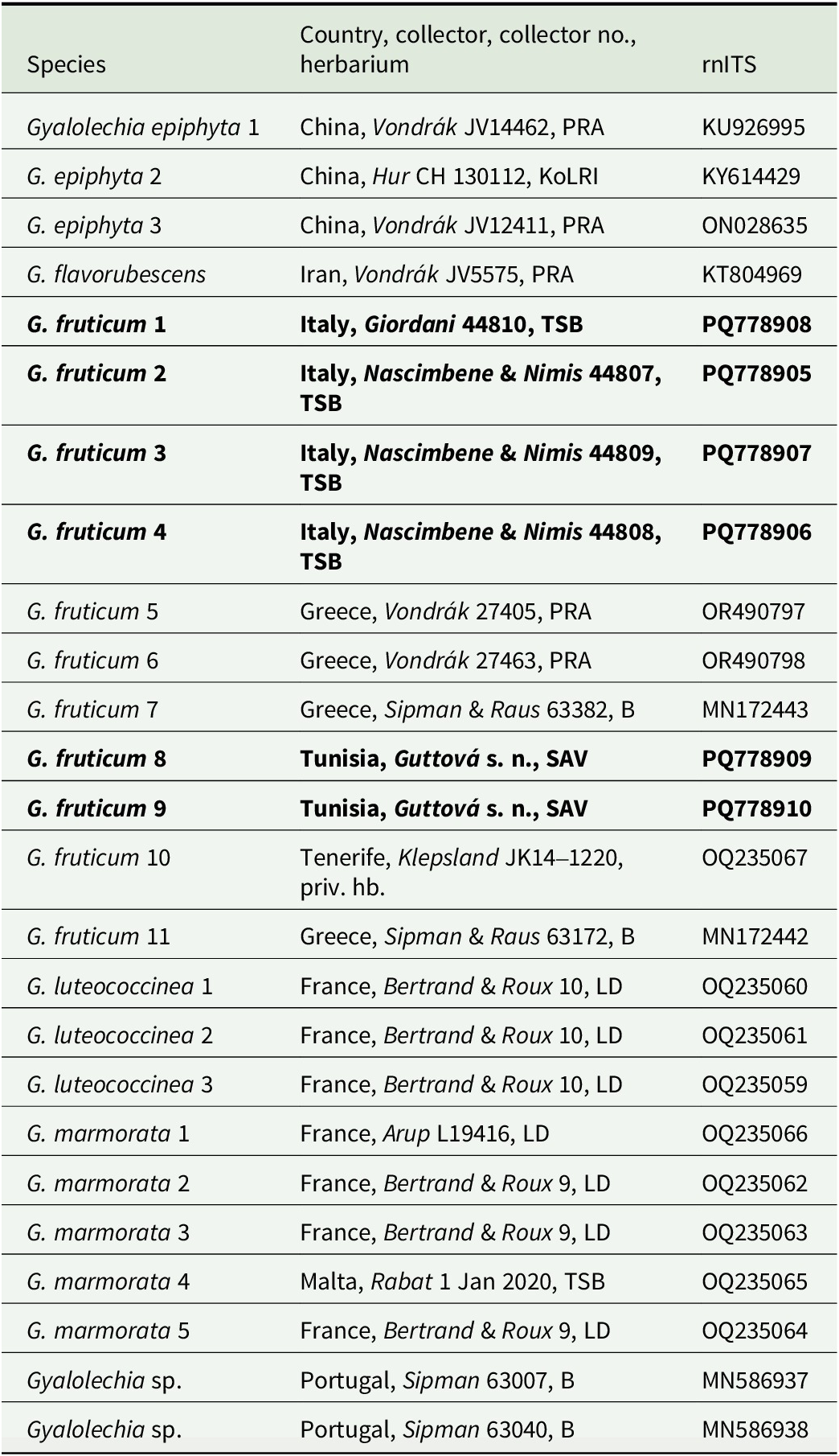
ITS sequences from 19 Gyalolechia specimens of G. luteococcinea, G. marmorata and related taxa were downloaded from GenBank (Table 1). The ITS sequences were aligned together with six newly produced sequences of our new species in Geneious v. 11.1.15 using the MAFFT option (auto). Unalignable ends, indels and ambiguously aligned parts were excluded from the alignment. Gyalolechia flavorubescens (Hudson) Søchting et al. was used as outgroup.
Voucher information and sequences of Gyalolechia species used in the analysis (Fig. 1). Newly produced sequences are in bold and the remainder were downloaded from GenBank.
Phylogenetic analysis
Phylogenetic relationships were inferred using maximum likelihood (ML) as implemented in IQ-TREE 2 (Minh et al. Reference Minh, Schmidt, Chernomor, Schrempf, Woodhams, von Haeseler and Lanfear2020), and Bayesian tree inference was carried out using Markov chain Monte Carlo (MCMC) as implemented in MrBayes v. 3.2 (Ronquist et al. Reference Ronquist, Teslenko, van der Mark, Ayres, Darling, Höhna, Larget, Liu, Suchard and Huelsenbeck2012). The most suitable likelihood model, SYM + G, was selected using the Bayesian information criterion (BIC) as implemented in the software jModelTest v. 2.1.4 (Guindon & Gascuel Reference Guindon and Gascuel2003; Darriba et al. Reference Darriba, Taboada, Doallo and Posada2012), evaluating only the 24 models available in MrBayes (Ronquist et al. Reference Ronquist, Teslenko, van der Mark, Ayres, Darling, Höhna, Larget, Liu, Suchard and Huelsenbeck2012). Three parallel runs with 20 000 000 generations, starting with a random tree and employing six simultaneous chains, were executed, five of which were incrementally heated with a temperature of 0.10. Analyses were diagnosed every 1000 generations in the last 50% of the tree sample and automatically halted when convergence was reached. Convergence was defined as an average standard deviation of splits (of frequency 0.1) between runs below 0.01. Every 2000th tree was sampled. A majority-rule consensus tree was constructed from the post-burn-in tree samples. The consensus trees were visualized using FigTree v. 1.4.4 (Rambaut Reference Rambaut2018) and redrawn in Adobe Illustrator. The maximum likelihood analyses used the same evolutionary models as those used in the Bayesian analyses. Branch support values were computed via 1000 non-parametric bootstrap replicates.
Results
Molecular analysis
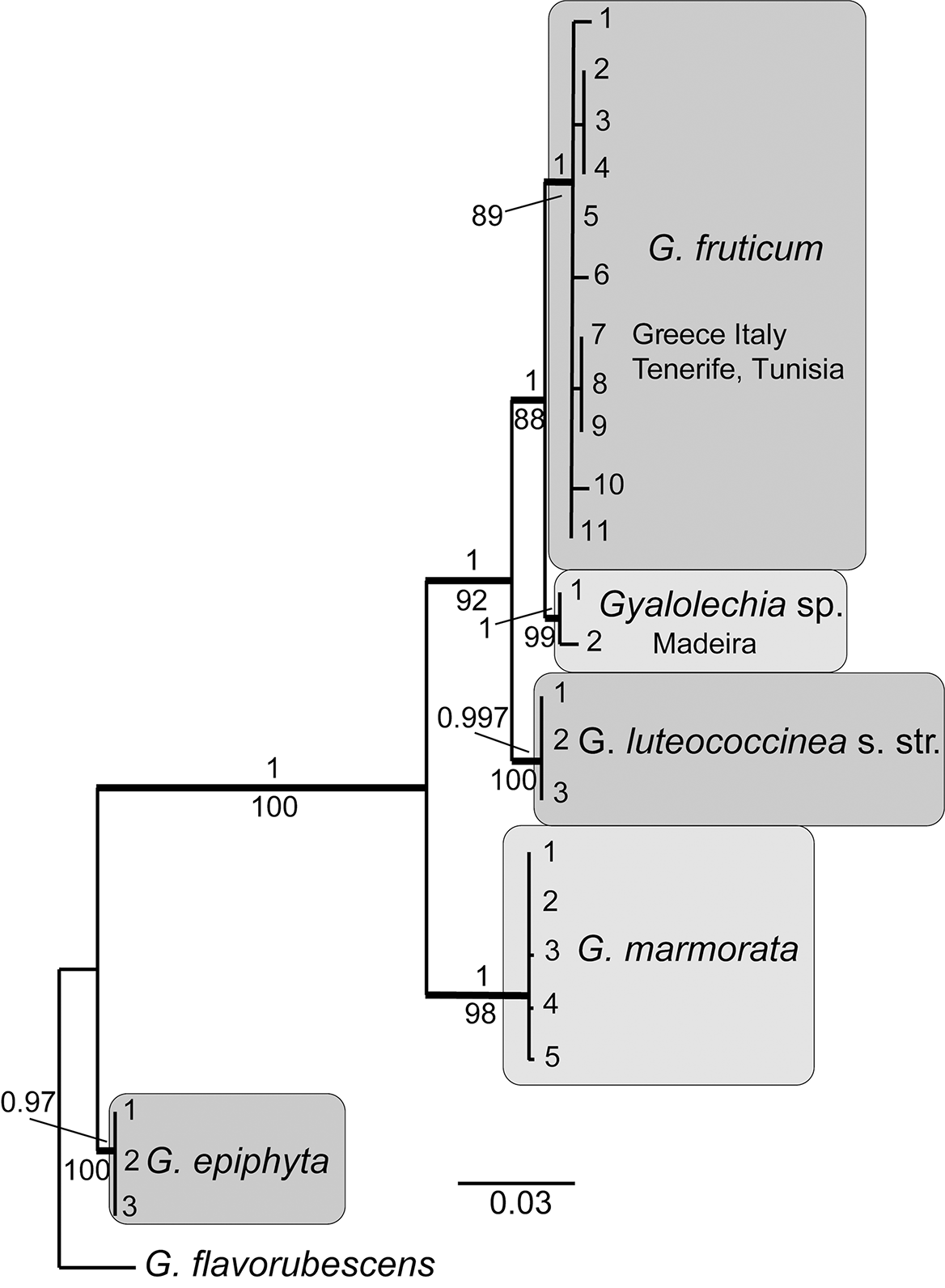
We generated six new nrITS sequences for this study and the alignment consisted of 25 sequences including the outgroup of 528 aligned nucleotide sites, of which 78 were parsimony-informative. The Bayesian analysis halted after 2 320 000 generations. The trees resulting from the Bayesian and maximum likelihood (ML) analyses were almost identical and did not differ significantly. In the ML tree (Fig. 1) the outgroup, Gyalolechia epiphyta (Lynge) Vondrák, and a branch holding G. luteococcinea, G. marmorata, an unknown epiphytic taxon from Madeira called Caloplaca cf. flavovirescens in the original paper by Sipman & Aptroot (Reference Sipman and Aptroot2020), and our new species G. fruticum are placed as sister clades. On the latter branch, G. fruticum is sister to the taxon from Madeira, both are sister to G. luteococcinea and finally G. marmorata is sister to all of them. All taxa are well supported. These results suggest the existence of at least one epiphytic species in the complex, represented by the samples from Greece, Italy, Tunisia and Tenerife. A second epiphytic species could be represented by the two samples from Madeira, although we have seen no material. This taxon could have been included in the new species since they both sit on a well-supported branch and the epithet cf. flavovirescens suggests similarities with the other epiphytic samples. We choose to provisionally keep them as two distinct taxa because there is a difference of 10–13 bases between the sequenced specimens of G. fruticum and those from Madeira, and the clades are distinctly separated from one another in the phylogenetic tree. The new species is formally described and discussed below.
Majority-rule consensus tree based on a maximum likelihood analysis of nrITS data showing the position of the new species Gyalolechia fruticum in relation to its close relatives. Branches with posterior probabilities (PP) ≥ 0.95 are shown in bold. Bootstrap values and PP are presented below and above the branches, respectively. Voucher information and GenBank Accession numbers are available in Table 1.
The mtSSU sequence from the type specimen is 99.8% similar to G. luteococcinea, the only available sequence in NCBI (OQ248479), and 98.7% and 98.8% similar to G. marmorata (OQ248480, OQ248481).
Secondary chemistry
The apothecia analyzed contain emodin and parietin as major compounds, and citreorosein, emodinal, emodic acid, fallacinal and fragilin as minor compounds. The thallus contains mainly fragilin but also low amounts of emodin and parietin that probably originate from apothecium initials embedded in the thallus.
Taxonomy
Gyalolechia fruticum Arup, Nimis & Vondrák sp. nov.
MycoBank No.: MB 857037
Differing from the calcicolous Gyalolechia luteococcinea in the epiphytic growth on littoral shrubs, and in molecular data.
Type: Italy, Sardegna, Prov. Sud Sardegna, St Anna Arresi, Loc. Porto Pino, 38°57′36″N, 08°36′39″E, alt. 10 m, sand dunes, on roots of Juniperus macrocarpa, 4 May 2024, coll. Giordani (TSB 44810, holotype). DNA sequences from the holotype: ITS (PQ778908), mtSSU (PQ778904); rbcL from the photobiont (PQ851754).
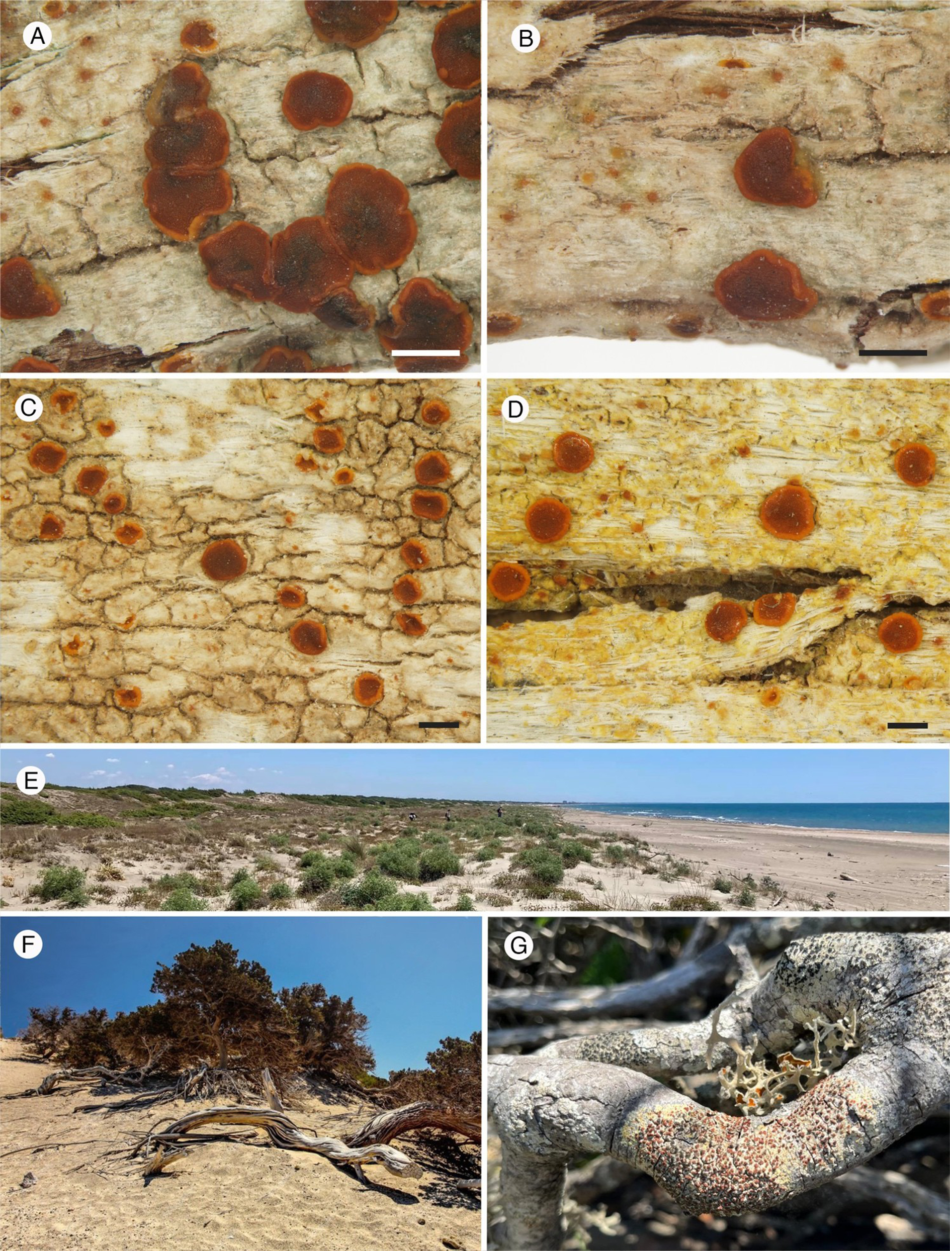
Morphology and habitat of Gyalolechia fruticum. A & B, the type specimen. A, with predominant apothecia. B, with predominant pycnidia (red dots). C, whitish thallus without anthraquinones (Vondrák 27405). D, yellowish thallus with anthraquinones (Vondrák 27463). E, the dunes of Castelporziano (Italy), with old shrubs in the background. F, old Juniperus macrocarpa with suitable microhabitats for G. fruticum (Greece, Chrissi) (source: freepic.com). G, G. fruticum with Seirophora villosa on a twig of Pistacia (Italy, Castelporziano). Scales = 0.5 mm. In colour online.
Anatomy of Gyalolechia fruticum in vertical sections. A, thallus and apothecium, algal cells forming colonies in a discontinuous layer between phorophyte bark tissue (below) and an epinecral layer (above). B, detail of thallus; vivid fungal cells (stained) are mostly restricted to algal colonies, whereas the epinecral layer is without living mycobiont cells. C, biatorine apothecium, with inspersed hypothecium and occasional algal colonies in lower exciple. D, pycnidium with distinct anthraquinone pigmentation around the ostiolum. E, apothecium in polarized light with glowing anthraquinones, but without POL+ crystals in the hypothecium. F, polarilocular ascospore (13 μm in length). G, ascus with ascospores. H, Teloschistes-type ascus. I, conidiophores and conidia. A, B & I, in lactoglycerol cotton blue; C–G, in water; H, in Lugol’s solution after K treatment. Scales: A, C–E = 100 μm; B = 50 μm; G & H = 10 μm; I = 20 μm. In colour online.

Thallus crustose, up to 3(–4) cm diam., at first very thinly episubstratal and c. 0.1 mm thick, then 0.2(–0.4) mm thick, bright orange in sun-exposed specimens to yellow (Fig. 2D) or almost white (Fig. 2A–C) in shade forms (but the peripheral areoles usually have a yellowish hue), continuous and smooth to rimose-areolate, often with a rough surface, usually surrounded by a pale prothallus (a dark, incomplete prothalline line was observed in a single specimen from Tenerife). Photobiont trebouxioid, the mature cells up to 15 μm wide. Based on the rbcL DNA barcode, the photobiont in the type specimen has the closest (99.5% identical) NCBI Blast match with Trebouxia decolorans (AJ969660, photobiont of Xanthoria parietina). The algal layer is c. 50 μm thick, discontinuous (Fig. 3A) in young, thin thalli, continuous in well-developed thalli, overlain by an up to 80 μm thick epinecral layer formed of cell remnants (Fig. 3B), which is, in yellow-orange thalli, inspersed with anthraquinone crystals, POL+ bright orange (POL+ white, glowing crystals, probably of calcium oxalates, present in a single specimen with a whitish thallus); true cortex and medulla absent.
Apothecia biatorine, at first semi-immersed, then sessile (Fig. 3A & C), circular to finally irregular in outline, (0.2–)0.3–0.8(–1.1) mm wide, with an initially deep orange, finally often dark rusty red to dark orange-red (rarely blackening with age), slightly concave to slightly convex, epruinose disc; proper margin slightly paler orange but never yellow (Fig. 2A–D), smooth, at first c. 0.1 mm thick and slightly raised, finally level with disc or sometimes excluded. Proper exciple prosoplectenchymatous, with orange anthraquinones at surface, colourless inside, but in the specimen from Tenerife, inspersed with anthraquinone crystals also inside. Excipular hyphae arranged in a fan-shaped way, the cells c. 2 μm wide and thin-walled in water, but lumina very thin and cell walls thickened and adglutinated in K; outermost cells of exciple almost spherical, up to 4 μm wide. Epithecium brownish orange, K+ purple, C−. Hymenium colourless, (40–)50–70(–80) μm high, not inspersed with oil droplets. Paraphyses simple or forked in upper part, not anastomosing, c. 1.5–2 μm thick at base, the apical cells (2–)2.5–3.5(–4) μm wide. Hypothecium colourless, 90–110 μm high, formed of a thin prosoplectenchymatous tissue below a strongly developed subhymenium, usually densely inspersed with very small oil droplets (Fig. 3C) not dissolving in K, without POL+ bright white crystals (Fig. 3E), or rarely with POL+ crystals. Asci 8-spored (Fig. 3G), Teloschistes-type (Fig. 3H), 40–60 × 12–15 μm. Ascospores polarilocular (Fig. 3F), hyaline, ellipsoid, (9–)10–14 × (4–)5–8(–9) μm, the septum 4–6 μm wide.
Pycnidia usually numerous, c. 0.1–0.2(–0.25) mm diam., half- to fully immersed in the thallus (Fig. 3D), visible as orange-red to dark red (rarely almost black) dots (Fig. 2A & B) due to concentrated anthraquinones around the ostiolum (K+ purple). Conidiophores of Umbilicaria-type (sensu Vobis Reference Vobis1980; Fig. 3I). Conidia short-bacilliform, 3–4 × 0.8–1(–1.3) μm.
Chemistry
Thallus and apothecia with unchlorinated anthraquinones. Parietin and emodin predominate in apothecia, whereas fragilin predominates in the thallus. Spot tests: thallus and apothecia K+ purple-red, C−, KC−, P−.
Etymology
The epithet refers to the occurrence of the new species on shrubs.
Similar species
Gyalolechia fruticum can be confused with other species which may occur in the same habitat: Gyalolechia flavorubescens also has a yellow thallus, but differs in having larger (up to 3 mm across), lecanorine apothecia with a prominent, yellow proper margin, an orange (never rusty red) disc reacting C+ red due to the presence of chlorinated anthraquinones in the epithecium, an oil-inspersed hymenium, and slightly larger spores. Occasional specimens of G. fruticum without a yellow thallus could be confused with Blastenia xerothermica Vondrák et al., or with Caloplaca aegatica Giralt et al., both of which differ from G. fruticum in having dark grey to black pycnidia reacting K−, without anthraquinones; the latter species, which probably belongs in Ikaeria (Sipman & Aptroot Reference Sipman and Aptroot2020), also differs in having broadly ellipsoid to subglobose ascospores.
Ecology and distribution
All samples were growing in coastal localities subject to humid maritime winds, mostly on the twigs of shrubs (Euphorbia balsamifera, Juniperus macrocarpa Sibth. & Sm., Phyllirea spp., Pistacia lentiscus L. and P. terebinthus L.) on sand dunes, but in one case also on wooden planks. The new species rarely occurs in very sun-exposed situations, preferring slightly shaded conditions with plenty of diffuse light; this was particularly evident in the case of specimens colonizing erect wooden planks on the dunes of the Castelporziano Estate (Italy), which were present only on the north-facing sides of the planks, while the south-exposed flanks were colonized by other lichens.
Frequent accompanying species, both in Greece and in Italy (Castelporziano), were Amandinea maritima Giralt et al., Bacidia parathalassica Llop & Gómez-Bolea, Bactrospora patellarioides (Nyl.) Almq, Caloplaca aegatica Giralt et al., Diploicia canescens (Dicks.) A. Massal., Lecidella elaeochroma var. flavicans (Ach.) Hazsl., L. elaeochroma var. juniperina Poelt & Nimis, Physcia adscendens H. Olivier, Pyrrhospora quernea (Dicks.) Körb., Ramalina canariensis J. Steiner and Xanthoria parietina (L.) Th. Fr. At Castelporziano (Italy), Ramalina lacera (With.) J. R. Laundon, Seirophora villosa (Ach.) Frödén and Tornabea scutellifera (With.) J. R. Laundon were also present and often abundant. The most typical lichen vegetation in which the new species occurs along the Tyrrhenian coasts of Italy was described by Nimis & Schiavon (Reference Nimis and Schiavon1986) as Teloschisto-Tornabeniopsidetum atlanticae, a rare community very much resembling the one described by Hernández Padron & Pérez de Paz (Reference Hernández Padrón and de Paz PL1980) for the Island of Hierro (Canary Islands), on twigs of Juniperus phoenicea L. subjected to humid winds from the ocean. In Italy, such vegetation is restricted to a small number of coastal, protected areas in the Mediterranean biome, having been largely destroyed by the touristic exploitation of beaches, with the consequent disappearance of well-developed, undisturbed sand dunes. The climate of the Italian stations can be characterized as Mediterranean-suboceanic (Nimis & Schiavon Reference Nimis and Schiavon1986), and has clear affinities with that of the Atlantic coasts of south-western Europe (Giacobbe Reference Giacobbe1949). The microclimatic conditions are generally dry, but with frequent spells of humid air from the sea. This is well in accordance with the peculiar ecology of the two most conspicuous accompanying species in the most undisturbed stands in Italy that was underlined by Nimis & Poelt (Reference Nimis and Poelt1987). These authors subdivided the lichen biota of Sardinia into several biogeographical groups, with the smallest group including only two species: Tornabea scutellifera and Seirophora villosa, designated as ‘species of fog-deserts’, well adapted to long, dry and warm periods but needing a periodic supply of high atmospheric moisture.
The lichen communities with G. fruticum observed in Rhodes, on coastal dunes with ancient Juniperus macrocarpa shrubs, were rather different from those in Italy. The two flagship lichens present at the Italian sites (Seirophora and Tornabea) were not observed, perhaps due to the slightly drier climate. Macrolichens were rarely observed, only Physcia adscendens, Xanthoria parietina and a small number of Ramalina canariensis thalli. However, microlichens co-occurring with G. fruticum formed a very specific maritime community. Frequent species included Alyxoria ochrocincta (Werner) Ertz, Arthonia albopulverea Nyl., A. tenellula Nyl., Catillaria servitii Szatala, Coppinsiella ulcerosa (Coppins & P. James) S. Y. Kondr. & Lőkös, Dirina ceratoniae (Ach.) Fr., Haloplaca sp., Lecanora lividocinerea Bagl., Melaspilea oleae J. Steiner, Naevia pinastri (Anzi) Thiyagaraja et al., Ocellomma picconianum (Bagl.) Ertz & Tehler, Polycauliona phlogina (Ach.) Arup et al., Rinodina furfuracea H. Magn., Scutula effusa (Rabenh.) Kistenich et al., Thelenella melanospora Etayo & H. Mayrhofer, Thelopsis isiaca Stizenb. and Toninia aff. populorum.
The distribution of the new species (Fig. 4) is clearly Mediterranean-Macaronesian, extending from the easternmost islands of Greece (Rhodes) to the Canary Islands, through Italy and Tunisia, and possibly mainland Spain (Almeria, El Ejido, 5 m, on twigs of Juniperus turbinata Guss.; see good images posted at http://liquenesdealmeria.blogspot.com/search/label/Caloplaca%20subochracea%20var.%20luteococcinea). In Italy, it has a mainly western, Tyrrhenian distributional pattern.
Distribution map of Gyalolechia fruticum. The triangle refers to a specimen identified from a photograph (see main text).

Additional specimens examined
Greece: Rhodes: Apolakkia, 36°04′11″N, 27°45′41″E, alt. c. 5 m, on wood of Juniperus macrocarpa, 18 iv 2023, Vondrák (PRA 27405); ibid., on bark of Pistacia lentiscus, Vondrák (PRA 27422); ibid., 36°04′43″N, 27°45′42″E, on bark and wood of Juniperus macrocarpa, 15 iv 2023, Vondrák (PRA 27463, 27479).—Italy: Toscana: Prov. Livorno, Parco Nazionale Arcipelago Toscano, Pianosa, 42°35′2.3″N, 10°04′28″E, alt. 15 m, on bark, 2005, Tretiach & Muggia (TSB 38194). Lazio: Prov. Roma, Torvaianica, 41°36′46″N, 12°28′44″E, alt. 4 m, on Juniperus, 1987, Nimis & Tretiach (TSB 9972); ibid., Presidential Estate of Castelporziano, 41°41′32″N, 12°22′45″E, alt. 20 m, on bark of Juniperus macrocarpa, 1985, Nimis (TSB 6369); ibid., 41°41′25.1″N, 12°21′59.2″E, alt. 11 m, coastal sand dunes, on wooden planks, N-exposed, 27 v 2024, Nascimbene & Nimis (TSB 44808); ibid., 41°41′32.5″N, 12°21′47.6″E, alt. 10 m, coastal sand dunes, on Juniperus macrocarpa, 27 v 2024, Nascimbene & Nimis (TSB 44807); ibid., 41°41′14.8″N, 12°22′17.7″E, alt. 10 m, coastal sand dunes, on Phillyrea, 27 v 2024, Nascimbene & Nimis (TSB 44809); Prov. Latina, Circeo National Park near Sabaudia, 41°14′39″N, 13°02′8.6″E, alt. 2–3 m, on Juniperus macrocarpa on coastal dunes, 1984, Nimis (TSB 4162). Puglia: Prov. Foggia, Tremiti Islands, San Domino, 42°06′44″N, 15°29′33″E, alt. c. 30 m, on Pistacia along the coast, 1986, Nimis (TSB 7589). Sicilia: Prov. Agrigento, Pelagie Islands, Linosa, Mt Nero, 35°51′31″N, 12°52′12″E, alt. 80 m, on bark of Pistacia, 1992, Nimis (TSB 17325); Prov. Trapani, Egadi Islands, Marettimo, 37°58′52″N, 12°03′29″E, alt. 30 m, on bark, 1988, Ottonello (TSB 12174).—Spain: Canary Islands: Tenerife, Anaga, between Chamorga and Roque Bermejo, 28°34′51″N, 16°08′27″W, alt. 255 m, on old Euphorbia balsamifera in Euphorbia habitat, xii 2014, J. Klepsland (JK14-1220).—Tunisia: Les Mogods, Corniche - La Grotte, alt. 27 m, 37°19.956′N, 9°50.593′E, on Juniperus twigs, 29 iii 2014, A. Guttová s. n. (SAV, as Gyalolechia flavorubescens in Guttová et al. (Reference Guttová, Vondrák, Schultz and El Mokni2015)). Nabeul: Cap Bon, Djebel Haouaria Mtn, 37°04.282′N, 11°02.456′E, alt. 367 m, on wood of Erica and Phillyrea branches growing on the cliff top, 26 iii 2014, A. Guttová s. n. (SAV, as Blastenia cf. hungarica in Guttová et al. (Reference Guttová, Vondrák, Schultz and El Mokni2015)).
Discussion and Conclusion
The new species is morphologically very similar to the calcicolous Gyalolechia luteococcinea, the only substantial differences being the higher frequency in the latter of a black prothalline line delimiting the thallus (which is, however, present in the epiphytic sample from Tenerife), and the presence of POL+ crystals (probably of calcium oxalates) in the hypothecium, most likely due to the growth on calcareous rocks (such crystals are, however, present in an epiphytic sample with a pale thallus from Greece). Other important differences are in the substratum, the new species being consistently epiphytic, and in the different molecular data.
The ITS sequences of G. fruticum differ from those of known related species in at least 15 positions, allowing convenient identification of the species by ITS-barcoding. There are numerous closely related species in Teloschistaceae that differ in fewer than five bases, as for example Flavoplaca communis (Vondrák et al.) Arup et al. and F. ruderum (Malbr.) Arup & Søchting (see Arup et al. Reference Arup, Søchting and Lorentzon2024). Some morphologically defined species are even undifferentiated by ITS; for example, Variospora aurantia (Pers.) Arup et al. and V. flavescens (Huds.) Arup et al., are phenotypically well separated (Šoun & Vondrák Reference Šoun and Vondrák2008), but are indistinguishable from each other in the ITS-barcode (T. Hauser & J. Vondrák, unpublished data). In this context, the differentiation of G. fruticum at the species level is sufficiently reliable.
Gyalolechia luteococcinea and G. fruticum are a further example of a species pair in which substratum requirements may have caused speciation, the substratum allowing their distinction in the simplest way, as discussed by, for example, Vondrák et al. (Reference Vondrák, Frolov, Košnar, Arup, Veselská, Halıcı, Malíček and Søchting2020) for Blastenia, Kantelinen et al. (Reference Kantelinen, Printzen, Poczai and Myllys2022) for Micarea, and Kosecka & Oset (Reference Kosecka and Oset2024) for the Varicellaria lactea–V. hemisphaerica species pair.
The typical habitats for G. fruticum are old-growth Juniperus macrocarpa stands on coastal sand dunes which have been heavily disturbed and partly damaged in many places along the Mediterranean coasts (Muñoz-Reinoso Reference Muñoz-Reinoso2003, Reference Muñoz-Reinoso2004; Picchi Reference Picchi2008). Due to the disappearance of its natural habitat (various undisturbed coastal shrubs), the new species is currently quite rare and probably further declining, apparently being highly sensitive to human disturbance: for example, in the Presidential Estate of Castelporziano near Rome, it is still rather frequent but only in the strictly protected area, while it seems to be completely absent in the adjoining area open to the public. Due to its conspicuousness, G. fruticum may represent an umbrella species for species-rich and threatened epiphytic communities growing on ancient shrubs along the Mediterranean-Macaronesian seashores.
Acknowledgements
Linda in Arcadia recommended an appropriate epithet for the new species. Jiří Machač assisted in taking photographs. Paolo Giordani (Genova) provided a sample from Sardinia, Juri Nascimbene (Bologna) helped in the collection of fresh specimens near Rome, and Andrea Moro (Trieste) retrieved old herbarium specimens, georeferenced the localities and prepared the distribution map. Jon Klepsland (Lier) provided material from Tenerife. JV was supported by a long-term research development grant RVO (grant no. 67985939).
Author Contributions
The first three authors contributed equally to the paper; SS carried out the molecular analysis of the samples from Greece and Italy; US carried out chemical analyses.
Author ORCIDs
Ulf Arup, 0000-0001-6612-8099; Pier Luigi Nimis, 0000-0003-3523-0183; Jan Vondrák, 0000-0001-7568-6711; Stanislav Svoboda, 0000-0001-9797-4984; Ulrik Søchting, 0000-0001-7122-9425.
Competing interests
The authors declare none.
Data Accessibility
Newly generated sequences have been deposited in GenBank. Nomenclatural novelties have been deposited in MycoBank.


 Open access