


Highlights
-
• MOG-IgG-positive severe optic neuritis (ON) showed the fastest and most complete visual recovery.
-
• AQP4-IgG-positive cases had the poorest prognosis; double-seronegative patients showed intermediate outcomes.
-
• Autoantibody testing is essential for prognosis and guiding management in non-MS ON.
Introduction
Optic neuritis (ON) is an acute inflammatory disorder of the optic nerve and a common cause of vision loss in adults, particularly young women between 15 and 45 years of age. Clinically, ON typically presents with sudden unilateral visual decline, ocular pain and dyschromatopsia. Although historically associated with multiple sclerosis (MS), ON is now recognized as a heterogeneous condition with diverse autoimmune etiologies. Reference Beck, Cleary and Anderson1
With advances in immunological diagnostics, two important ON-associated antibody-mediated diseases have been distinguished: aquaporin-4 immunoglobulin G (AQP4-IgG)-positive neuromyelitis optica spectrum disorder (NMOSD) and myelin oligodendrocyte glycoprotein antibody-associated disease (MOGAD). In addition, a considerable proportion of patients are classified as double seronegative (DN), lacking both AQP4-IgG and MOG-IgG, whose pathophysiology remains unclear. Reference Bennett, Costello and Chen2
The global incidence of ON varies widely, from 0.56 to 5.36 per 100,000 individuals annually, with idiopathic ON being the most common form. Prevalence data for ON show considerable variability, with reported rates ranging from 5.5 to 115.3 cases per 100,000 people. Reference Abbass, Shaia and Shukla3 In contrast to Western populations, where MS remains the predominant cause of ON, Asian populations more commonly exhibit ON secondary to NMOSD and MOGAD, accounting for approximately 3.4%–43.5% and 10.2%–27.6% of cases, respectively. A recent meta-analysis highlighted a significantly greater prevalence of both AQP4-IgG- and MOG-IgG-positive ON in Asian cohorts than in non-Asian populations, underscoring important geographical and ethnic differences in ON etiologies. Reference Hassan, Stern and Flanagan4–Reference Filippatou, Mukharesh, Saidha, Calabresi and Sotirchos6 In India, epidemiological data remain limited, although recent estimates suggest an incidence of approximately 1.33 per 100,000. Reference Saxena, Phuljhele, Menon, Gadaginamath, Sinha and Sharma7 The proportion of DN cases among patients presenting with non-MS ON varies considerably, ranging from 0% to 79%, largely reflecting heterogeneity in cohort selection and the diagnostic assays utilized. Reference Montcuquet, Collongues and Papeix8–Reference Vanikieti, Janyaprasert and Lueangram11 Notably, data from the Optic Neuritis Treatment Trial demonstrated that, over a 15-year follow-up, approximately 50% of patients were ultimately classified as having idiopathic ON. 12
Visual outcomes in ON vary considerably based on the underlying etiology, therapeutic responsiveness and timeliness of intervention. Among the subtypes, ON associated with AQP4-IgG is consistently linked to poorer visual prognosis. During acute episodes, up to 30% of affected individuals may experience complete vision loss with no light perception, and nearly 70% present with a severe logMAR visual acuity (VA) of ≤1. The risk of severe VA is markedly greater in AQP4-IgG-positive ON patients than in MOG-IgG-associated ON patients. Reference Filippatou, Mukharesh, Saidha, Calabresi and Sotirchos6,Reference Liu, Zhou and Wang13–Reference De Lott, Bennett and Costello15 In contrast, ON related to MOGAD typically results in a more favorable trajectory, with most patients achieving good visual recovery. In a cohort of 87 MOG-IgG-positive patients, the median final logMAR VA was 0.17, and only a small proportion (6%) had a logMAR VA of 1 or worse. Although generally responsive to corticosteroids (CSs), MOGAD-ON often relapses upon tapering, indicating a pattern of steroid dependence. Comparative studies consistently highlight superior visual outcomes in MOG-IgG-associated ON patients relative to AQP4-IgG-positive ON patients. Reference Chen, Flanagan and Jitprapaikulsan16–Reference Jeyakumar, Lerch, Dale and Ramanathan18
In ON associated with the DN, the clinical behavior is heterogeneous, and recovery trajectories are less well understood. Attacks in the DN ON result in disability that is comparable to, and may even exceed, that observed in AQP4-IgG–positive disease. Although the severity at nadir is comparable across groups, long-term functional outcomes in double-seronegative NMOSD patients are notably poorer than those in MOGAD patients. Reference Sato, Callegaro and Lana-Peixoto9,Reference Kitley, Waters and Woodhall19–Reference Chu, Shi and Zhu21 Visual recovery outcomes are intermediate between MOGAD and AQP4-positive NMOSD, with a reported median BCVA of 0.3 logMAR in DN ON patients versus 0.1 logMAR in MOGAD patients and 1.7 logMAR in AQP4-positive patients. Reference Jitprapaikulsan, Chen and Flanagan22 Although DN ON can follow either a monophasic or relapsing course, unlike AQP4-positive NMOSD, which typically relapses, it may still lead to disability comparable to that observed in AQP4-NMOSD. Reference Wu, Geraldes, Juryńczyk and Palace23
To add to the existing knowledge base, we conducted a longitudinal study of severe ON patients without MS stratified by serostatus (AQP4-IgG, MOG-IgG and DN). The primary objective was to compare visual outcomes and recovery trajectories across these groups via time-to-event analysis.
Materials and methods
Patients and study design
This retrospective cohort study was conducted at the Department of Neurology, Kasturba Medical College, Manipal, a tertiary care center in southern India. We screened the electronic health records of patients diagnosed with ON between January 1, 2016, and April 30, 2024. The inclusion criteria were as follows: (i) a confirmed diagnosis of ON; (ii) age ≥ 18 years; (iii) presentation with severe visual impairment, defined as a logMAR BCVA of 1 or worse Reference Merle, Olindo and Bonnan24 and (iv) availability of serum antibody results for both AQP4-IgG and MOG-IgG. The exclusion criteria included (i) visual loss attributed to other ocular pathologies (e.g., glaucoma, cataract), (ii) ON associated with MS, (iii) systemic autoimmune causes (e.g., lupus, sarcoidosis, Sjögren’s syndrome) and (iv) incomplete medical records.
Clinical and data collection
Demographic, clinical and treatment-related data were extracted from electronic medical records. This included demographic and clinical variables such as age, sex, number of prior ON episodes, VA at baseline and nadir. BCVA at each visit was assessed via the Snellen chart and subsequently converted to logMAR values via standard protocols. Nonnumeric visual assessments were converted as follows: no light perception = 3.0 logMAR; light perception = 2.3; hand movements = 2.0 and counting fingers = 1.7. Reference Moussa, Bassilious and Mathews25,Reference Tiew, Lim and Sivagnanasithiyar26 If preattack VA data were unavailable, a baseline VA of logMAR 0.0 was assigned.
Acute treatment modalities included intravenous CSs (IV methylprednisolone) alone or in combination with intravenous immunoglobulin or plasma exchange therapy (PLEX), which were administered at the discretion of the treating physician. Long-term immunosuppressive therapy included oral CSs (short (<6 months or prolonged duration ≥6 months), azathioprine, mycophenolate mofetil or rituximab, depending on serostatus and clinical recurrence.
The records of 176 patients with ON were reviewed. After the exclusion of 134 patients due to incomplete antibody data (n = 15), incomplete medical records (n = 12), non-severe visual loss (n = 14), multiple sclerosis–associated ON (n = 78), ON secondary to ON secondary to systemic autoimmune inflammatory diseases (n = 15), 42 patients fulfilled the inclusion criteria and were analyzed. The participants were stratified into three groups on the basis of serological test results: AQP4-IgG-positive, MOG-IgG-positive and DN.
Outcomes
The primary outcomes were as follows:
-
Good visual recovery was defined as a ≥ 66.77% improvement in BCVA from nadir.
-
Complete visual recovery was defined as the return of BCVA to preattack baseline.
These definitions were adapted from Bonnan et al., who incorporated the percent change in VA as a functional measure. Reference Bonnan, Valentino and Debeugny27 Visual recovery was assessed at 1, 3, 6 and 12 months (±4 weeks) post-onset. Recovery patterns were analyzed via Kaplan–Meier survival curves to estimate the cumulative probability of achieving visual improvement over time.
Statistical analysis
Descriptive statistics were used to summarize patient characteristics. Continuous variables were reported as medians and interquartile ranges (IQRs), whereas categorical variables were expressed as frequencies and percentages. Between-group comparisons were conducted using Fisher’s exact test (for categorical data) and the Mann–Whitney U test or independent t test (for continuous variables), as appropriate.
The time to visual recovery was assessed via Kaplan–Meier survival curves, and differences across groups were analyzed via the log-rank test. Cox proportional hazards regression was used to estimate hazard ratios (HRs) for achieving good and complete recovery, with the MOG-IgG group used as the reference. A p value < 0.05 was considered to indicate statistical significance. All analyses were performed via SPSS software (version 25.0; IBM Corp, Armonk, NY).
Results
Demographic data
A total of 42 patients with severe ON were included. Patients were classified into three groups according to their antibody status: AQP4-IgG-positive (n = 17), MOG-IgG-positive (n = 10) and DN (n = 15). The median age of the participants was 41.5 years (range: 18–65), with the DN group being the youngest (median: 30 years), followed by AQP4 (43 years) and MOG-IgG (49.5 years). A female predominance was observed in the AQP4-IgG (94.1%) and DN (60%) groups, whereas the MOG-IgG group exhibited nearly equal sex distribution (40% female).
Most patients were experiencing their first ON episode. Bilateral ON was observed in seven patients in the AQP4-IgG group, eight patients in the MOG-IgG group and four patients in the DN group. The baseline VA was 0.0 logMAR in most patients. The median VA at nadir was comparable across groups: 1.7 logMAR in both the AQP4-IgG and DN groups and 1.4 in the MOG-IgG group.
All patients with confirmed AQP4-IgG or MOG-IgG seropositivity received acute-phase treatment, whereas only 86.7% of patients in the DN group underwent similar management. Furthermore, long-term immunosuppressive therapy was more commonly initiated in seropositive patients than in seronegative patients. Table 1 summarizes the demographic and treatment-related characteristics across the three subgroups.
Baseline characteristics of all patients at presentation

AQP4 = aquaporin-4; CS = corticosteroid; IgG = immunoglobulin G; IQR = interquartile range; IVIG = intravenous immunoglobulin; ON = optic neuritis; MOG = myelin oligodendrocyte glycoprotein; VA = visual acuity.
Visual recovery in the entire cohort
Good recovery
Kaplan–Meier analysis demonstrated that visual recovery commenced within the first month post-attack, suggesting that some patients experienced early improvement in BCVA. Approximately 55% of patients achieved good visual recovery within 3 months, with an additional 25% recovering gradually thereafter. By 12 months, nearly 80% had achieved good recovery. The recovery trajectory followed a steady upward pattern, with most gains occurring between 3 and 6 months and tapering off beyond 10 months.
Complete recovery
In contrast, fewer than 35% of patients achieved complete visual recovery by 12 months. Recovery began early but plateaued after month 6, with minimal improvements noted after month 9 (Figure 1B). No patients experienced ON recurrence during the follow-up period.
Kaplan–Meier survival analysis illustrating overall visual recovery in the entire cohort of optic neuritis patients. (A) Time to good visual recovery (defined as ≥66.77 % improvement in best corrected visual acuity [BCVA]) demonstrated that approximately 75% of patients achieved this outcome within 12 months. (B) Time to complete visual recovery (defined as return to baseline BCVA) reveals that fewer than 30% achieved this endpoint, with minimal improvement beyond 6 months.

Visual outcomes by antibody subtype
Good recovery
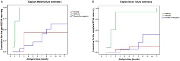
The visual recovery patterns varied significantly between the subgroups (Figure 2A). MOG-IgG-positive patients who achieved the most rapid improvement, at approximately 70%, achieved good recovery within 1 month, and all achieved this outcome by month 2.
Kaplan–Meier survival curves stratified by antibody subtype. (A) Good visual recovery occurred most rapidly and universally among MOG-IgG-positive patients, whereas AQP4-IgG-positive and double seronegative patients demonstrated slower and less complete improvement. (B) Complete visual recovery was most frequently achieved in the MOG-IgG-positive group; the double seronegative group showed intermediate recovery, and the AQP4-IgG-positive patients had the poorest outcomes. BCVA = best corrected visual acuity; NMOSD = neuromyelitis optica spectrum disorder; MOGAD = myelin oligodendrocyte glycoprotein antibody-associated disease.

In contrast, the NMOSD group exhibited a slower and less favorable recovery trajectory. The cumulative incidence of good recovery plateaued at approximately 40% by the third month, with no significant gains observed throughout the remainder of the 12-month follow-up period. These findings suggest that more than half of NMOSD patients fail to achieve good visual recovery, and while initial improvement may occur, it tends to stagnate relatively early.
The DN group showed a more gradual but steady recovery, with approximately 65% achieving good BCVA by 12 months. The recovery curve for this group was steady and progressive, lacking the early plateau observed in patients with NMOSD, indicating continued improvement beyond the initial 3-month period. Log-rank tests confirmed significant differences across groups (p = 0.001).
To further evaluate differences in recovery outcomes among the groups, Cox proportional hazards regression analysis was performed, with the MOG-IgG-positive group used as the reference group. The analysis revealed a significantly reduced likelihood of achieving good BCVA recovery in both the AQP4-IgG-positive group (hazard ratio [HR]: 0.16; 95% confidence interval [CI]: 0.05–0.64; p = 0.001) and the DN group (HR: 0.24; 95% CI: 0.09–0.78; p = 0.001) compared with the MOGAD group. When the DN was used as reference, AQP4-IgG had a comparable recovery trajectory (HR = 0.92, p = 0.10), indicating that there was no statistically significant difference between these two subgroups.
Complete recovery
Figure 2B illustrates the cumulative incidence of complete BCVA recovery across the three diagnostic subgroups. Patients in the MOG-IgG-positive group exhibited the most favorable recovery pattern, with nearly all patients achieving complete visual recovery within 3 months. This rapid recovery trajectory underscores the typically benign visual prognosis associated with MOGAD. In contrast, the DN group demonstrated a more gradual and stepwise course of improvement, with approximately 35% reaching complete recovery by 12 months. The AQP4-IgG-positive group had the least favorable outcomes, with only 15% of patients attaining complete BCVA recovery. The recovery curve in this group plateaued at 6 months, suggesting limited potential for further improvement beyond this point and highlighting the chronic visual morbidity commonly associated with NMOSD. The log-rank test did not reach statistical significance (p = 0.09).
Cox proportional hazards analysis, in which the MOG-IgG-positive group was used as the reference, revealed a lower likelihood of complete visual recovery in both the AQP4-IgG-positive (HR: 0.18; 95% CI: 0.10–2.14; p = 0.16) and DN groups (HR: 0.56; 95% CI: 0.09–2.64; p = 0.34). Furthermore, when the DN group was used as the reference, the AQP4-IgG-positive group continued to show a trend toward poorer recovery (HR: 0.61; 95% CI: 0.07–3.42; p = 0.42).
Discussion
This study assessed visual outcomes in patients with severe ON without MS stratified by antibody status into AQP4-IgG–positive, MOG-IgG–positive and DN groups. Among the three patients, MOG-IgG-positive patients had the most favorable prognosis, with rapid and complete recovery in nearly all patients. In contrast, AQP4-IgG-positive individuals experienced the poorest outcomes, while DN patients showed intermediate recovery. These findings align with prior research and further validate the prognostic relevance of antibody status in ON. Reference Jitprapaikulsan, Chen and Flanagan22,Reference Demuth, Guillaume and Bourre28–Reference Pandit, Sato and Siritho30
The superior outcomes in MOGAD are consistent with its underlying pathophysiology, which primarily involves demyelination of myelin sheaths, allowing for substantial remyelination and functional recovery. In contrast, AQP4-IgG-mediated disease triggers complement-mediated astrocytic injury, leading to secondary neuronal loss and extensive, often irreversible, tissue damage that underlies the poorer visual prognosis in NMOSD. Reference Saadoun, Waters, Owens, Bennett, Vincent and Papadopoulos31 DN ON appears to encompass a clinically heterogeneous spectrum. While some DN patients exhibit MOGAD in the recovery trajectory, others align more closely with the NMOSD phenotype, suggesting possible unidentified autoimmune mechanisms or postinfectious etiologies. Reference Wu, Geraldes, Juryńczyk and Palace23,Reference Petzold, Fraser and Abegg32
Consistent with prior research, although the severity of visual loss at nadir was comparable across subtypes, recovery patterns varied markedly. Reference Chu, Shi and Zhu21,Reference Akaishi, Nakashima and Takeshita33–Reference Zhao, Tan and Chan35 MOG-IgG–positive ON patients demonstrated the most rapid and substantial improvement, typically within the first two months, suggesting that early visual recovery predicts favorable long-term outcomes. In contrast, patients with DN ON exhibited a slower but progressive recovery, often extending beyond six months. The AQP4-IgG–positive patients showed minimal improvement after the third month, highlighting the importance of early recognition and prompt, aggressive treatment to prevent irreversible damage. These findings align with emerging evidence that neuronal injury in the DN ON may approach the extent observed in AQP4-IgG–associated disease and exceed that observed in MOGAD or MS, underscoring the biological heterogeneity and therapeutic implications across these subtypes. Reference Hoshina, Seay, Vegunta, Stulberg and Wright36
Our findings contribute additional perspective to the comparison between DNs and AQP4-IgG–positive ON. While the likelihood of attaining good BCVA appeared comparable – and in some cases slightly more favorable – in the DN group, the proportion achieving complete recovery remained lowest among AQP4-IgG–positive patients. These results are consistent with those of previous studies, further supporting the concept of distinct and subtype-specific visual recovery profiles in patients with inflammatory optic neuropathies. Reference Ishikawa, Kezuka and Shikishima37–Reference Cakan, Demirel and Cimen39
Martinez-Hernandez et al. reported that among patients with clinically isolated ON, those positive for AQP4-IgG had the worst prognosis, whereas both the MOG-IgG-positive and DN groups presented more favorable visual recovery. Reference Martinez-Hernandez, Sepulveda and Rostásy40 In contrast, a separate study involving 23 DN ON patients suggested that double seronegativity may be associated with poor visual outcomes, comparable to those observed in AQP4-IgG–associated ON patients. Reference Deschamps, Gueguen and Lecler41 Similarly, another cohort study revealed no significant difference in visual recovery between the AQP4-IgG–positive and DN ON groups. Reference Gomes de Brito Ventura Lís, Solla and Silva42
Our findings are broadly consistent with these observations and extend the current understanding by incorporating longitudinal time-to-event analysis. Across all subtypes, the most rapid visual improvement occurred within the first three months. The MOG-IgG–associated subtype showed the fastest and most consistent recovery, with all patients achieving good outcomes within two months. Compared with the AQP4-IgG–positive group, the DN group demonstrated a more favorable recovery pattern. When complete recovery was assessed, the DN subgroup continued to outperform the AQP4-IgG–positive subgroup. Although the difference in complete recovery did not reach statistical significance, this may reflect a limited sample size rather than the absence of a true effect, given the clinically meaningful trends observed.
A recent study reported that MOG-IgG–associated ON was characterized by the most rapid and favorable visual recovery, whereas the AQP4-IgG–positive and DN subtypes presented significantly lower HRs for good visual recovery (0.187, p = 0.013 and 0.189, p = 0.005, respectively) than MOG-IgG–positive patients did. Reference Laosirirat, Apiwattanakul and Aungsumart43 Similarly, another cohort study demonstrated significant variation in recovery across subtypes, with good visual outcomes (logMAR BCVA ≥ 0.30) most frequently observed in MOG-IgG–positive patients (90.2%), followed by the DN (50.9%) and AQP4-IgG–positive (42.9%) groups (P < 0.001). Reference Yang, Wu and Song44 While some studies suggest that DN patients recover similarly to MOGAD, others have reported outcomes comparable to AQP4-IgG-positive ON. Overall, the visual prognosis of DN ON tends to be less favorable than that of MOG-IgG–associated cases. These discrepancies highlight the imperative for comprehensive antibody testing, specifically anti-AQP4 and anti-MOG assays, using cell-based techniques in all patients presenting with ON of unclear etiology to ensure accurate diagnosis and guide appropriate management.
An international multicenter study reported comparable median post-PLEX BCVA across AQP4-IgG–positive ON, MOGAD and double-seronegative ON (0.176, 0 and 0.10 logMAR, respectively). Reference Chen, Flanagan and Pittock45 Although these findings differ from the poorer visual outcomes observed in the AQP4-IgG–positive subgroup in our cohort, the discrepancy is likely attributable to differences in acute treatment practices. In the cited study, PLEX was implemented early according to a standardized protocol, whereas in our setting, PLEX was not routinely incorporated into acute management. High-dose intravenous methylprednisolone (MPS) formed the cornerstone of initial therapy, consistent with standard practice, where PLEX is reserved for cases with inadequate steroid response and may not be universally accessible. Furthermore, the retrospective design of our study precluded uniform treatment escalation; in many cases, antibody testing was completed only after the acute episode, limiting the feasibility of targeted PLEX administration.
The evidence suggests that the initiation of high-dose intravenous MPS within the first week is critical for achieving clinically meaningful visual recovery (≥0.3 logMAR) and that the efficacy of PLEX is greatest when it is administered within two weeks of symptom onset. Reference Kemchoknatee, Singhakul, Arjkongharn, Chainakul, Tangon and Srisombut46,Reference Kleiter, Gahlen and Borisow47 Differences in treatment timing, influenced in part by incomplete referral documentation, and variability in escalation protocols may have contributed to the observed recovery outcomes. These findings underscore real-world outcomes under corticosteroid-based management and reinforce the critical role of early PLEX access in optimizing visual recovery for NMOSD-related ON patients.
A key strength of this study lies in its longitudinal design, which enables a detailed assessment of both the timing and trajectory of visual recovery across ON subtypes. By distinguishing between “good” and “complete” recovery, this analysis provides a more nuanced understanding of functional outcomes beyond simple restoration of baseline vision. These findings have practical relevance for clinicians when counseling patients regarding prognosis and expected recovery timelines. Notably, the evaluation of good recovery offers a clinically meaningful measure of improvement rather than relying solely on the achievement of full BCVA restoration.
This study has several limitations. First, the retrospective nature of the study limits control over potential confounders such as treatment timing and standardization of therapeutic protocols. Second, ancillary investigations – including optical coherence tomography and visual field analysis – were not uniformly available, precluding detailed structure–function correlation. Third, the relatively small sample size, particularly within subgroups, may have limited the statistical power to detect subtle differences, especially in complete recovery rates. In addition, PLEX was not routinely employed as part of acute management during the study period, which may have influenced visual outcomes in severe cases.
Future studies should include larger, multicenter cohorts with standardized data collection, ideally via a national registry, to improve understanding of visual recovery in non–MS ON. Prospective studies are particularly important to better define the clinical course and long-term outcomes of seronegative ON.
Conclusion
This study provides important longitudinal insights into visual recovery patterns among patients with non-MS-related ON, stratified by autoantibody status. Our findings demonstrate marked heterogeneity across subtypes and emphasize the need for early, targeted management to optimize outcomes. MOG-IgG–associated ON had the most favorable prognosis, with most patients achieving good or complete recovery within two to three months. In contrast, AQP4-IgG–associated ON had the poorest outcomes, whereas DN ON exhibited an intermediate recovery profile. Recovery trends across all subtypes suggest that most of the functional visual improvement occurs within the first three to four months following onset, with limited gains beyond six months. These results provide a valuable clinical context for counseling patients regarding visual prognosis and expected recovery timelines.
Acknowledgment
The authors thank the patients and their families for their participation in this study. I am deeply grateful to my wife, Dr Shivangi Tiwari, for her constant encouragement, patience, and support throughout the course of this work. Her unwavering belief and understanding were invaluable in completing this manuscript.
Author contributions
All the authors contributed to the study conception and design. P.B. was involved in material preparation, writing, generation of figures, data collection and analysis. A.N.P. and A.R.P. were involved in conceptualization, review and editing of the manuscript. All the authors read and approved the final manuscript.
Funding statement
No funding was received to assist with the preparation of this manuscript.
Competing interests
The authors have no competing interests to declare that are relevant to the content of this article.
Ethical standards
Ethical approval was granted by the Kasturba Medical College and Kasturba Hospital Institutional Ethics Committee (IEC1 – 229–2024), and we conducted this study in accordance with the ethical principles of the Declaration of Helsinki.
Data availability
Deidentified patient data used for this study are available upon reasonable request.



 Open access

