Hypothyroidism is a condition where the thyroid gland produces insufficient thyroid hormones, leading to a slowed metabolism(Reference Kostoglou-Athanassiou and Ntalles1–Reference Kochman, Jakubczyk and Bargiel3). The thyroid primarily produces thyroxine (T4) and triiodothyronine (T3)—T4 is exclusively synthesised in the thyroid, while T3 is produced both directly and via the conversion of T4 in peripheral tissues(Reference Taylor, Albrecht and Scholz4). Reduced T3 levels in hypothyroidism impair Na+/K + ATPase regulation, affecting metabolic equivalents and causing symptoms such as fatigue, weight gain, cold intolerance, anxiety, constipation, hypothermia, decreased appetite and locomotion(Reference Kostoglou-Athanassiou and Ntalles1,Reference Fatourechi2) . Hypothyroidism affects 1–2 % of the population. Women are ten times more likely to develop hypothyroidism than men. Clinical hypothyroidism affects 0·5–1·9 % of women compared with <1 % of men, while subclinical hypothyroidism occurs in 3–13·6 % of women and 0·7–5·7 % of men(Reference Kostoglou-Athanassiou and Ntalles1,Reference Mulder5) . Studies highlight the critical role of reactive oxygen species (ROS) in thyroid hormone production, making the thyroid highly vulnerable to oxidative damage(Reference Kochman, Jakubczyk and Bargiel3,Reference Vanderpump, Luster, Duntas and Wartofsky6–Reference Pereira, Rosa and Safi12) . This damage is exacerbated by increased lipid peroxidation and mutations in NADPH oxidase, which impair the body’s antioxidant defences(Reference Kochman, Jakubczyk and Bargiel3). Increased oxidative stress not only impairs thyroid function but also contributes to weight gain, anxiety and reduced locomotor activity. Excess fat in overweight or obese individuals produces higher levels of free radicals, unstable molecules that damage cells and disrupt metabolic processes, leading to further weight gain(Reference Bonomini13). In the brain, oxidative stress damages cells and interferes with neurotransmitter function, contributing to heightened anxiety and mood disturbances(Reference Bauer, Heinz and Whybrow14). Additionally, oxidative damage to proteins and lipids within the nervous system impairs motor control, reducing muscle function and coordination, which leads to decreased physical activity. Together, these effects create a cascade of dysfunctions that exacerbate hypothyroidism symptoms and overall health decline(Reference Zegers-Delgado, Blanlot and Calderon15).
Lifelong levothyroxine therapy, though relatively inexpensive, can impose a financial burden over time. Moreover, its prolonged use is associated with adverse effects, including cardiovascular complications such as shortened systolic time intervals, increased atrial premature beats and potential left ventricular hypertrophy. It may also impact bone health by reducing bone density and mass. Additionally, hepatic effects include elevated serum levels of key enzymes like glutathione-S-transferase, γ-glutamyl transferase and alanine transaminase(Reference Bartalena, Bogazzi and Martino16,Reference Rovet and Ehrlich17) .
The rationale of the study is to develop an easily adaptable cost-effective lifestyle intervention to prevent hypothyroidism progression by targeting oxidative stress—an essential factor in thyroid hormone synthesis and gland vulnerability(Reference Kochman, Jakubczyk and Bargiel3). We propose intermittent fasting (IF) and vitamin E (Vit. E) as alternative strategies due to their potent antioxidant and free radical-scavenging properties. IF enhances cellular defences by activating internal antioxidant systems, reducing oxidative stress, regulating blood sugar and delaying cellular ageing(Reference Moro, Tinsley and Bianco18–Reference Sharsher, Ahmed and Metwally24), while Vit. E protects against lipid peroxidation. By assessing their individual and combined effects on oxidative stress, antioxidant function, total protein content, nitrite levels, body weight, anxiety-like behaviour and locomotor activity, this study advocates for sustainable, non-drug strategies to manage hypothyroidism(Reference Seven, Seymen and Hatemi21,Reference Napolitano, Fasciolo and Di Meo25,Reference Ye, Zhong and Du26) . It underscores oxidative stress modulation as a key approach to slowing disease progression and improving thyroid health.
Methods
Drug solution and dosing
propylthiouracil (PTU) was purchased from Tokyo Chemical Industry and Vit. E from Central Drug House, India. Other chemicals were of analytical grade. PTU stock solution (1 mg/ml) and administered 1 ml orally by oral gavage to an animal daily for 24 days to induce hypothyroidism(Reference Bilici, Ural and Saçik27–Reference Silva, Ocarino and Serakides29). Vit. E stock solution (66 mg/ml in 10 % alcohol) was prepared and administered 1 ml intraperitoneally(Reference Seven, Seymen and Hatemi21). The injection site was cleaned with alcohol to prevent infection. All solutions were prepared and filtered aseptically.
Experimental animals
Female Wistar albino rats (8–10 weeks old, 120–150 g) were obtained from Chakraborti Enterprise, Narkeldanga, Kolkata, India. The animals were housed in polypropylene cages lined with paddy husk as bedding material. A 12-hour light/dark cycle was maintained, with the temperature set at 23 ± 3°C and relative humidity kept at 60 ± 10 %. The Institutional Animal Ethics Committee (IAEC), registered under 994/GO/Re/S/06/CPCSEA and approved the procedures (ref. no. 235/24/IAEC/Pharmacy/2022). The investigation adhered to CPCSEA guidelines under the Government of India.
Experimental design
The experiment was performed on thirty female Albino Wistar rats (8–10 weeks old, 120–150 g). Six animals with normal thyroid profiles were randomly assigned to each experimental group according to G-Power analysis. There were five groups named as euthyroid, hypothyroidism control, Vit. E + IF, Vit. E and IF. The euthyroid group received 1 ml of distilled water daily for 24 days. The hypothyroidism control group received 1 ml of 1 mg/ml PTU solution daily for 24 days(Reference Bilici, Ural and Saçik27–Reference Silva, Ocarino and Serakides29). Vit. E received intraperitoneal Vit. E at 66 mg/ml and IF group underwent IF once in a week for 16 h (4 pm to 8 am) but Vit. E + IF group went under both interventions simultaneously. The hypothyroidism was induced in all experimental groups (except the euthyroid) in conjunction with IF and Vit. E.
Body weight
The animals’ weight was monitored weekly using a digital weighing machine following the experimental animal model. The weight check was performed between 8·00 am to 10·00 am(Reference Singh, Bansal and Bhandari30).
Evaluation of locomotor activity
The locomotor activity of each female Wistar albino rat was assessed using an Actophotometer (Bio Craft Scientific Systems Pvt. Ltd., Agra, India). Measurements were conducted for a duration of 5 min per session, between 8:00 am and 10:00 am, on a weekly basis(Reference Silva, Vidigal and Galvao28).
Evaluation of anxiety
To evaluate the anxiety levels of the experimental rats, the elevated plus maze was utilised, as Sharma AC et al. outlined in 1992(Reference Sharma and Kulkarni31).
Isolation of the thyroid gland from the experimental rats
Firstly, the experimental rats were killed by the overdose of ketamine (300–360 mg/kg) + xylazine (30–40 mg/kg) intraperitoneally and had their thyroids removed. Thyroid tissue was weighed, homogenised in nine volumes of 0·1M phosphate buffer and centrifuged at 15 000 rpm for 5 min at 2–8°C(Reference Son, Kim and Kwon32). The resulting supernatant was used for biochemical analysis using a Shimadzu UV–visible spectrophotometer.
Quantitative estimates of the antioxidants in the blood serum and thyroid homogenates
Antioxidant levels of catalase (CAT), superoxide dismutase (SOD), glutathione (GSH) and glutathione peroxidase (GPx) were assessed in blood serum and thyroid homogenates using a UV spectrophotometer(Reference Sharma and Kulkarni31,Reference Son, Kim and Kwon32) . Lipid peroxidation was measured by quantifying malondialdehyde (MDA) content in both serum and thyroid homogenates(Reference Shree, Singh and Choudhary35,Reference Das, Kashyap and Singh36) .
Quantitative estimation of nitrite levels in blood serum and thyroid homogenates
To determine the amount of nitrite present, the standard curve was created using sodium nitrite, and nitrite content was measured in nanomoles per milligram of protein(Reference Singh and Bodakhe33,Reference Shree, Singh and Choudhary35) .
Quantitative estimates of the total protein content in the blood serum and thyroid homogenates
The total protein was measured content in the blood serum and thyroid homogenates as per the standard procedure. 4·0 ml of alkaline copper solution was added to 0·1 ml of the tissue lysate and allowed to stand for 10 min. 0·4 ml of phenol reagent was further added very rapidly, mixed quickly and incubated at room temperature for 30 min for colour development. The absorbance of the resultant colour product was measured at 610 nm against blank. Bovine serum albumin (0·2–2·0 mg/ml) was used as a standard for establishing the calibration curve(Reference Choudhary, Shree and Singh34,Reference Das, Kashyap and Singh36) .
Statistical analysis
The findings of all statistical analyses were presented as mean ± s d using the Graph Pad Prism (version 5·0). The means of the various groups were compared using a one-way ANOVA, followed by the Newmann–Keuls test for thyroid profile (T3 and T4), SOD, CAT, GPx, GSH, MDA, nitrite and total protein content; similarly, the data were analysed using a two-way ANOVA, followed by Bonferroni test for weight, anxiety (elevated plus maze) and locomotor count. The criterion for statistical significance was set at ∗ P < 0·05, ∗∗ P < 0·01 and ∗∗∗ P < 0·001.
Results
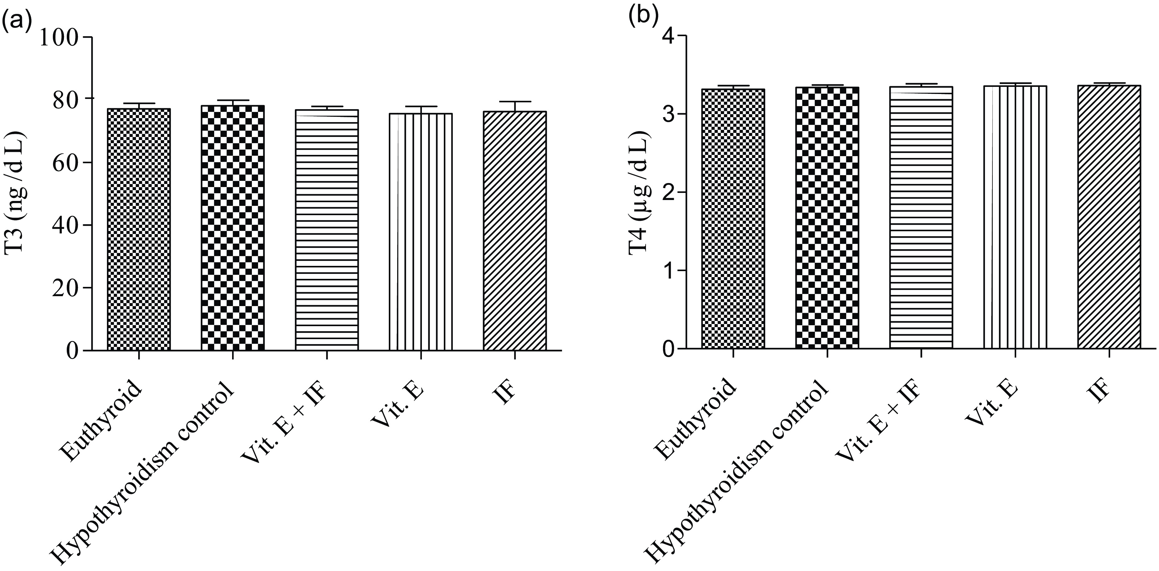
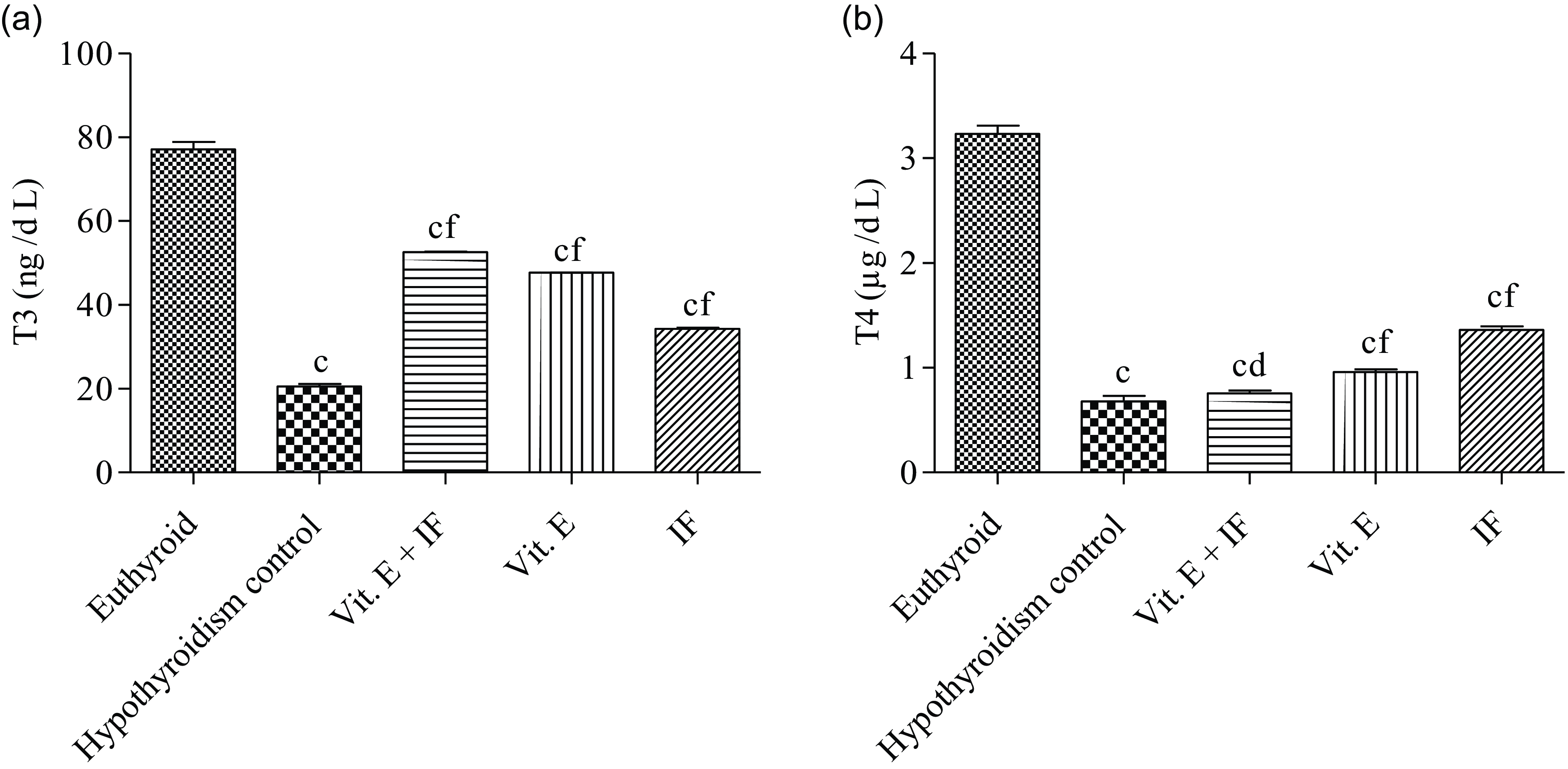
Effect of proposed therapies on blood serum (a) T3 (triiodothyronine), (B) T4 (thyroxine)
Figure 1 represents the baseline data of thyroid hormones T3 and T4. The result was nonsignificant among all groups euthyroid, hypothyroidism control, IF + Vit. E, Vit. E and IF (Figure 2). The result demonstrated that the hypothyroidism control animals had a significant decrease (P < 0·001) in the T3 and T4 levels in the blood serum as compared with the euthyroid group animals. The Vit. E + IF therapy experimental rats showed a significant increase in the levels of T3 (P < 0·001) and T4 (P < 0·05) as compared with the hypothyroidism control experimental rats and a marked decrease (P < 0·001) in the T3 and T4 levels upon comparison with the euthyroid group. The Vit. E experimental rats showed a marked incline (P < 0·001) in the levels of T3 and T4 as compared with the hypothyroidism control animals and a significant decrease (P < 0·001) in the T3 and T4 levels upon comparison with the euthyroid group. The IF experimental animals exhibited a notable rise (P < 0·001) in the levels of T3 and T4 as compared with the hypothyroidism control and a decline (P < 0·001) in, T3 and T4 levels on comparison with euthyroid experimental rats.
Baseline data for thyroid hormones (a) T3 (triiodothyronine), (b) T4 (thyroxine). Values are expressed as mean ± sd (n 6). Data were analysed by using the one-way ANOVA, followed by the Newman–Keuls multiple comparison test and expressed as aP < 0·05, bP < 0·01, cP < 0·001 when compared with euthyroid group and dP < 0·05, eP < 0·01, fP < 0·001 when compared with hypothyroidism control group.

Effect of proposed therapies on blood serum (a) T3 (triiodothyronine), (b) T4 (thyroxine). Values are expressed as mean ± sd (n 6). Data were analysed by using the one-way ANOVA, followed by the Newman–Keuls multiple comparison test and expressed as aP < 0·05, bP < 0·01, cP < 0·001 when compared with euthyroid group and dP < 0·05, eP < 0·01, fP < 0·001 when compared with hypothyroidism control group.

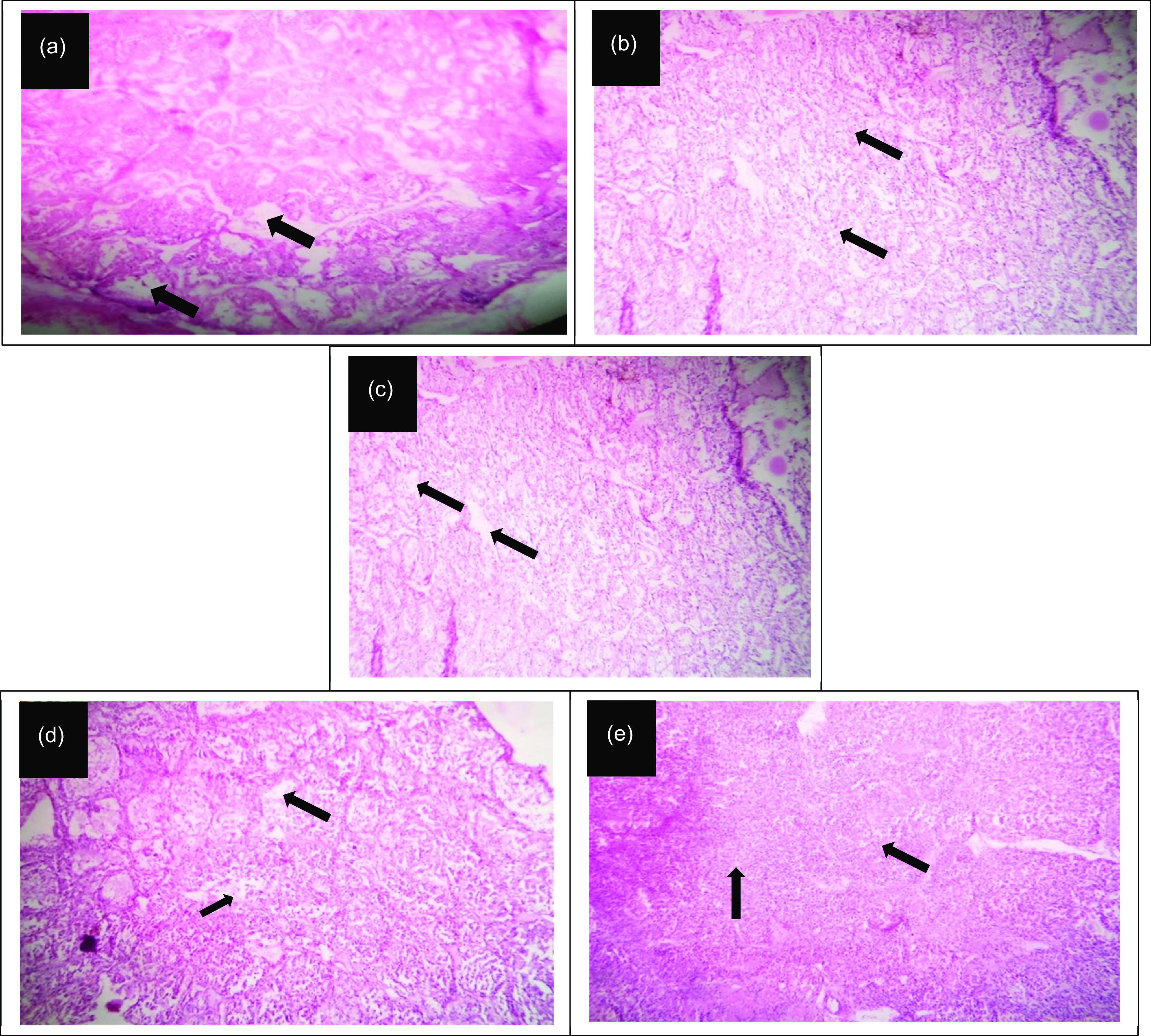
Effect of proposed therapies on the restoration of the histological changes in thyroid gland induced by propylthiouracil exposure
Figure 3(a)–(e) demonstrates the histological changes in the various experimental groups. Euthyroid in 2a depicts a photomicrograph of the thyroid gland of a rat showing no significant changes in thyroid follicles and follicle epithelium. Hypothyroidism control in 2b demonstrates a photomicrograph of the thyroid gland of a rat showing degeneration of follicle epithelium and lumen of follicles containing degenerated epithelium with pyknotic nuclei, eosinophilic colloid and infiltration of inflammatory cells. The photomicrograph of the thyroid gland of a rat of Vit. E + IF in Fig. 2(c) manifests a repair of the thyroid gland vasculature marked with reduced degeneration of follicle epithelium. Vit. E in Fig. 2(d) displays a photomicrograph of the thyroid gland of a rat showing curtailed hypertrophies of interfollicular cells. Thyroid follicles showing vacuolar degenerated follicle epithelium and loss of colloid. Finally, histological data of IF in 2e illustrates a photomicrograph of the thyroid gland of a rat showing restoration of the thyroid vasculature which is evident with the reduced hypertrophies of interfollicular cells.
Effect of proposed therapies on the restoration of the histological changes in thyroid induced by propylthiouracil exposure. (a) Euthyroid, (b) hypothyroidism control, (c) Vit. E + IF, (d) Vit. E and (e) IF. IF, intermittent fasting.

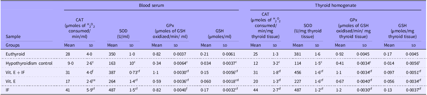
Effects on antioxidants CAT, SOD, GPx and GSH
Blood serum
In the hypothyroidism control rats, the levels of antioxidants, such as CAT, SOD, GPx and GSH were found to decrease notably (P < 0·001) as compared with the euthyroid group (Table 1). The Vit. E + IF experimental rats showed a significant increase (P < 0·001) in some of the antioxidant levels like CAT, SOD, GPx and GSH as compared with the hypothyroidism control group. Upon comparison of the antioxidant levels with the euthyroid group rats, a marked increase (P < 0·001) was observed in the levels of GPx and GSH, and nonsignificant changes were demonstrated in the CAT and SOD level. The Vit. E and IF experimental rats showed marked incline in the antioxidant levels of CAT (P < 0·01, P < 0·001), SOD (P < 0·001, P < 0·001), GPx (P < 0·001, P < 0·001) and GSH (P < 0·05, P < 0·001), respectively, as compared with hypothyroidism control animals. Upon comparison of the antioxidant levels of the Vit. E experimental animals with that of the euthyroid group, a marked decrease (P < 0·001) was observed in the levels of CAT, SOD, GPx and GSH while IF experimental animals exhibited a marked decrease (P < 0·001) in the levels of CAT and SOD and nonsignificant changes in the GPx and GSH level when compared with the euthyroid group.
Effects on blood serum and thyroid homogenate CAT, SOD, GPx and GSH level in the different experimental groups of propylthiouracil-induced hypothyroidism animal model

Values are expressed as mean ± sd (n 6). CAT, catalase; SOD; GPx, glutathione peroxidase; SOD, superoxide dismutase; GSH, glutathione. Data were analysed by using the one-way ANOVA, followed by the Newman–Keuls multiple comparison test and expressed as aP < 0·05, bP < 0·01, cP < 0·001 when compared with euthyroid group and dP < 0·05, eP < 0·01, fP < 0·001 when compared with hypothyroidism control group.
Thyroid homogenate
In the hypothyroidism control animals, the levels of antioxidants, such as CAT, SOD, GPx and GSH were found to decrease notably (P < 0·001) as compared with the euthyroid group (Table 1). The Vit. E + IF, Vit. E and IF therapy experimental rats showed a significant increase (P < 0·001) in the antioxidant levels like CAT, SOD, GPx and GSH as compared with the hypothyroidism control group. Upon comparison of the antioxidant levels of Vit. E + IF animals with the euthyroid, a marked increase (P < 0·001) was observed in the levels of CAT, SOD, GPx and a decrease (P < 0·001) in the level of GSH. While, upon the comparison of the antioxidant levels of Vit. E therapy animals with the euthyroid, a marked decrease (P < 0·001) was observed in the levels of CAT, SOD, GPx and GSH. Upon comparison of the antioxidant levels of IF with the normal, a marked decrease (P < 0·01) was observed in the levels of GSH and a notable increase (P < 0·001) in the CAT, SOD, GPx.
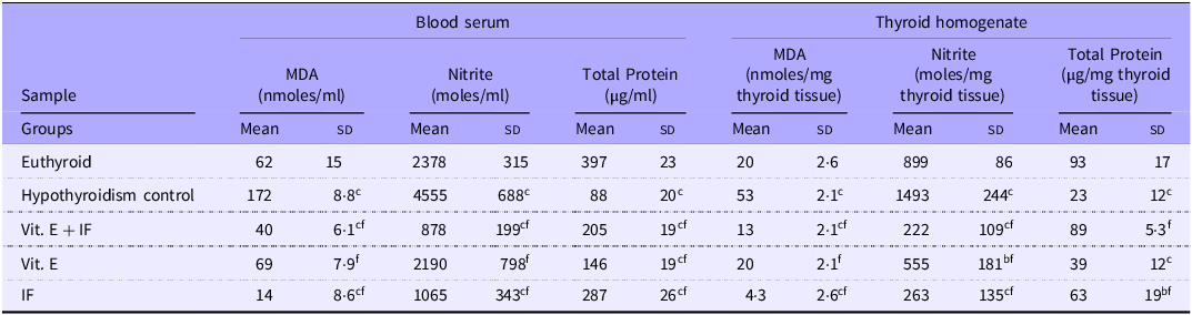
Effects on MDA, nitrite and total protein
Blood serum
The results showed that the hypothyroidism control animals had a significant increase (P < 0·001) in the MDA and nitrite content in the blood serum as compared with the euthyroid group (Table 2). The Vit. E + IF, Vit. E and IF therapy experimental rats showed a significant decrease (P < 0·001) in the levels of lipid peroxidation quantified by the levels of MDA and nitrite as compared with the euthyroid and hypothyroidism control animals. The level of nitrite in Vit. E therapy animals showed nonsignificant changes when compared with euthyroid group experimental rats. The hypothyroidism control animals showed a significant decrease (P < 0·001) in the total protein content in the blood serum as compared with the euthyroid group. The Vit. E + IF, Vit. E and IF experimental rats showed a significant decrease (P < 0·001) in the total protein content of blood serum as compared with the euthyroid group animals and an increase (P < 0·001) when compared with hypothyroidism control.
Effect of proposed therapies on blood serum and thyroid homogenate MDA, nitrite level and total protein content

Values are expressed as mean ± sd (n 6). MDA, malondialdehyde; IF, intermittent fasting.
Data were analysed by using the one-way ANOVA, followed by the Newman–Keuls multiple comparison test and expressed as aP < 0·05, bP < 0·01, cP < 0·001 when compared with euthyroid group and dP < 0·05, eP < 0·01, fP < 0·001 when compared with hypothyroidism control group.
Thyroid homogenate
The results indicate that the hypothyroidism control animals had a significant increase (P < 0·001) in the MDA and nitrite content in the thyroid homogenate as compared with the euthyroid group (Table 2). The Vit. E + IF and IF experimental rats showed a significant decrease (P < 0·001) in the levels of lipid peroxidation quantified by the levels of MDA and nitrite, respectively, as compared with the euthyroid and hypothyroidism control group. The Vit. E experimental rats showed a marked decline in the levels of MDA (P < 0·001) when compared with hypothyroidism control and nonsignificant changes as compared with euthyroid. On the other hand, the level of nitrite in thyroid homogenate decreased (P < 0·01, P < 0·001) as compared with the euthyroid and hypothyroidism control group respectively.
The results exhibit the remarkable remodelling of the total protein content in the thyroid homogenate of the various experimental groups (Table 2). The hypothyroidism control animals showed a significant (P < 0·001) decrease in the total protein content in the thyroid homogenate as compared with the euthyroid. The Vit. E + IF experimental rats showed a significant increase (P < 0·001) in the total protein content of thyroid homogenate as compared with the hypothyroidism control animals and nonsignificant when compared with the euthyroid group. The Vit. E therapy experimental rats showed a marked decline in the levels of total protein content (P < 0·001) as compared with euthyroid, and non-significant changes were observed when compared with hypothyroidism control. The IF experimental animals, exhibited a notable fall (P < 0·01), in the levels of total protein content when compared with the euthyroid group and an increase (P < 0·001) when compared with hypothyroidism control experimental rats.
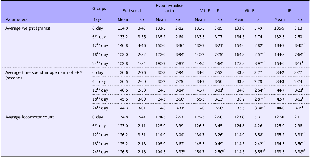
Effects of proposed therapies on weight
Administration of PTU to the hypothyroidism control group animals revealed a significant (P < 0·001) increase in the weight from the twelfth day till the end of the protocol as compared with euthyroid group (Table 3). In Vit. E + IF group there was a decrease (P < 0·001) in the weight from sixth day till the end of the protocol as compared with euthyroid and hypothyroidism control group rats in a time-dependent manner. In Vit. E and IF group, there was a notable decrease (P < 0·001) in the weight from 12th to 24th day as compared with the euthyroid group. The Vit. E experimental rats demonstrated a significant incline (P < 0·001) when compared with hypothyroidism control group while the IF group showed a marked decline (P < 0·001) in the weight of the experimental rats when compared with the hypothyroidism control rats.
Effect of proposed therapies on the weight, anxiety and locomotor activity

Values are expressed as mean ± sd (n 6). IF, intermittent fasting; EPM, elevated plus maze. Data were analysed by two-way ANOVA, followed by Bonferroni’s post hoc test and expressed as a P < 0·05, b P < 0·01, c P < 0·001 when compared with euthyroid group and d P < 0·05, e P < 0·01, f P < 0·001 when compared with Group II hypothyroidism control group.
Effects of proposed therapies on anxiety
The hypothyroidism control experimental rats revealed a significant decrease (P < 0·01) in the time spent in the open arm of elevated plus maze from the 12th day till the end of the protocol as compared with the euthyroid group (Table 3). The Vit. E + IF experimental rats showed that a significant increase (P < 0·001) in the time spent in the open arm was observed from 18th to 24th day of the protocol as compared with the euthyroid and hypothyroidism control group. The experimental rats of Vit. E and IF group showed a significant increase (P < 0·001) in the time spent in the open arm from 12th to 24th day when compared with the hypothyroidism control experimental rats. The Vit. E experimental animals showed a decreased open arm count (P < 0·001) as compared with euthyroid group in a time-dependent manner whereas non-significant changes were observed upon the comparison of the euthyroid group to IF group experimental rats.
Effects of proposed therapies on locomotor activity
The experimental rats of the hypothyroidism control group showed a significant (P < 0·001) decrease in locomotor activity from the 12th day till the end of the protocol as compared with euthyroid group rats (Table 3). The locomotor activity of Vit. E + IF group rats significantly increased (P < 0·001) from 12th to 24th day as compared with euthyroid and hypothyroidism control rats in a time-dependent manner. The Vit. E group rats showed a notable increase in locomotor activity (P < 0·001) on 18th and 24th day as compared with the hypothyroidism control group and decreased locomotor count (P < 0·001) as compared with euthyroid group animals in a time-dependent manner. However, on 12th day locomotor activity was decreased (P < 0·001) as compared with euthyroid and found nonsignificant when compared with the hypothyroidism control group. The locomotor activity of the IF group notably increased (P < 0·001) on 12th day, 18th day and 24th day as compared with the euthyroid and hypothyroidism control group in a time-dependent manner.
Adverse events
There was no any adverse event happened during the whole study.
Discussion
The underactive state of the thyroid gland is known as hypothyroidism in which it is not able to produce a sufficient amount of thyroid hormone. In this study, the experimental rats were administered PTU (1mg/ml orally) which has been formerly studied to induce hypothyroidism via inhibiting the thyroid peroxidase activity. Such a downregulation blocks the incorporation of iodine into thyroglobulin lowering circulating levels of thyroid hormones(Reference Shiroozu, Taurog and Engler37). This study incorporated the usage of IF and Vit. E administration to attenuate the hypothyroidism condition. The female albino Wistar rats were regularly checked for their weight, locomotor activity and anxiety during the study and backed by evaluation of various parameters like thyroid hormone profile, histopathology of the thyroid gland, antioxidant (CAT, GSH, GPx, SOD) level, MDA (indicator for lipid peroxidation), nitrite level, total protein content in blood serum and thyroid homogenate.
Former studies have elucidated the direct role of ROS in the formation of thyroid hormones and the functioning of the thyroid gland. However, the thyroid gland is highly susceptible to oxidative damage whereby any dysregulation in the ROS levels happens to damage the thyroid hormone production and functioning enhancing hypothyroid conditions(Reference Kochman, Jakubczyk and Bargiel3). MDA is a parameter to measure lipid peroxidation. Nitrite is a stable end product of NO metabolism, which converts hypochlorous acid into the more potent chlorinating species. MPO and inducible nitric oxide synthase are localised in the primary granules of leukocytes. When oxidative stress is increased within the cells MPO and inducible nitric oxide synthase generate hypochlorous acid and NO, respectively. As a result, the MPO-dependent oxidation of nitrite increases, forming a reactive nitrogen species which further leads to an increase in oxidative burden(Reference Erdamar, Demirci and Yaman38). Literatures have revealed that the thyroid hormone levels regulate the total protein synthesis and metabolism(Reference Alfadda, Benabdelkamel and Masood39,Reference Ito and Miyauchi40) . Protein deficiency leads to downregulation of the hypothalamus–pituitary axis resulting in the decreased synthesis and release of thyroid-stimulating hormone causing hypothyroidism(Reference Pavlović-Hournac and Delbauffe41). When PTU was given to hypothyroidism control experimental rats, it probably inhibited the synthesis of protein which in turn decreased the thyroxine-binding globulin(Reference Kochman, Jakubczyk and Bargiel3,Reference Seven, Seymen and Hatemi21–Reference Sharsher, Ahmed and Metwally24,Reference Lewicki, Lesniak and Bertrandt42) . Thus, a hypothyroid condition results in low total protein levels which is also evidenced from our study.
IF was utilised in the study owing to its property to enhance the antioxidant defense systems via possible activation of the Nrf2 pathway. This pathway enhances the production of enzymes like GPx and SOD which help to neutralise ROS(Reference Lettieri-Barbato, Minopoli and Caggiano43). Moreover, the role of IF in downregulating ROS via managing the mitochondrial function and autophagy has been well documented(Reference Shabkhizan, Haiaty and Moslehian44,Reference Grundler, Mesnage and Goutzourelas45) . IF has also been known to reduce the cellular stress response which acts as another important regulator of ROS production(Reference Sharsher, Ahmed and Metwally24,Reference Diab, Dimachkie and Zein46) . Vit. E was utilised in this study due to its inherent antioxidant potential. Moreover, it also supports the conversion of T4 (inactive thyroid hormone) to T3 (active form) which also supports the reduction in hypothyroid conditions(Reference Seven, Seymen and Hatemi21,Reference Napolitano, Fasciolo and Di Meo25,Reference Ye, Zhong and Du26) . As evidenced by the results, the adoption of the IF regime has led to the best activation of the antioxidant defence systems while reducing the MDA and nitrite activities providing a reduction in the hypothyroid conditions via possible management of the ROS and stress responses(Reference Lettieri-Barbato, Minopoli and Caggiano43). Vit. E plays a vital role in facilitating protein synthesis for the growth of follicular cells(Reference Napolitano, Fasciolo and Di Meo25,Reference Mutaku, Many and Colin47) . However, there are no studies that report the role of IF in regulating protein levels. This gap has been filled by our study which reports a positive regulatory role of IF in total protein synthesis. Furthermore, the histopathological studies conducted provide clear proof of widespread mechanobiological alterations in the cellular structure of thyroid follicles marked by degeneration of thyroid follicles with the appearance of pyknotic nuclei in follicles epithelium, in the hypothyroidism control. The Vit. E and IF monotherapy groups demonstrate restoration of the cellular structure in the thyroid gland thus providing sufficient evidential data regarding the preventive impacts of such a diet regimen.
Underactive thyroid affects weight, movement and exploration. IF may help reduce oedema and weight gain from salt and water accumulation. Studies suggest IF promotes natriuresis, regulating the renin–angiotensin–aldosterone system and lowering blood pressure(Reference Jeong, Yu and Bak48,Reference Tsaban49) . Fasting promotes cellular responses that prioritise using ketone energy over glucose and lower free radical production(Reference Tsaban49). On the other hand, Vit. E has been studied to reduce the overall inflammation in the body thereby reducing the weight which justifies our results thus stating the impact of the dual therapy regime in the management of weight(Reference Saboori, Shab-Bidar and Speakman50–Reference Bauer, Heinz and Whybrow52).
Hypothyroidism contributes to anxiety by disrupting thyroid hormone levels, which are essential for regulating neurotransmitters like serotonin that maintain mood stability. Low thyroid hormone levels can destabilise serotonin, leading to symptoms of anxiety(Reference Bauer, Heinz and Whybrow52). In addition, hypothyroidism is linked to reduced antioxidant levels, worsening oxidative stress—a known trigger for anxiety. Antioxidants play a critical role in mitigating this stress, and studies show that low antioxidant levels are prevalent in hypothyroid patients(Reference Bathla, Singh and Relan53–Reference Siegmann, Muller and Luecke58). IF and Vit. E-enhanced antioxidant levels have been shown to significantly reduce anxiety, highlighting their protective benefits. IF has emerged as a potential method to manage anxiety by addressing oxidative stress. It may boost antioxidant effects and stimulate brain-derived neurotrophic factor, a protein that enhances stress resilience and mood. By reducing oxidative stress and increasing brain-derived neurotrophic factor, IF offers a promising approach to alleviating anxiety in hypothyroid individuals(Reference Berthelot, Etchecopar-Etchart and Thellier59).
Studies have elucidated that an underactive thyroid reduces metabolism, thus restricting movement and physical activity, thereby causing a reduction in locomotor activity. It is also responsible for muscle weakness, fatigue and joint pain. PTU administration results in a reduced level of thyroid hormones, which lowers the rate of metabolism in the body, thereby restricting movement. Moreover, the generation of ROS by PTU tends to damage the cellular components, impacting the contractile function and movement capacity(Reference Smith, Grondin and LeMaster60). Our results depict a positive role of IF in enhancing the locomotor activity of the experimental rats, possibly due to the enhancement of ghrelin production in the plasma(Reference Akcali, Uslu and Onsinejad61). Moreover, IF and Vit. E happen to enhance muscle coordination and function due to its inherent antioxidant action, which is in synchronisation with the results obtained in this study(Reference Kandeil, Mohammed and Radi62,Reference Zadeh-Ardabili, Rad and Khazaài63) .
Our study has demonstrated the positive impacts of IF and Vit. E in the behavioural parameters, which is primarily due to the ROS scavenging effects of IF and Vit. E. Vit. E has the potential to bypass the BBB, which leads to stronger impacts in effects associated with the CNS, whereas IF monotherapy has surpassed the Vit. E + IF therapy effects in the activation of the antioxidant defence systems in the periphery.
The physiological impacts driven by our proposed therapies have not only benefited the thyroid gland functioning but have also impacted locomotion, anxiety-like behaviour and weight management. Thus, a profound impact of IF and Vit. E supplementation on a regular basis can be a better therapeutic intervention to help prevent the occurrence of incurable metabolic disorders like hypothyroidism. This study is entirely based on non-drug therapy and has yielded highly significant results. Moreover, it can be extended to human subjects without any drawbacks, as the therapy is safe and non-invasive.
Limitation of the study
This study had several limitations. Due to its short duration, we were unable to determine how long hypothyroidism can be prevented through IF and Vit. E administration. The study lasted only 24 days, which makes it difficult to compare its outcomes with the lifetime cost and potential side effects of levothyroxine treatment. Additionally, Vit. E was administered via the intraperitoneal route rather than through dietary supplementation, which may not fully replicate its natural physiological effects. We also did not assess key metabolic parameters such as lipid profile, liver function tests and glucose levels, which could have provided a more comprehensive understanding of the interventions. Furthermore, a sham injection was not performed for the control group, which may have introduced procedural bias. Lastly, our study did not include a levothyroxine-treated control group, limiting direct comparisons between standard therapy and our proposed interventions. These limitations should be considered when interpreting the findings and designing future studies.
Acknowledgements
The authors sincerely thank UGC-SAP, UGC-MRP and AYUSH-EMR for providing the required instrumental facilities.
This research received no specific grant from public, commercial or not-for-profit funding agencies.
N. K., A. D. and S. H. B. have framed the study. N. K., A. D., N. K., U. J. and G. S. have conducted the in vivo and experimental analyses. N. K. and A. D. have written the manuscript. S. H. B. and D. P. P. have analysed all the experimental data and have proofread the manuscript.
The authors declare that they do not have any conflict of interest.