

1. Introduction
First-episode psychosis (FEP) is characterized by the appearance of psychotic symptoms for the first time in an individual’s life. More than 3% of the general population suffers a psychotic episode at some point in life [Reference Perälä, Suvisaari, Saarni, Kuoppasalmi, Isometsä and Pirkola1]. The incidence rate is 21.4 per 100,000 person-years, and the average age when the first episode occurs is 30.5 years. However, epidemiologic data must be cautiously interpreted because of the heterogeneity of existing studies [Reference Jongsma, Gayer-Anderson, Lasalvia, Quattrone, Mulè and Szöke2].
There is enough evidence to indicate the need to improve and develop tools for early diagnosis and interventions in FEP to improve treatment response, reduce dysfunction and improve the quality of life of patients [3–Reference Rosa, González-Ortega, González-Pinto, Echeburúa, Comes and Martínez-Àran6].
There is a lack of knowledge regarding why some people fully recover from their symptoms while others tend toward deterioration and chronicity. More longitudinal investigations examining the biological markers of FEP are needed [Reference Fond, Albis, Jamain, Tamouza, Arango and Fleischhacker7].
One of the principal instruments used in biomarker research for FEP is magnetic resonance imaging (MRI) [Reference Northoff and Qin8]. Many studies have used MRI scans to obtain information about the main volumetric changes in the brain during psychosis over time (the possible secondary effects of medication, the loss of grey matter, etc.) [Reference Wright, Rabe-Hesketh, Woodruff, David, Murray and Bullmore9]. However, structural changes are not specific and cannot be used as predictors in individual cases, so more sensitive instruments are needed [10–Reference Cavelti, Kircher, Nagels, Strik and Homan13].
Another way to view the brain is with functional MRI (fMRI). Given recent theoretical models of psychosis, fMRI could provide an important means of understanding what happens in the brain during the processing of different stimuli [14–Reference Smieskova, Roiser, Chaddock, Schmidt, Harrisberger and Bendfeldt19]. This knowledge could contribute to an understanding of the biological issues underpinning the longitudinal course of psychosis.
To the best of our knowledge, there is only one systematic review focusing on psychotic patients in general [Reference Kani, Shinn, Lewandowski and Öngür20] and no systematic reviews of longitudinal fMRI studies in FEP patients. Hence, the purpose of this review is to systematically gather, analyse and review longitudinal fMRI studies in FEP patients with a specific focus on the relevance of methodology, the different paradigms used during scans and the mode of presentation. We think this review could show different methodological biases and activation results to explore their relationships and contribute to the methodological optimization of fMRI studies. In addition, we expect to elucidate the longitudinal changes in the brain and to shed some light on treatment effects in FEP.
2. Materials and methods
This review was methodologically outlined as a protocol prepared according to the PRISMA guidelines [Reference Gray21, Reference Moher, Liberati, Tetzlaff, Altman and PRISMA Group22].
2.1. Inclusion criteria
A longitudinal design involving at least two fMRI scans performed at different times with any interval between them.
Functional data, including activation or functional connectivity (FC) results.
Task-activated or resting-state fMRI paradigms.
Clinical studies with control groups.
Psychotic spectrum diagnoses (schizophrenia, schizophreniform disorder, bipolar disorder, schizoaffective disorder, brief psychotic episode and psychosis not otherwise specified) verified by structured clinical interviews.
First-episode samples, defined as individuals who had been diagnosed with a psychotic disorder for the first time in their lives and were assessed and scanned for the first time within the first 18 months after symptom onset.
The results, of every step, in the inclusion process of fMRI longitudinal studies of FEP.
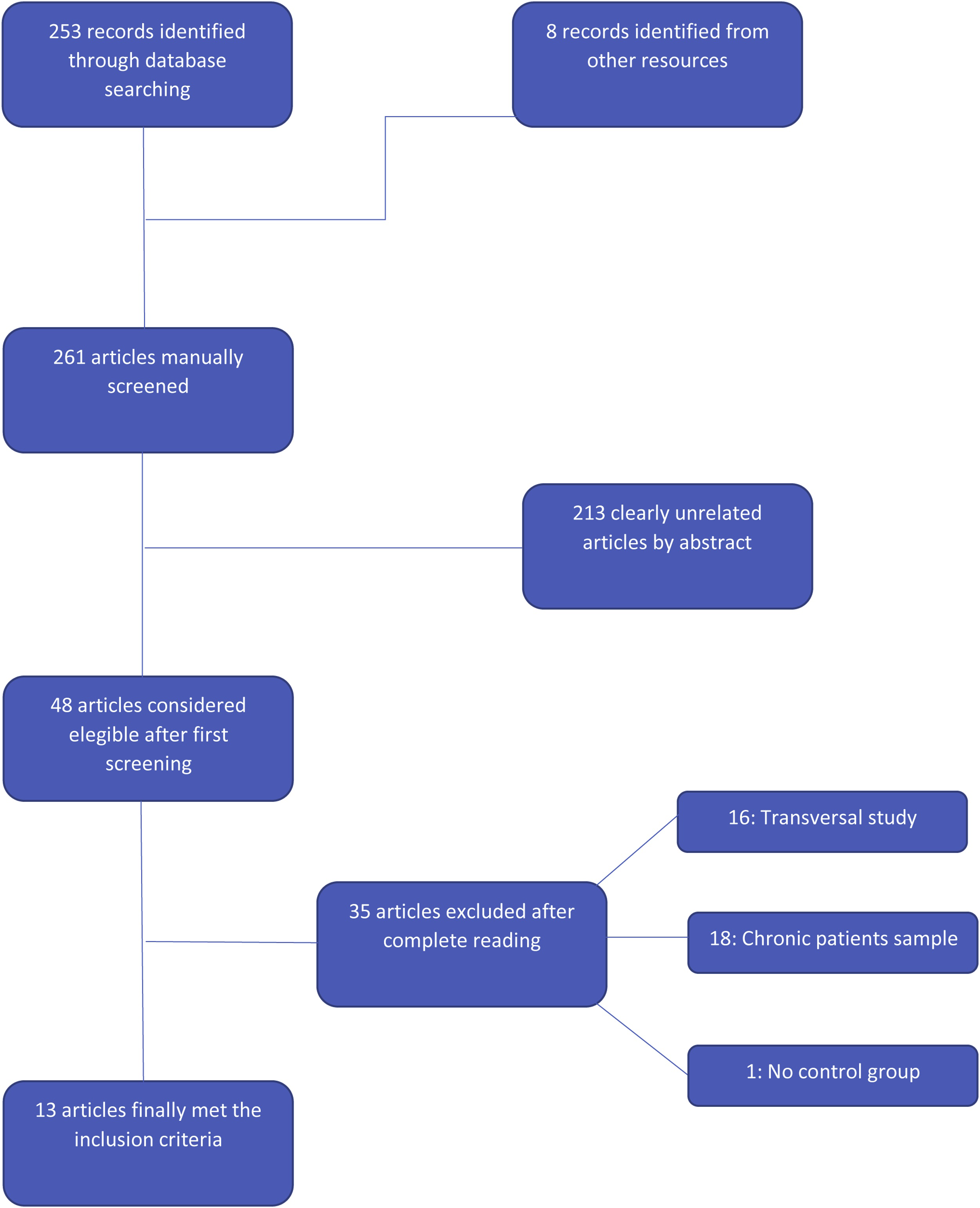
Longitudinal studies of fMRI in First Episode Psychosis.

FEP: First Episode Psychosis; FES: First Episode Schizophrenia; NOS: Not Otherwise Specified; HC: Healthy Control; DUI: Duration of Untreated Illness; ROI: Region of Interest; SCID I: Structured Clinical Interview for DSM-IV; BPRS: Brief Psychiatric Rating Scale; PANSS: Positive And Negative Scale Syndrome; MINI: Mini International Neuropsychiatric Interview; GAF: Global Functioning Scale; SUMD: Scale to Assess Unawareness of Mental Disorder; HAM-D: Hamilton Rating Scale for Depression; SDS: Schedule for the Deficit Syndrome; SAPS: Scale for the Assessment of Positive Symptoms; SANS: Scale for the Assessment of Negative Symptoms; SCAN: Schedule of Clinical Assessment in Neuropsychiatry; DLPFC: Dorsolateral Prefrontal Cortex; ACC: Anterior Cingulate Cortex; OFC: Orbito Frontal Cortex; ATP: Anterior Temporal Pole; IPS: Intraparietal Sulcus; IPL: Inferior Parietal Lobule; MPFC: Medial Prefrontal Cortex; DMPFC: Dorsomedial Prefrontal Cortex.
2.2. Exclusion criteria
Morphometric (MRI, DTI…) results.
Epidemiological studies, reviews and meta-analyses.
No psychosis spectrum diagnoses.
Inclusion of chronic patients, defined as those who have had one or more previous psychotic disorder diagnosed in their lifetime, or those who were scanned more than 18 months after the onset of symptoms.
2.3. Study search, selection and data extraction
A systematic search was conducted by two independent researchers (CG-V and PS-M) using the query (“longitudinal” and “fMRI” and “schizophrenia”) combined with (“longitudinal” and “fMRI” and “psychosis”) in the PubMed, MEDLINE and Web of Science (WOS) databases on April 2019. There was potential for an overlap in the final number of studies found due to the multiple databases employed.
These automatic searches were complemented by manual reviews of the references of the eligible articles after the final screening.
The selection of these datasets was performed hierarchically. An initial screening was performed based on the title and abstract, and a second screening was performed based on the full article reading.
Data were extracted using a table that addressed the following points: 1) author and year of publication, 2) sample data, including number of cases, mean age, percentage of males and diagnoses, 3) clinical assessment scales, 4) medication, 5) duration of illness and untreated period, 6) number of scans and intervals between them, 7) approach used (region of interest (ROI) or global), 8) paradigm and task applied, 9) results, according to hypo/hyper activation at baseline or follow-up and 10) comments regarding the discussion.
Given the very few articles that were finally included and the small resulting sample size, we decided to include both task and resting-state paradigms in the same table. The table summarizes the methodology of the studies, and their possible relationship with results will be examined in the discussion.
2.4. Neuroimaging acquisition and data processing
The MR field strength reported in the selected articles was either 1.5 or 3 T. Scans were performed by neuroimaging experts. There was high heterogeneity in the software tools used for data processing and brain mapping. Activation and connectivity studies were both considered.
3. Results
3.1. Included studies and sample description
The results of every step in the inclusion process are summarized in Fig. 1. A total of 13 clinical studies were included [23–Reference Sarpal, Robinson, Lencz, Argyelan, Ikuta and Karlsgodt35]. By aggregating the included samples, a total of 290 FEP patients and 401 healthy volunteers were assessed. The mean age of the patients was 24.55 years old, and the mean age of the controls was 29.9 years. Males comprised more than half of the study population (62% in both experimental and control groups). Every participant was scanned twice, with an average interval of 4.76 months between scans.
Diagnoses were made according to the DSM-IV-TR or ICD-10 criteria in every included article. Nine out of 13 studies used the Positive and Negative Symptom Scale (PANSS) for psychotic symptom measurement. The Brief Psychotic Rating Scale (BPRS) was used instead of the PANSS in 4 studies. The Global Assessment of Functioning (GAF) was used in 4 studies for global function measurement. Other clinical scales were used in only one study each and included the following: the Hamilton Depression Scale (HAM-D), the Mini International Neuropsychiatric Interview (MINI), the Scale to Assess Unawareness of Mental Disorder (SUMD) to evaluate insight, the Scale for the Assessment of Positive Symptoms (SAPS), the Scale for the Assessment of Negative Symptoms (SANS) and the Schedule of Clinical Assessment in Neuropsychiatry (SCAN).
3.2. Methodological issues
The articles included in this review showed important methodological heterogeneity.
Regarding article design, only 3 out of the 13 included articles used a double-blinded medication approach; these studies were interested in investigating the differences in different antipsychotic effects [Reference Reske, Kellermann, Habel, Jon Shah, Backes and von Wilmsdorff24, Reference Ikuta, Robinson, Gallego, Peters, Gruner and Kane25, Reference Sarpal, Robinson, Lencz, Argyelan, Ikuta and Karlsgodt35]. The remaining 10 studies implemented naturalistic designs with longitudinal follow-up of clinically selected samples treated according to a psychiatrist’s criteria.
In all studies, the main diagnosis was first-episode schizophrenia (FES), although 4 studies [Reference Niendam, Ray, Iosif, Lesh, Ashby and Patel27, Reference Nielsen, Rostrup, Wulff, Bak, Broberg and Lublin31, Reference Bergé, Carmona, Salgado, Rovira, Bulbena and Vilarroya32, Reference Keedy, Reilly, Bishop, Weiden and Sweeney34] included also patients with schizoaffective disorder. Nine out of 13 studies reported medication naïve patients at baseline, but all of the patients were receiving psychopharmacological treatment at follow-up assessment. Different antipsychotics were managed in the pharmacological treatments. Most of studies applied atypical antipsychotic medications, but 1 study implemented typical antipsychotic treatment (haloperidol) in a half of its patients [Reference Reske, Kellermann, Habel, Jon Shah, Backes and von Wilmsdorff24]. Antipsychotic doses are fully described in Table 1.
Second, the ROI approach was implemented in 12 of the 13 included studies. Specifically, the striatum, but also the prefrontal cortex, basal ganglia, hippocampus and limbic system, were the most investigated brain areas. The global approach was used in 1 study [Reference Li, Lui, Yao, Hu, Lv and Huang29], and there were 2 articles investigating specific functional networks involving emotional processing (the anterior cingulate cortex, pre-post central gyri and inferior frontal and temporal areas) [Reference Reske, Kellermann, Habel, Jon Shah, Backes and von Wilmsdorff24] and motor learning areas (the dorsal prefrontal cortex and striatum) [Reference Keedy, Reilly, Bishop, Weiden and Sweeney34].
Third, a resting-state paradigm was used in 4 of 13 studies. In these studies, images are obtained using no task in particular, and the results reflect the spontaneous fluctuation of the blood oxygen level-dependent (BOLD) signal. In contrast, 9 studies presented some kind of visual stimuli during fMRI scans to develop task paradigms. Two of them presented pictures of human facial emotional expressions to induce a mood or discriminate emotions [Reference Reske, Kellermann, Habel, Jon Shah, Backes and von Wilmsdorff24, Reference Bergé, Carmona, Salgado, Rovira, Bulbena and Vilarroya32]. Four other articles used a cognitive control paradigm [Reference Snitz, MacDonald, Cohen, Cho, Becker and Carter23, Reference Ikuta, Robinson, Gallego, Peters, Gruner and Kane25, Reference Niendam, Ray, Iosif, Lesh, Ashby and Patel27, Reference Keedy, Reilly, Bishop, Weiden and Sweeney34]. For this purpose, Snitz et al. [Reference Snitz, MacDonald, Cohen, Cho, Becker and Carter23] developed an overcome prepotency task in which the subject received visual targets (arrows pointing to left or right) and had to respond with ipsilateral or contralateral button that would be pressed depending on the target’s colour. In the Ikuta et al. study [Reference Ikuta, Robinson, Gallego, Peters, Gruner and Kane25], subjects completed the Multi-Source Interference Task (MSIT) to assess attentional control by the identifying targets with interference between the target number and the key. In the Keedy et al. study [Reference Keedy, Reilly, Bishop, Weiden and Sweeney34], subjects tracked a white dot along its relocations over the horizontal meridian. Two other studies implemented a reward paradigm to explore the benefits of medication for the brain’s reward system by changing the certainty of monetary gains according to the participants’ correct responses to target detection tasks [Reference Wulff, Nielsen, Svarer, Rostrup, Glenthøj and Pinborg26, Reference Nielsen, Rostrup, Wulff, Bak, Broberg and Lublin31]. Finally, one study developed a visual working memory task by presenting a memory set of 5 digits for 5 s, followed by series of target discrimination tasks [Reference van Veelen, Vink, Ramsey, van Buuren, Hoogendam and Kahn30].
Fourth, in terms of the presentation of the results, 3 of the 13 studies reported FC results [Reference Li, Lui, Yao, Hu, Lv and Huang29, Reference Anticevic, Hu, Xiao, Hu, Li and Bi33, Reference Sarpal, Robinson, Lencz, Argyelan, Ikuta and Karlsgodt35], presenting BOLD signal changes in networks that involve different brain structures. The other 10 studies presented the results according to the BOLD activation level differences in seed regions.
3.3. Longitudinal changes
Taken together, most of the included studies reported findings of hypoactivation in several brain areas during the basal assessment. Different studies found hypoactivation in the prefrontal cortex (PFC) areas [Reference Snitz, MacDonald, Cohen, Cho, Becker and Carter23, Reference Reske, Kellermann, Habel, Jon Shah, Backes and von Wilmsdorff24, Reference Niendam, Ray, Iosif, Lesh, Ashby and Patel27, Reference Li, Lui, Yao, Hu, Lv and Huang29, Reference van Veelen, Vink, Ramsey, van Buuren, Hoogendam and Kahn30, Reference Anticevic, Hu, Xiao, Hu, Li and Bi33, Reference Keedy, Reilly, Bishop, Weiden and Sweeney34] and limbic regions, specifically the amygdala and hippocampus [Reference Reske, Kellermann, Habel, Jon Shah, Backes and von Wilmsdorff24, Reference Li, Lui, Yao, Hu, Lv and Huang29, Reference Bergé, Carmona, Salgado, Rovira, Bulbena and Vilarroya32, Reference Keedy, Reilly, Bishop, Weiden and Sweeney34]. Hypoactivation was also found in regions such as the basal ganglia, particularly the striatum [Reference Reske, Kellermann, Habel, Jon Shah, Backes and von Wilmsdorff24, Reference Wulff, Nielsen, Svarer, Rostrup, Glenthøj and Pinborg26, Reference Nielsen, Rostrup, Wulff, Bak, Broberg and Lublin31], anterior cingulate cortex (ACC) [Reference Snitz, MacDonald, Cohen, Cho, Becker and Carter23, Reference Reske, Kellermann, Habel, Jon Shah, Backes and von Wilmsdorff24], thalamus [Reference Reske, Kellermann, Habel, Jon Shah, Backes and von Wilmsdorff24, Reference Li, Lui, Yao, Hu, Lv and Huang29], bilateral precuneus [Reference Li, Lui, Yao, Hu, Lv and Huang29] and posterior cingulate cortex (PCC) [Reference Keedy, Reilly, Bishop, Weiden and Sweeney34] (Fig. 2).
On the other hand, 5 studies reported some kind of hyperactivation at baseline. One study reported hyperconnectivity around medial prefrontal cortex (MPFC) regions [Reference Anticevic, Hu, Xiao, Hu, Li and Bi33]. Two studies reported hyperactivation of the basal ganglia [Reference Ikuta, Robinson, Gallego, Peters, Gruner and Kane25, Reference Hu, Zong, Zheng, Pantazatos, Miller and Li28]. One article reported hyperactivation of the cerebellum in a patient group [Reference Reske, Kellermann, Habel, Jon Shah, Backes and von Wilmsdorff24], but only during sadness processing. Finally, one study found hyperconnectivity between the right orbitofrontal cortex (ROFC) and the dorsolateral prefrontal cortex (DLPFC) [Reference Li, Lui, Yao, Hu, Lv and Huang29]. Just one included article reported no differences between patients and healthy control groups [Reference Sarpal, Robinson, Lencz, Argyelan, Ikuta and Karlsgodt35].
A summary of the main baseline findings in comparison with healthy control subjects is presented. Y = number of studies reporting this activation. X = grouped areas according to the main results from sources. Multiple studies report some hypoactivation (Blue) results in several brain areas, but specially in PFC, occipital cortex, cingulate cortex, basal ganglia and limbic system (For interpretation of the references to colour in this figure legend, the reader is referred to the web version of this article).

A summary of longitudinal changes in comparison with baseline results is presented. Y = number of studies reporting this activation. X = grouped areas according to the main results from sources. Increased activation (Red) when compared to baseline was found in most of the included studies. PFC and basal ganglia are the most frequently activated areas after treatment. (For interpretation of the references to colour in this figure legend, the reader is referred to the web version of this article).
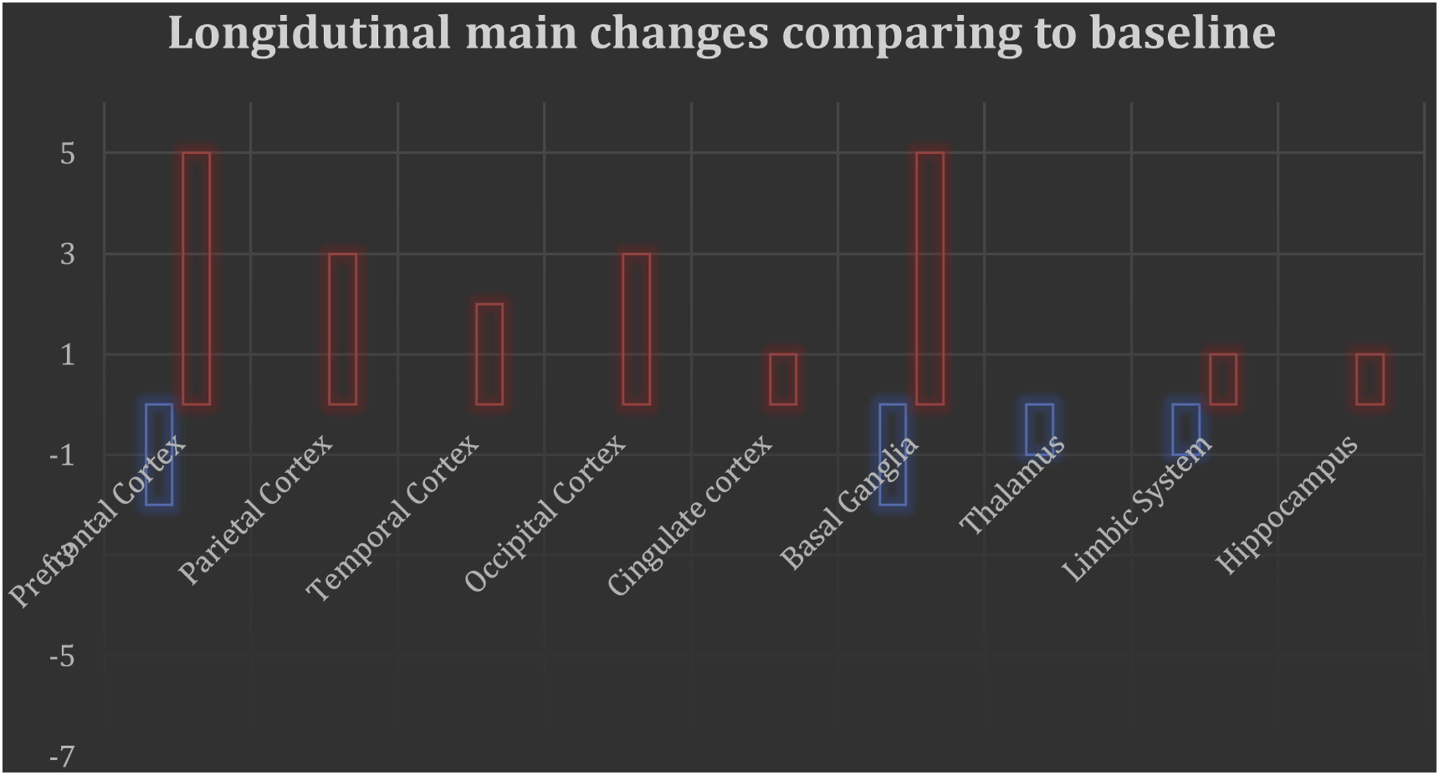
At follow-up, the main result was increased activation in those hypoactivated regions [Reference Snitz, MacDonald, Cohen, Cho, Becker and Carter23, Reference Wulff, Nielsen, Svarer, Rostrup, Glenthøj and Pinborg26, 29–Reference Sarpal, Robinson, Lencz, Argyelan, Ikuta and Karlsgodt35], which could be interpreted as normalization. Most of studies reported increased activation in PFC (OFC, DLPFC and MPFC) and basal ganglia but also in cingulate cortex, limbic system, parietal cortex, temporal cortex and thalamus. Some studies reported marginally decreased activations respect to pretreatment scans in the DLPFC [Reference Keedy, Reilly, Bishop, Weiden and Sweeney34], right accumbens [Reference Sarpal, Robinson, Lencz, Argyelan, Ikuta and Karlsgodt35], MPFC [Reference Anticevic, Hu, Xiao, Hu, Li and Bi33] and parietal cortex areas [Reference Li, Lui, Yao, Hu, Lv and Huang29, Reference Sarpal, Robinson, Lencz, Argyelan, Ikuta and Karlsgodt35]. Only one study reported as a main result a decrease in activation in the basal ganglia and thalamus [Reference Ikuta, Robinson, Gallego, Peters, Gruner and Kane36]. A summary of the main longitudinal changes can be found in Fig. 3.
Based on the differences in results, different explanatory hypotheses were proposed by the original authors. The dopamine hypothesis of schizophrenia was used in one study [Reference Hu, Zong, Zheng, Pantazatos, Miller and Li28]. Two studies linked the imaging results with the aberrant salience hypothesis [Reference Wulff, Nielsen, Svarer, Rostrup, Glenthøj and Pinborg26, Reference Sarpal, Robinson, Lencz, Argyelan, Ikuta and Karlsgodt35], which could be considered a version of the dopamine hypothesis. Meanwhile, one study considered the expanded dopamine hypothesis [Reference Keedy, Reilly, Bishop, Weiden and Sweeney34], and another proposed the dopamine and serotonin hypothesis [Reference Li, Lui, Yao, Hu, Lv and Huang29]. One study was interpreted to support the disconnection syndrome [Reference Anticevic, Hu, Xiao, Hu, Li and Bi33]. One study was interpreted to support the neurodevelopmental hypothesis for schizophrenia [Reference Niendam, Ray, Iosif, Lesh, Ashby and Patel27]. The other 6 studies proposed design-specific interpretations related to treatment response.
4. Discussion
In the present review, we examined longitudinal fMRI studies in FEP samples and found general hypoactivation in pretreatment scans that was reduced ("normalized") in the follow-up and after the administration of antipsychotic treatment. Regarding the implicated areas, there is a dependent relationship between results, seed regions and tasks. The main hypoactivated areas were the PFC (OFC, DLPFC and MPFC), the basal ganglia (caudate nucleus and striatum), the limbic system (the amygdala and insula), the hippocampus and the ACC. We also found hypoconnectivity in the included FC studies, which also tended to normalize at follow-up scans.
Notably, there are few papers reporting longitudinal follow-ups of FEP patients, so we considered all those that were available, including resting-state and task-activation designs. Of the 13 included studies, 4 used resting-state scans, and the other 9 studies presented different paradigms (mood induction, emotion discrimination, reward anticipation, working memory and cognitive control) in visual modalities.
Despite the commonalities, there are some different contributions that deserve to be discussed. Two of the three longest (more than 12 months between scans) articles [Reference Anticevic, Hu, Xiao, Hu, Li and Bi33, Reference Li, Lui, Yao, Hu, Lv and Huang37] were based on resting-state scans, but they reported different results, which may in part be due to their different ROI. Anticevic et al. [Reference Anticevic, Hu, Xiao, Hu, Li and Bi33] studied the PFC as an ROI, and reported hypo- and hyperconnectivity at baseline in the MPFC and DLPFC regions, respectively. Li et al. [Reference Li, Lui, Yao, Hu, Lv and Huang29] reported several hypoconnected areas across the brain in their global approach to image processing. Niendam et al. [Reference Niendam, Ray, Iosif, Lesh, Ashby and Patel27] studied the DLPFC using a task, with a long interval between scans (15 months) and they reported the absence of deterioration during the first 2 years of the illness in a very young patients sample.In contrast, Anticevic et al. reported stability of the DLPFC disfunctions starting with the youngest sample that we included in this review. The interpretation of these results is based on the neurodevelopmental hypothesis of schizophrenia, in which deficits emerge before illness onset.
According to the employed task, Snitz et al. [Reference Snitz, MacDonald, Cohen, Cho, Becker and Carter23], Ikuta et al. [Reference Ikuta, Robinson, Gallego, Peters, Gruner and Kane25], and Niendam et al. [Reference Niendam, Ray, Iosif, Lesh, Ashby and Patel27] implemented cognitive control paradigms and reported different findings (hypoactivation in the DLPFC and ACC versus hyperactivation in the basal ganglia and thalamus). Consequently, differences in the ROI approach (DLPFC and ACC versus the basal ganglia and thalamus), longer intervals between scans and different medication states at baseline (naïve versus prior antipsychotic exposure) must be considered for interpretation. The two studies that implemented emotional paradigms [Reference Reske, Kellermann, Habel, Jon Shah, Backes and von Wilmsdorff24, Reference Bergé, Carmona, Salgado, Rovira, Bulbena and Vilarroya32] found hypoactivation of its ROIs. They used different task and time intervals, but both reported some selective treatment effects resulting in the normalization of aberrant activation in some emotional processing areas, and both link their results with positive improvement in symptoms; and the stability of dysfunctions is more associated with negative symptoms. An important contribution comes from the Danish group, which has published two articles [Reference Wulff, Nielsen, Svarer, Rostrup, Glenthøj and Pinborg26, Reference Nielsen, Rostrup, Wulff, Bak, Broberg and Lublin31] using the same task paradigm and the same antipsychotic medication (amisulpride). The results vary according to the ROI, but they are consistent with the basal ganglia hypoactivation at baseline and the normalization after treatment.
We also found that the time between symptom onset and the first scan was not normally specified in our selected studies. Only the most recent articles included this important information for FEP samples categorization. According to the sample size, the largest study [Reference Hu, Zong, Zheng, Pantazatos, Miller and Li39] (n = 42) only reported hyperactivation of its ROI (striatum) at baseline that increased after treatment. This is one of the two articles [Reference Hu, Zong, Zheng, Pantazatos, Miller and Li28, Reference Li, Lui, Yao, Hu, Lv and Huang37] that used amplitudes of low frequency fluctuations (ALFF), fractional ALFF (fALFF) and regional homogeneity (ReHo) indices to show spontaneous brain activity during resting-state fMRI scans. They linked these results to the compensatory effects of treatment.
Regarding the use of medication at baseline, Sarpal et al. [Reference Sarpal, Robinson, Lencz, Argyelan, Ikuta and Karlsgodt35] scanned subjects with prior exposure to antipsychotics; their study is the only one in our review to report no significant differences at baseline between patients and control subjects. It also linked symptom improvement to increased connectivity in cortical areas and decreased connectivity in subcortical areas, normalizing the salience system.
Most of the studies linked their results with clinical variables. Nielsen et al. [Reference Nielsen, Rostrup, Wulff, Bak, Broberg and Lublin31] reported clinical improvement as patients obtained normalization as a brain reward system. Van Veelen et al. [Reference van Veelen, Vink, Ramsey, van Buuren, Hoogendam and Kahn30] reported normalization at the DLPFC after treatment in only a subgroup of patients (responders) versus persistent hypoactivation at the DLPFC in non-responders. Finally, only Keedy et al. [Reference Keedy, Reilly, Bishop, Weiden and Sweeney34] reported some adverse effects of medication resulting from hypoactivation at the DLPFC after treatment administration.
Other reviews have studied the longitudinal results of fMRI in chronic schizophrenia samples [Reference Kani, Shinn, Lewandowski and Öngür20]. In these reviews the most consistently reported finding is “normalization” or increased activation in frontal cortical regions, a finding that was consistent with our results. Decreased prefrontal activation (hypofrontality) has been reported in other neuroimaging reviews of chronic patients and is linked with negative symptoms and antipsychotic response [Reference Liemburg, Knegtering, Klein, Kortekaas and Aleman40]. In a recent study [Reference Cadena, White, Kraguljac, Reid, Maximo and Nelson41] of the relationship between BOLD signals and glutamate levels in the salience network (SN) and the default mode network (DMN) during resting-state scans in a sample of schizophrenia patients, the authors found a positive correlation between glutamate levels and activation in the HC group and in the SZ medicated group (positive but decreased at baseline), but a negative relationship in the SZ non-medicated groups. After 6 weeks, all the SZ patients were receiving antipsychotic treatment, and they all showed a positive correlation between glutamate levels and BOLD signals, but with significantly lower activation compared to the HC group in the SN (ACC and bilateral insula) and DMN (precuneus). Regarding other key brain areas, Blasi et al. [Reference Blasi, Popolizio, Taurisano, Caforio, Romano and Di Giorgio42] studied longitudinal changes in a schizophrenia sample and found hyperactivation in the left amygdala and hypoactivation in the right ventrolateral prefrontal cortex (VLPFC) during emotional processing in the SZ group at baseline compared to healthy controls. Both activation alterations were normalized after 8 weeks of treatment with olanzapine. Attending to the hippocampal network, Duan et al. [Reference Duan, Gan, Yang, Cheng, Gao and Shi43], studied FC in a sample with schizophrenia over the course of 24 months. They found hypoconnectivity between the left hippocampal network and the bilateral cerebellum at baseline. After 2 years, decreased FC in the left hippocampal network and increased FC in the right hippocampal network was observed. The authors proposed the disconnection syndrome as an explanatory hypothesis.
As opposed to the very few longitudinal fMRI studies in FEP samples, there are many longitudinal studies that analyse structural neuroimaging results. In a recent review [Reference Dietsche, Kircher and Falkenberg44], some studies reported significant grey matter loss in the frontal regions, thalamus and total brain volume. They also reported progressive cortical thinning in the superior and inferior frontal cortex and superior temporal cortex. White matter (WM) remained unclear due to contradictory results of recent studies reporting higher WM with longer periods of antipsychotic exposure. In spite of everything, linking structural and functional neuroimaging findings remains a challenge for researching.
In terms of limitations, it should be noted that the included studies present some important biases in sample selection. First, the sample size of most of the fMRI studies in general, but the longitudinal studies in particular, was small, typically approximately 20 subjects per group. Second, longitudinal course information such as symptom onset or its duration is frequently poorly described, which posed problems when categorizing articles as studies of FEP, high-risk individuals or chronic psychotic patients. Third, the included articles selected groups of patients with different ages, but they are balanced with healthy control groups by age and gender. Fourth, there were different medication and dose selections. Typical and atypical antipsychotics were used for treatment, and we consider that a potential area of confusion when interpreting the results. In general, antipsychotic treatment helps to normalize the BOLD signal in most cerebral regions, even in the first weeks of treatment [Reference Abbott, Jaramillo, Wilcox and Hamilton45]. Fifth, although all the included patients were on non-affective psychosis spectrum, there could be an important heterogeneity in the phenotypes of the included studies. This bias could be fixed by selecting more homogeneous samples with a specific phenotype.
Another very relevant methodological issue is paradigm selection. The type of paradigm selected is a key to understanding fMRI results [Reference Cao, Chén, Chung, Forsyth, McEwen and Gee46]. Different tasks have been reported to activate different regions [Reference Zhou, Wang, Zuo, Zhang, Wang and Jiang47]. In general, task-activated studies reported hypoactivations at baseline, while resting-state studies found both hypo and hyperactivations in basal assessments.
Our reviewed task studies mainly reported hypoactivations in the involved areas by their specific task and modality. When the task involves emotional processing, hypoactivations in the PFC, OPFC, ACC, amygdala and ventro-limbic region are reported. In the reward learning task, decreased activation of the striatum was found. In the visual cognitive control task, the involved areas were the DLPFC, ACC, left intraparietal sulcus (IPS), left lentiform nucleus, PCC, bilateral insula and visual cortex. A general decrease in activation was found in these areas.
Our group found important differences in activation (hyperactivation at baseline) and involved brain areas (notably the amygdala) in hallucinatory patients with an emotional auditory paradigm [Reference Aguilar, Corripio, García-Martí, Grasa, Martí-Bonmatí and Gómez-Ansón48], again suggesting a decisive influence of the selected paradigm.
On the other hand, resting-state studies reported both hypoactivations and hyperactivations at baseline. The differences in the areas’ activation could be explained by the methodological approach used. Hypoconnectivity between the left hippocampal network and the bilateral posterior lobe of the cerebellum was reported when the ROI was the hippocampal network. MPFC and lateral prefrontal cortex (LPFC) hypoconnectivity was found when the ROI was the prefrontal cortex, and increased activation of the left caudate nucleus and putamen was found in a striatum ROI approach.
Despite these important differences, we decided to combine task-activation and resting-state paradigms because of the lack of longitudinal studies found for each one. Recent studies have reported a convergent hypoactivation effect for the task-activation and resting-state studies in the fronto-temporal pathway that involves regions such as the DLPFC, OFC and superior temporal gyrus (STG) [Reference Mwansisya, Hu, Li, Chen, Wu and Huang49].
Regarding the resting-state paradigm, an important issue must be considered. In this paradigm, the investigation subject has to “not to think in something in particular” and wait until the end of the scan. Thus, there is no way to control the internal mental processes that could be modulating brain activation images during the scan [Reference Smitha, Akhil Raja, Arun, Rajesh, Thomas and Kapilamoorthy50].
Another important factor to consider is the difficulty of precisely determining the time interval between symptom onset and the first clinical assessment and treatment. Psychotic episodes sometimes start with negative symptoms, which are often hard to place along a timeline. We must also consider the different intervals between scans. In general, when the aim of the study is to follow the course of the illness, there is a longer period between scans (12 months or longer); in contrast, when the aim of the study is to demonstrate a specific treatment effect, we found shorter intervals (from 1 to 3 months).
In line with the observed heterogeneity in methodology, different interpretations have been proposed for the frequently reported result of normalization after treatment. Methodological homogeneity could help to establish a common theoretical framework. At the moment, the salience model offers an interesting framework for linking biological, clinical and neuroimaging findings. It has to be considered that longitudinal brain studies involving presentation modes other than visual or cognitive paradigms could show interesting results. We think that the development of simpler and more homogeneous paradigms, perhaps involving other modes of presentation, could help us to link this biological and clinical knowledge.
Our review has some limitations. First, there was a small number of longitudinal fMRI studies with FEP samples, so considered all that were available, including task-activation and resting-state paradigms. Second, small sample sizes are very common in functional neuroimaging studies due to the difficulty of recruiting and following an FEP sample over time and the high cost of assessment with fMRI scans. Third, there was no standardization of medications or doses, which could clearly affect the results. Fourth, there was considerable methodological heterogeneity in the included studies. Greater homogeneity of methods is needed to integrate different studies. Fifth, greater clinical heterogeneity in FEP patients than chronic psychosis patients has been reported [Reference Quattrone, Di Forti, Gayer-Anderson, Ferraro, Jongsma and Tripoli51]. This finding is important because some clinical symptoms (negative symptoms) early in the course of the illness may predict more severe mid-term outcomes [Reference Mezquida, Cabrera, Bioque, Amoretti, Lobo and González-Pinto52]. Sixth, regarding the follow-up, longer periods are needed to allow a naturalistic observation, which could help provided a prognosis beyond the treatment response.
5. Conclusions
To the best of our knowledge, this is the first review of longitudinal fMRI studies of FEP patients. We summarized the problems of longitudinal neuroimaging follow-ups and their methodological heterogeneity. The main finding was the normalization of brain activity after treatment. Although fMRI seems a promising tool for finding a biological marker of the illness, the mechanisms that distinguish which FEP patients will convert to chronic disease from those who will obtain a full recovery from symptoms have not been clarified. More longitudinal studies with larger samples and simpler and more replicable paradigms could help us to improve our knowledge of this illness and fill the gap between research and clinical practice.
Funding
This study was supported by grants from the Consellería de Educación (PROMETEO/2016/082) and Carlos III Health Institute (ISCiii PI17/00402) cofunded by The European Union through FEDER funds.


Comments
No Comments have been published for this article.