




Introduction
Acute appendicitis is an inflammatory condition of the vermiform appendix in the distal segment of the human gastrointestinal tract, which persists as one of the leading factors behind emergency surgeries in developed countries(Reference Bhangu, Søreide and Di Saverio1,Reference Ferris, Quan and Kaplan2) . Acute appendicitis has remained as the leading cause of emergency abdominal surgery worldwide(Reference Wickramasinghe, Xavier and Samarasekera3). The data from the Global Burden of Disease study reveals that the absolute number of annual cases has risen by nearly 39% over the last three decades, compounding to an estimated 17.7 million cases annually(Reference Wickramasinghe, Xavier and Samarasekera3). In countries undergoing rapid economic growth in regions like South Asia, the Middle East, and parts of South America(Reference Guan, Liu and Pan4,Reference Yang, Guo and Gu5) this surge has been particularly pronounced, where age-standardized incidence rates have seen an average year-on-year increase of 0.58%(Reference Yang, Guo and Gu5). Alleviating the financial and human resource burden imposed by the disease has an extraordinary social value(Reference Ferris, Quan and Kaplan2). The aetiological origins and the molecular processes by which inflammation arises in the appendix remain unclear(Reference D’Souza and Nugent6). Dietary shifts towards a western diet pattern (WDP) including low fibre and high levels of ultra-processed foods have been identified as one of the primary drivers for this surge(Reference Salminen, Paajanen and Rautio7). Thus, dietary patterns and nutrient intake have been previously identified as one of the critical factors associated with the onset of appendicitis(Reference Carr8). The formation of fecaliths (hardened stool) that can block the appendiceal lumen has been associated with the development of acute appendicitis(Reference Salminen, Paajanen and Rautio7). Fecaliths in the appendiceal lumen are referred as appendicoliths and are considered one of the main criteria in the clinical diagnosis of acute appendicitis(Reference Balthazar, Megibow, Siegel and Birnbaum9). In addition, acute appendicitis has been related to changes in the microbiome(Reference Vitetta, Chen and Clarke10). However, the modification of nutritional and other environmental factors affects a large percentage of the population, much larger than the incidence of acute appendicitis, which is around 1% of the global population. In other words, the pathogenesis of acute appendicitis involves individual genetic profiles predisposing to the onset of the disease(Reference Ferris, Quan and Kaplan2). Investigating how genetic factors and subsequent interactions with dietary patterns influence appendicitis pathogenesis is essential to develop preventive strategies(Reference Dimberg, Rubér and Skarstedt11). This review aims to critically assess the nutrition–microbiome–genetic axis in the pathogenesis of acute appendicitis.
Dietary and nutritional patterns associated with appendicitis
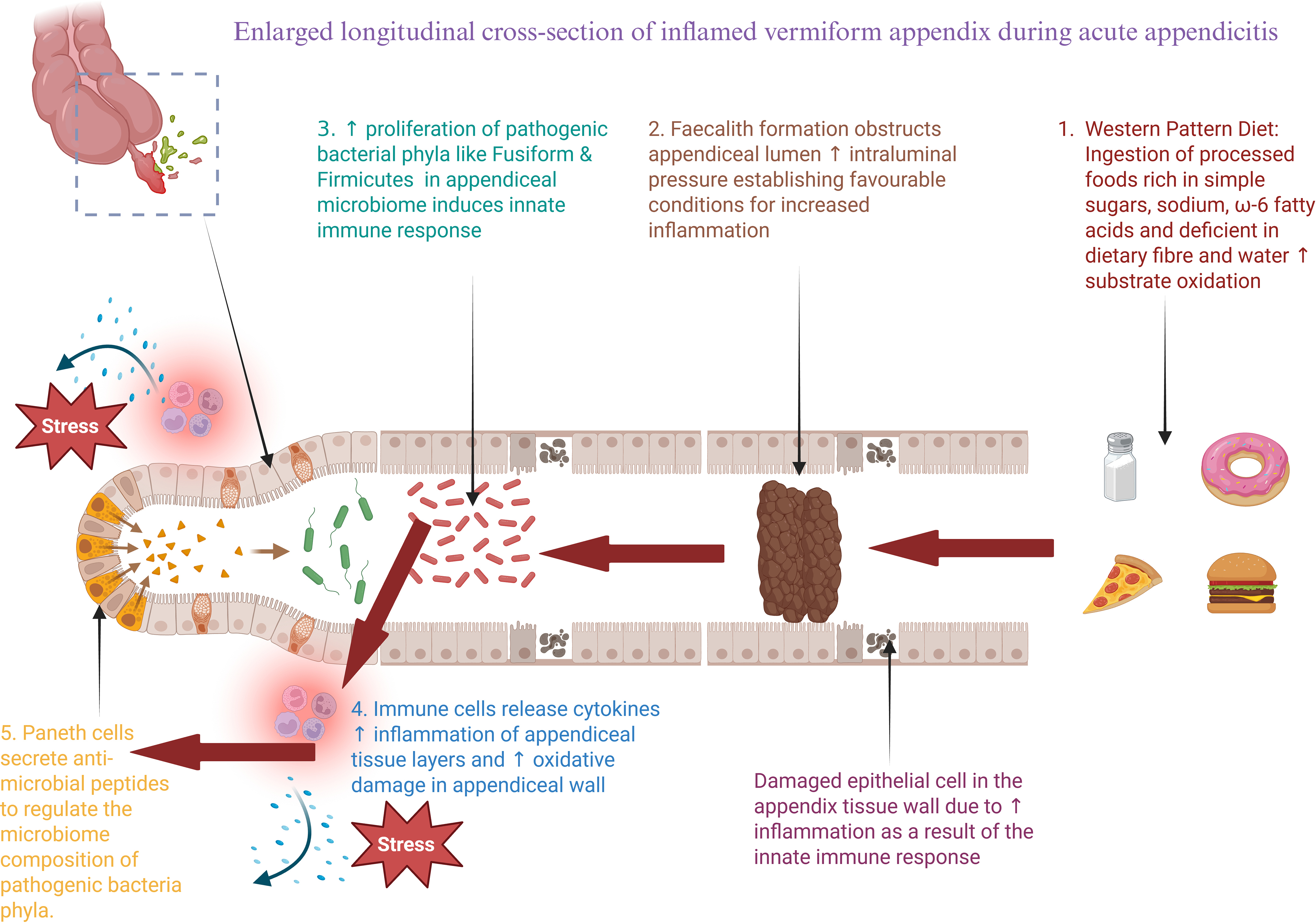
Dietary patterns in agrarian societies are commonly high in fibre which, in turn, may prevent the formation of appendicoliths that can block the appendiceal lumen, thus, protecting against the development of acute appendicitis(Reference Salminen, Paajanen and Rautio7). In contrast, rapid economic growth and the increase of urban populations have been associated with a dietary shift towards more processed and less fibrous foods characteristic of the WDP. The westernization of the diet in developing countries appeared as a key contributing factor in the pathogenesis of acute appendicitis and other distal gut disorders like inflammatory bowel disease (Fig. 1)(Reference Vitetta, Chen and Clarke10,Reference Ryoo, Hwang and Wright12) . For comprehensive reviews on the WDP the readers are referred to other sources(Reference Bouchard-Mercier, Rudkowska and Lemieux13–16).
The onset of appendiceal inflammation has been linked to the western diet pattern (WDP) resulting in increased fecalith formation and substrate oxidation accompanied by the proliferation of pathogenic bacteria (Firmicutes & Fusobacterium), the loss of epithelial integrity and the activation of the innate immune system.

Figure 1. Long description
A diagram of the process of acute appendicitis. The diagram shows the sequence of events leading to appendicitis. It starts with the ingestion of processed foods rich in simple sugars, sodium, omega-6 fatty acids, and deficient in dietary fiber and water, leading to substrate oxidation. This is followed by fecalith formation that obstructs the appendiceal lumen, increasing intraluminal pressure and creating favorable conditions for inflammation. The proliferation of pathogenic bacterial phyla like Fusiform and Firmicutes in the appendiceal microbiome induces an innate immune response. Immune cells release cytokines, increasing inflammation of the appendiceal tissue layers and causing oxidative damage in the appendiceal wall. Damaged epithelial cells in the appendix tissue wall due to increased inflammation result from the innate immune response. Paneth cells secrete anti-microbial peptides to regulate the microbiome composition of pathogenic bacteria phyla. Stress is shown to impact the process at multiple points.
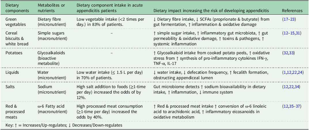
Multiple studies exploring the relationship between dietary habits and the risk of developing acute appendicitis have predominantly focused on dietary fibre consumption, particularly leafy green vegetables, as summarized in Table 1 (Reference Arnbjörnsson17–Reference Alzahrani, Al-Sehli and Alsharif24). Differences in daily dietary fibre intake in appendicitis patients compared with healthy controls were observed in two independent studies(Reference Adamidis, Roma-Giannikou and Karamolegou18,Reference Damanik, Fikri and Nasution20) . Additionally, the results from a binomial linear regression analysis of fruits and vegetables in the UK Biobank(Reference Sudlow, Gallacher and Allen25) platform highlighted how the absence of fruit intake raised the respective odds of appendicitis by 12% and 16%(Reference Ryoo, Hwang and Wright12). A recent meta-analysis involving 4 case studies demonstrated that a lower fibre intake was evident in acute appendicitis patients(Reference Sudlow, Gallacher and Allen25). From the same investigation, a meta-regression of ecological studies supported a pattern towards low acute appendicitis incidence with high fibre intake(Reference Pitesa, Spiekermann and Paterson26). Fruits like tomatoes and green vegetables have been previously suggested to induce anti-inflammatory effects from the appendiceal microbiota in the literature(Reference Carr8,Reference Barker, Morris and Nelson27) . Short-chain fatty acids (SCFAs) are some of the main metabolites produced by gut microbiota via the anaerobic fermentation of indigestible polysaccharides (i.e. such as dietary fibre and resistant starch) in the large intestine(Reference Cai, Fan and Zhang28). The anaerobic colonic fermentation of indigestible fibre from the cellulose cell walls of green vegetables has been shown to generate antioxidant SCFAs that subsequently down-regulated appendiceal inflammation and reduced the likelihood of acute appendicitis(Reference Ríos-Covián, Ruas-Madiedo and Margolles29). For example, probiotic supplements that enhance the synthesis of SCFAs were suggested as potential therapeutic applications for reducing the likelihood of appendicitis related to anti-inflammatory and anti-oxidative properties minimizing tissue damage(Reference Cai, Fan and Zhang28,Reference Elwan, Abdennour and Akl30) .
Dietary components and respective physiological effects on acute appendicitis pathogenesis

Table 1. Long description
A table with six columns and six rows, including a header row. The columns are labeled ‘Dietary components’, ‘Metabolites or nutrients’, ‘Dietary component intake in acute appendicitis patients’, ‘Dietary impact increasing the risk of developing appendicitis’, and ‘References’. The rows detail various dietary components such as green vegetables, cereal biscuits & white bread, potatoes, liquids, salts, and red & processed meats, along with their respective metabolites or nutrients, intake in patients, dietary impact, and references. Each row provides specific information on how different dietary components affect the risk of developing appendicitis.
IFN-γ = Interferon-gamma; IL-17=Interleukin-17; TNF-α=Tumour Necrosis Factor-alpha; SCFA=Short Chain Fatty Acids.
Additionally, constipation is increasingly recognized as a significant clinical risk factor in the pathogenesis of paediatric acute appendicitis(Reference Iwata, Yamaguchi and Itoh38). The existing literature evaluating appendicitis risks suggest that insoluble fibre provides superior mechanical protection relative to its soluble counterpart(Reference Arnbjörnsson17). This protective efficacy is primarily attributed to its non-viscous structural properties; insoluble fractions resist fermentation, increase faecal bulk, and accelerate hindgut transit times. Physiologically, this accelerated transit could mitigate luminal stasis and subsequent fecalith compaction within the restricted appendiceal lumen, a critical event in luminal obstruction(Reference Burkitt39). This mechanically driven aetiology diverges from the paradigm of gastrointestinal health, wherein soluble fibre is recognized for its beneficial role(Reference Barber, Kabisch and Pfeiffer40,Reference Gill, Rossi and Bajka41) . Soluble fibre dictates digesta rheology by immobilizing water to form a cohesive, hydrated viscous gel essential for smooth bolus transit(Reference Gunness and Gidley42). The scarcity of epidemiological evidence directly coupling soluble fibre to reduced appendicitis risk may reflect a fundamental gap in the literature regarding digesta hydration. In conditions of physiological dehydration, the gel matrix of soluble fibre is disrupted, and this otherwise protective prebiotic substrate may transform into a highly desiccated, hyper-viscous mass, exacerbating luminal stasis and contributing to the generation of appendicoliths(Reference Babington43).
The low consumption of water was also identified as a risk factor for appendicitis (Table 1)(Reference Ryoo, Hwang and Wright12,Reference Nurafitha, Syahputra and Novirianthy22) . A case-control study involving paediatric patients aged 6 to 15 years old demonstrated that 84.2% (48/57) of the appendicitis patients did not meet the Recommended Daily Intake (RDI) of water, whilst only 42.1% (24/57) control participants had insufficient water intake that did not meet the criteria(Reference Alzahrani, Al-Sehli and Alsharif24). Similarly, a cross-sectional study involving 916 post-operative appendicitis patients demonstrated that 69.8% (639/916) drank ≤6 glasses of water per day(Reference Nurafitha, Syahputra and Novirianthy22). Low water intake may lead to systemic hypohydration, which is the primary driver of constipation, as the colon intensifies the reabsorption of luminal water to maintain homeostatic fluid balance. The resulting desiccated faecal matter predisposes the individual to infrequent defecation and the formation of appendicoliths particularly in paediatric populations(Reference Bhangu, Søreide and Di Saverio1,Reference Nurafitha, Syahputra and Novirianthy22) . The confluence of low fluid and fibre intake appears to exacerbate colonic stasis, thereby increasing the clinical incidence of obstructive appendiceal disease. Thus, while speculative, the synergistic effects of insufficient water and dietary fibre intake may represent a significant contributing factor in the pathogenesis of acute appendicitis.
The expansion of urban centres acts as a catalyst for the nutrition transition characterized by the rise of commercial food systems that prioritize the accessibility and rapid delivery of ultra-processed foods over traditional dietary patterns(Reference Popkin, Adair and Ng44,Reference Monteiro and Cannon45) . Ultra-processed carbohydrate-rich foods were positively correlated with a higher incidence of acute appendicitis in post-industrial developed societies(Reference Vitetta, Chen and Clarke10,Reference Kim, Kim and Lim46,Reference Monteiro, Cannon and Levy47) . A binomial regression analysis performed upon dietary parameters examined against acute appendicitis using the UK Biobank(Reference Sudlow, Gallacher and Allen25) database showed an elevated appendicitis risk of 8% and 11% for individuals who consumed white bread and cereal biscuits, respectively (Table 1)(Reference Ryoo, Hwang and Wright12). According to the Australian Food Composition Database, the total sugar content for white bread and cereal biscuits (3.1% and 8.3%, respectively) is similar or higher than the sugar threshold of 5 per 100 grams of solid food product established by Food Standards Australia and New Zealand(48–50). A higher absorption of simple sugars from processed carbohydrates can lead to a proliferation in pathogenic bacteria in the gut, which increases gut permeability and oxidative damage, allowing pathogens and toxins to enter the bloodstream raising systemic inflammation and appendicitis risk(Reference Ryoo, Hwang and Wright12–Reference Drake, Sonestedt and Ericson15,Reference Loh and Blaut31) . In addition, a significant positive correlation was reported between potato consumption and appendicitis(Reference Barker, Morris and Nelson27). A direct cause linking a potato-heavy diet to appendiceal inflammation remains an open area of study, but several overlapping mechanistic pathways seem to be related. In short, potatoes primarily influenced appendicitis risk through a potential increase in faecal viscosity and prolonged colonic transit time promoting the desiccation and compaction of faecal matter into appendicoliths(Reference Brender, Weiss and Koepsell51,Reference Zampieri, Cinquetti and Murri52) . Potatoes have been well established as rich sources of soluble fibre and resistant starch that confer anti-inflammatory properties via SCFA production in the lower gut(Reference Burgos, Zum Felde, Andre, Campos and Ortiz53). However, the popular cooking method of deep-frying potato with skin, as opposed to peeling and boiling, elevate the intake of glycoalkaloids α-chaconine and α-solanine. Potato-skin glycoalkaloids have known toxicological thresholds that induce localized gastrointestinal irritation, mucosal inflammation, and alterations in epithelial permeability(Reference Schrenk, Bignami and Bodin54). These glycoalkaloids are known to enhance the disruption of intestinal integrity thus increasing circulating levels of pro-inflammatory cytokines such as IFN-γ, TNF-α, and IL-17 in individuals predisposed to inflammatory bowel disease(Reference Patel, Schutte and Sporns32,Reference Pramod, Venkatesh and Mahesh33) . In addition, skin-on potatoes could play a secondary role by immunogenic proteins (lectins) that stimulate localized appendiceal lymphoid tissue. Solanum Tuberosum Agglutinin (potato lectin) reach the lower bowel intact binding to cell-bound immunoglobulins (such as IgE) on mast cells and basophils and triggering non-allergic degranulation and the release of histamine and pro-inflammatory mediators(Reference Iablokov, Sydora and Foshaug55). In the appendix this localized hypersensitivity or tissue irritation can induce lymphoid follicular hyperplasia which is the second major cause of luminal obstruction beside fecaliths(Reference Zampieri, Cinquetti and Murri52). Hence, a high intake of deep-fried skin-on potatoes has been associated with glycoalkaloids and lectins resulting in pro-inflammatory effects on the gut and increase appendicitis risk in humans.
Another investigation from the same authors using the UK Biobank(Reference Sudlow, Gallacher and Allen25) showed that regular addition of salt to food in meals increased the risk by 12%(Reference Ryoo, Hwang and Wright12). A high level of sodium is another common feature of ultra-processed foods and may also be indicative of a wider dietary pattern that increases the risk of developing acute appendicitis in predisposed individuals(Reference Ryoo, Hwang and Wright12,Reference Nurafitha, Syahputra and Novirianthy22) . An increase in sodium levels upon the intake of fast food and instant noodle products was related to changes in the gut microbiome and immune cells, leading to inflammation and decreased functionality of the immune system(Reference Nurafitha, Syahputra and Novirianthy22,Reference Pramod, Venkatesh and Mahesh33) . About noodles, an Indonesian study described that 93% of appendicitis patients consumed instant noodles ≥4 times per month(Reference Ryoo, Hwang and Wright12,Reference Nurafitha, Syahputra and Novirianthy22,Reference Elijovich, Laffer and Sahinoz34) .
Like other distal gut diseases such as colon cancer and inflammatory bowel disease, red and processed meats have been hypothesized to exacerbate inflammation in the vermiform appendix, as shown in Table 1 (Reference Ryoo, Hwang and Wright12,Reference Peeters, Houben and Cools23,Reference Alzahrani, Al-Sehli and Alsharif24) . A cohort study of 4852 vegetarian or non-vegetarian participants with an appendectomy history showed that people with vegetarian diets had a 50% lower chance of requiring an emergency appendectomy in their lifetime compared to those who consumed meat(Reference Appleby, Thorogood and McPherson35). Similarly, in a retrospective study of 130 appendicitis patients in a rural context, only 20 (15.4%) patients out of 130 were exclusively vegetarian, which was below the percentile of the overall population of 32.8% classified as solely vegetarian(Reference Lohar, Asger Calcuttawala and Nirhale36,Reference Shridhar, Dhillon and Bowen56) . Therefore, this observation led to the interpretation that individuals who displayed dietary patterns other than solely vegetarian were at a greater risk of being diagnosed with appendicitis(Reference Lohar, Asger Calcuttawala and Nirhale36,Reference Shridhar, Dhillon and Bowen56) . Moreover, an observational study that compared an acute appendicitis patient cohort (254 cases) with an equal number of healthy controls found that daily meat consumption significantly increased the risk of developing acute appendicitis(Reference Peeters, Houben and Cools23). Consistent with the previous findings, an investigation of the UK Biobank database(Reference Sudlow, Gallacher and Allen25) also highlighted that eating processed meat everyday increased the odds of acute appendicitis diagnosis by up to 40%(Reference Ryoo, Hwang and Wright12). In contrast, in the same study, low consumption of oily fish (less than once a week) was found to increase the risk of appendicitis by 10%(Reference Ryoo, Hwang and Wright12,Reference Sudlow, Gallacher and Allen25) .
The pathogenesis and severity of appendicitis have been linked to dietary fat content and type(Reference Prieto, Wang and Halbach57,Reference Shommu, Blackwood and Jenne58) . Currently, the literature suggests that the WDP is characterized by an imbalance in the ratio of excess omega-6 PUFAs to omega-3 PUFAs, attributed to frequent red and processed meat consumption(Reference Naja, Hwalla and Itani14,Reference Jeong, Moon and Cho37) . The abundance of linoleic acid (LA) may enhance the conversion to arachidonic acid (AA) and up-regulate the production of pro-inflammatory eicosanoids during oxidative metabolism and could accelerate appendicitis pathogenesis(Reference Ryoo, Hwang and Wright12,Reference Jeong, Moon and Cho37) . Fatty acid degradation gene pathways were up-regulated during appendicitis and were positively correlated with the disease severity. The biomarkers in plasma included lower concentrations of 2-hydroxybutanoate and octadecadienoic acid coupled with elevated levels of isopropanol and o-acetylcarnitine(Reference Shommu, Blackwood and Jenne58). Furthermore, the composition of appendicoliths obtained from paediatric appendicitis patients identified palmitic acid and stearate as main constituents(Reference Prieto, Wang and Halbach57). These are constituents of a WDP that increase the likelihood of appendicitis(Reference Ryoo, Hwang and Wright12,Reference Murru, Manca and Carta59) . Palmitic acid is a SFA commonly found in full-fat dairy products, red meat, corn oil, palm oil and ultra-processed foods. It increases acute appendicitis risk by inducing gut dysbiosis, triggering TLR4-mediated inflammatory pathways, promoting fecalith formation and the mechanical obstruction of the appendiceal lumen(Reference Ryoo, Hwang and Wright12,Reference Murru, Manca and Carta59) . Additionally, appendicoliths were shown to have a higher omega-6 to omega-3 fatty acid ratio, which is also testament to a WDP linked to higher risk of gastrointestinal inflammation and appendicitis(Reference Naja, Hwalla and Itani14,Reference Murru, Manca and Carta59) . This strongly suggests that high dietary levels of lipids, particularly saturated fat, present in the nutritional profile exacerbate the likelihood and severity of acute appendicitis(Reference Ryoo, Hwang and Wright12,Reference Prieto, Wang and Halbach57) . In addition, the presence of redox-active metals such as iron, manganese and zinc in appendicoliths has also been implicated in the production of reactive oxygen/nitrogen species (RONS), which results in oxidative stress disrupting intracellular processes and exacerbating inflammation that ultimately may manifest as acute appendicitis(Reference Prieto, Wang and Halbach57).
The quantification of diet-derived metabolites in body fluids has been used to describe acute paediatric appendicitis(Reference Yu, Xiang and Wu60). Dietary amino acids glutamine, isoleucine, leucine and valine from amino acid metabolism, and betaine were identified as potential indicators of appendicitis in children(Reference Yu, Xiang and Wu60). Essential amino acids isoleucine, leucine, and valine (commonly referred as branched chain amino acids-BCAA-) are solely derived from external dietary sources(Reference Yu, Xiang and Wu60). Hence, an increase in serum BCAA in acute paediatric appendicitis can only be explained by elevated protein catabolism to BCAA, decreased BCAA catabolism and/or reduced gluconeogenesis from BCAA(Reference Yu, Xiang and Wu60–Reference Würtz, Soininen and Kangas62). An up-regulation of the anaerobic glycolytic pathway is observed in acute paediatric appendicitis as energy requirement significantly raises 3-hydroxybutyric acid and lactic acid levels that can be used to classify the severity of appendicitis(Reference Demircan, Cetin and Uguralp63). A high energy demand during acute inflammation requires the betaine metabolite carnitine to promote β-oxidation of fatty acids, which subsequently reduces serum betaine concentration in acute paediatric appendicitis patients(Reference Xie, Waters and Schirra64).
The global surge in appendicitis cases has been associated with dietary shifts partially associated with the population migration from rural to urban environments. The main nutritional changes that seemed to contribute to the pathogenesis of acute appendicitis included decreased dietary fibre, omega-3 fatty acids and water consumption, together with increased levels of red meat and saturated fat, the regular addition of salt and the high reliance on processed food, including ultra-processed foods. Collectively, these changes in dietary patterns associated with appendicitis align with the western diet (WDP). However, caution needs to be applied in these findings since the links between diet and appendicitis were largely from observational studies. Thus, the potential causality of the WDP pattern on acute appendicitis is not strong. These dietary changes, in turn, have a strong influence on the development of the microbiome, which may distinctively characterize appendicitis patients as discussed in the next section.
Diet, microbiome and appendicitis
The dietary patterns associated with increased acute appendicitis risk may be mediated, in part, by specific shifts in the large intestine and appendiceal microbiome. Several studies have highlighted how the inflammation of the appendix was associated with an altered appendiceal microbiome, including a proliferation of Fusobacterium and other oral pathogens, suggesting a potential role in the aetiology and severity of acute appendicitis(Reference Vitetta, Chen and Clarke10,Reference Guinane, Tadrous and Fouhy65–Reference Blohs, Mahnert and Brunnader70) . In two of the studies, the enrichment in Fusobacterium was found to be concomitant with a decline in the phylum Bacteroides(Reference Swidsinski, Dörffel and Loening-Baucke66,Reference Salö, Marungruang and Roth69) . In addition, a study involving 60 paediatric acute appendicitis patients showed that the increase in Fusobacterium was correlated with the increase in other pathogens, namely Porphyromonas and Parvimonas, implicated in tissue necrotizing activities that led to perforation and a breakdown of the appendiceal wall in complicated appendicitis cases(Reference Blohs, Mahnert and Brunnader70). Vitetta and co-workers compared the microbial populations in non-inflamed relative to inflamed appendices showing an increase in Fusobacteria together with a decrease in Proteobacteria (Table 2)(Reference Vitetta, Chen and Clarke10).
Relative abundance of bacterial phyla in normal or inflamed appendices and large intestine

Table 2. Long description
A table comparing bacterial phylum types in inflamed and non-inflamed appendixes and large intestine. The table has six rows and five columns. Column headers are: Bacterial phylum type, Inflamed appendix, Non-inflamed appendix, Large intestine, Phylum change from non-inflamed appendix to inflamed appendix, Phylum change from non-inflamed appendix to large intestine. Row 1: Actinobacteria, 1~3 percent, 1~11 percent, 9 percent, Decrease, Similar. Row 2: Bacteroidetes, 8~10 percent, 4~12 percent, 25 percent, Similar, Increase. Row 3: Firmicutes, 55~75 percent, 30~70 percent, 35 percent, Increase, Similar/Decrease. Row 4: Fusobacteria, 1~29 percent, 1~10 percent, 0 percent, Increase, Decrease. Row 5: Proteobacteria, 5~22 percent, 14~46 percent, 1 percent, Decrease, Decrease.
Adapted from Vitetta et al., 2019(Reference Vitetta, Chen and Clarke10), Guinane et al., 2013(Reference Guinane, Tadrous and Fouhy65).
Fusobacteria spp were identified as key constituents in the epithelial and submucosal infiltrates in 63% of patients with appendicitis(Reference Swidsinski, Dörffel and Loening-Baucke66,Reference Rogers, Brower-Sinning and Firek68,Reference Randal Bollinger, Barbas and Bush71) . A study of intraluminal appendiceal samples reported that Fusobacterium necrophorum was predominantly identified in catarrhal appendicitis and Fusobacterium nucleatum was mainly found in gangrenous appendicitis(Reference Schülin, Schlichting and Blod72). This expansion has been associated with dietary patterns. Dietary fibre deprivation is a determinant of Fusobacterium overgrowth, potentially affecting the colonic and appendiceal mucosa(Reference Desai, Seekatz and Koropatkin73). In the absence of fermentable substrates, beneficial butyrate-producing taxa decline sharply, prompting an increased vulnerability of the colonic mucus layer and pathogen susceptibility(Reference Desai, Seekatz and Koropatkin73,Reference Parada Venegas, De la Fuente and Landskron74) . Current pathophysiological models of appendicitis frequently employ a broad definition of dietary fibre, thereby overlooking specific fibre fractions. However, the evidence suggests that it is primarily the depletion of soluble, highly fermentable fibre that drives the loss of these protective, butyrate-producing taxa(Reference Chen, Chen and Tian75). Soluble fractions, such as inulin and pectins, serve as the primary substrates for butyrate-producing commensal bacteria within the Bacillota (formerly Firmicutes) phylum, particularly Faecalibacterium prausnitzii and Roseburia spp.(Reference Gill, Rossi and Bajka41,Reference Holscher76) . While an omission of insoluble fibre induces the physical stasis implicated in appendiceal obstruction, a deficit in soluble fibre may specifically lead to the depletion of anti-inflammatory butyrate-producing bacteria. The lack of SCFAs (in particular, butyrate) compromises mucosal barrier integrity and disrupts the immunological suppression required to prevent opportunistic pathogen overgrowth(Reference Parada Venegas, De la Fuente and Landskron74,Reference Chen, Chen and Tian75) . This is a critical step for Fusobacterium (which is highly muco-adhesive) to attach to the appendiceal or colonic epithelium, potentially facilitating the inflammatory cascade leading to acute appendicitis. Secondly, Fusobacterium species are proteolytic and thrive in a protein-rich environment(Reference Mehta, Nishihara and Cao77).
The relatively high consumption of meat products (i.e. red and processed meat) and saturated fat with low-fibre diets associated with acute appendicitis and characteristic of the WDP can lead to a rapid enrichment in the Fusobacterium population(Reference Liu, Tabung and Zhang78). The high consumption of red and processed meats provides the necessary sulphur-containing amino acids and heme that favour the increase of these pro-inflammatory bacteria such as Fusobacterium and Proteobacteria(Reference David, Maurice and Carmody79). However, in inflamed appendices the increase in Fusobacterium was paired with a decline in Proteobacteria(Reference Balthazar, Megibow, Siegel and Birnbaum9). This may suggest the occurrence of a competitive exclusion model, where a Western-style, fibre-depleted diet facilitates the degradation of the mucosal biofilm, allowing opportunistic, sulphur-reducing Fusobacteria to outcompete facultative anaerobes (i.e. Proteobacteria) in the increasingly anaerobic environment of the obstructed appendix.
The main dietary factor affecting microbial populations is fibre. Dietary fibre and resistant starch increase anaerobic fermentation producing short-chain fatty acids (SCFAs) in the large intestine(Reference Cai, Fan and Zhang28). Acetate, propionate and butyrate are SCFAs in the gastrointestinal tract that are hypothesized to be involved in anti-inflammatory activity, epithelial energy source provision, immune response modulation and the protection of intestinal mucosal integrity(Reference Ríos-Covián, Ruas-Madiedo and Margolles29). The quantification of bacterial fermentation-derived SCFA in body fluids has also been used to describe acute appendicitis. Four metabolites, including 3-hydroxybutyric acid, acetate, formate, and lactic acid, were identified as potential indicators of appendicitis in children(Reference Yu, Xiang and Wu60). A cross-sectional study investigating bacterial and fungal diversity in the stool samples of 60 adults with or without a history of appendectomy demonstrated that the appendicitis group had a lower diversity of gut bacteria species, including a decrease in anti-oxidative SCFA-producing microbes (i.e. Roseburia, Barnesiella, Butyricicoccus, Odoribacter, and Butyricimonas species)(Reference Cai, Fan and Zhang28). Additionally, a prospective cohort study involving rectal swab samples from 60 acute appendicitis patients and 20 healthy controls, showed a decline in commensal SCFA-producing bacteria composition particularly the Blautia, Faecalibacterium and Lachnospiracea genera(Reference Lee, Sulit and Frizelle80). Thus, SCFA-producing microbiota in the appendiceal microbiome seem to play a role in down-regulating the inflammatory mechanisms that culminate in appendicitis(Reference Loh and Blaut31).
The vermiform appendix has been postulated as a microbial reservoir that is involved in gut microbiome homeostasis, via the production and maintenance of protective biofilms that facilitate reinoculation of the gastrointestinal tract, following adverse events such as diarrhoea(Reference Randal Bollinger, Barbas and Bush71,Reference Laurin, Everett and Parker81) . Vitetta and co-workers suggested that the appendix may have a role in regularly re-inoculating the colonic microbiome upon minor and transient disturbances(Reference Balthazar, Megibow, Siegel and Birnbaum9). A recent review of the therapeutic application of probiotics has tentatively suggested that probiotics may have potential as adjunctive therapy for improving the management of acute appendicitis via promotion of the release of anti-inflammatory cytokine IL-10 and down-regulation of the levels of pro-inflammatory TNF-α and IL-6 cytokines(Reference Petruzziello, Saviano and Ojetti82).
Other bacterial pathogens have been described in the literature in association with an increased acute appendicitis risk. These include Campylobacter jejuni, Clostridium, Klebsiella and Prevotella that were significantly increased, whilst that of Streptococcus was reduced in paediatric appendicitis patients compared with the control group(Reference Megraud, Tachoire and Latrille83–Reference Bi, Yang and Li87). However, there is no direct evidence of the link between these pathogens and dietary shifts.
In summary, while the current knowledge on appendiceal microbiota does not fully explain the aetiology of acute appendicitis, the existing literature reveals links between diet and changes in specific microbial phyla worth further investigation. The WDP trends observed in acute appendicitis patients (e.g. low dietary fibre and high red and processed meat) may contribute to the shifts in the microbiome observed in inflamed compared to non-inflamed appendices such as an increased abundance of Fusobacteria together with a lower level of Proteobacteria. It is possible that the host immune response in appendicitis patients is more effective towards specific bacteria (e.g. Proteobacteria) paving the way for other pathogenic bacteria (e.g. Fusobacteria) to become dominant and subsequently leading to a higher risk of acute appendicitis. This, in turn, would suggest a role in genetic predisposition in the development of appendicitis. For example, genetic mutations in the NOD2 gene (a receptor expressed at the intestinal mucosa that regulates inflammatory activity against pathogenic bacteria(Reference Noguchi, Homma and Kang88)) frequently found in appendicitis patients may help open an ecological niche for Fusobacteria. The gene variants relevant to the nutrition-microbiome-genetic axis in the development of appendicitis are evaluated in the next section.
Genetic risk factors in appendicitis
The pathogenesis of acute appendicitis is essentially connected to an inflammatory response to compounds or bacteria present in gastrointestinal tract contents(Reference Peeters, Martens and D’Onofrio89). However, only a minority of the population exposed to similar environments (e.g. diet and other risk factors) develop the pathology, suggesting that some genetic or epigenetic drivers must be involved. Thus, this section studies the genetic polymorphisms associated with the nutrition-microbiome axis in acute appendicitis(Reference Salminen, Paajanen and Rautio7,Reference Peeters, Martens and D’Onofrio89–Reference Kiss, Minderjahn and Reismann91) .
The immune response observed in acute appendicitis is mainly mediated by T helper 2 (Th2) cells, a subset of CD4+ T lymphocytes that orchestrate antibody-mediated (e.g. IgE) response to extracellular parasites (helminths) and allergens. The appendicular content from appendicitis patients was associated with an elevated Th2 cytokine profile characteristic of food allergic responses(Reference Carvalho, Barros and Coelho92–Reference Hasassri, Jackson and Ghawi94). This was further supported by histopathological findings that identified the presence of eosinophilic infiltration, mastocyte degranulation and muscular oedema highly characteristic of an allergy response, in inflamed epithelial appendices(Reference Carvalho, Barros and Coelho92,Reference Carvalho, Carolino and Coelho93,Reference Cevizci, Sogut and Cayir95,Reference Carvalho, Carolino and Coelho96) .
One of the clinically relevant findings from genome-wide association studies (GWAS) conducted by the Global Biobank Meta-Analysis Initiative database (a consortium of 23 biobanks across 4 continents, including 32 706 acute appendicitis cases) is the identification of the single nucleotide polymorphism (SNP) HLA-C rs2524046, which is associated with both an increased risk of acute appendicitis and coeliac disease(Reference King, Jeong and Underwood97). Coeliac disease is an autoimmune, non-IgE-mediated disorder triggered by gluten ingestion from carbohydrate sources in the gastrointestinal tract, leading to small intestinal inflammation and impaired nutrient absorption(Reference Sarsu, Yılmaz and Bayram98). A potential association with the low consumption of water as a risk factor for appendicitis has been identified with the variant (rs9953918) within an intron of NEDD4L(Reference Itani, Stokes and Thomas99). NEDD4L encodes an enzyme involved in regulating epithelial sodium ion channels and fluid balance(Reference Jiang, Kawabe and Rotin100–Reference Cuellar-Partida, Lundberg and Kho102). Reduced NEDD4L activity has been reported in patients with acute appendicitis, suggesting that dysregulated ion transport may increase luminal sodium ion and water reabsorption, subsequently leading to luminal obstruction and appendiceal inflammation(Reference Itani, Stokes and Thomas99).
In addition, GWAS of gastrointestinal inflammation traits, including food allergies and acute appendicitis, have identified 6 loci in or near IL-6, IL-10 and IL-13, NOD2, CCL22 and CTLA4 (Reference Salminen, Paajanen and Rautio7,Reference Peeters, Martens and D’Onofrio89–Reference Kiss, Minderjahn and Reismann91,Reference Ricaño-Ponce, Peeters and Matzaraki103) . IL-13 is a key driver of Th2-mediated immune responses, promoting IgE synthesis and playing a central role in food allergies(Reference Salminen, Paajanen and Rautio7). The IL-13 rs1800925 T/T genotype has been specifically linked to an increased appendicitis risk(Reference Salminen, Paajanen and Rautio7). Unlike IL-13, IL-6 is often a marker of the severity of the immune response. Elevated levels of IL-6 have been reported during anaphylaxis(Reference Boyce, Assa’ad and Burks104), where it facilitates B cell immunoglobulin and T cell differentiation in the acute inflammatory response(Reference Megraud, Tachoire and Latrille83,Reference Jiang, Kawabe and Rotin100) . Several genetic polymorphisms in IL-6 (rs1800795, rs1800796) and IL-6R (rs7529229) have been investigated in relation to appendicitis(Reference Cuellar-Partida, Lundberg and Kho102,Reference Zhou, Kanai and Wu105) . The rs1800795 variant has been associated with disease severity(Reference Trimpert, Wesche and de Groot101). However, subsequent studies have not consistently replicated associations for these variants, and their role as clinically significant genetic risk factors remains uncertain.
In contrast, IL-10 is an anti-inflammatory cytokine involved in the development of oral tolerance to food proteins by inhibiting macrophage activity(Reference Chatterjee, Chiasson and Bounds106–Reference Rubér, Andersson and Petersson108). The rs1800896 polymorphism has been associated with reduced IL-10 levels and persistent milk allergy(Reference Satitsuksanoa, Jansen and Głobińska109,Reference Jacob, Pastorino and Okay110) . Similarly, elevated IL-10 concentrations have also been reported in plasma and peritoneal fluid of appendicitis patients compared with baseline controls(Reference Peeters, Martens and D’Onofrio89,Reference Chatterjee, Chiasson and Bounds106,Reference Daneshmandi, Ghasemi and Pourfathollah111) . The up-regulation of IL-10 is partially dependent on NOD2, a receptor expressed in the intestinal mucosal barrier that regulates immune responses to pathogenic bacterial stimuli(Reference Noguchi, Homma and Kang88,Reference Sarsu, Yilmaz and Bayram107) . While not directly involved in acute appendicitis, the NOD2 variant rs2066845 is a known risk factor for Crohn’s Disease(Reference Sarsu, Yilmaz and Bayram107). Research suggests that NOD2 variants may be linked to alterations in the gut microbiome, potentially contributing to gut dysbiosis (unbalanced bacteria) as a precursor to the development of an allergic response(Reference Taico Oliva, Musa and Kopulos112).
Variants in two additional genes – CLTA4 and CCL22 – have been associated with diet-related immune responses and acute appendicitis. CTLA4 encodes cytotoxic T lymphocyte-associated protein 4, an inhibitor of T cell activity and proliferation that down-regulates inflammation and immune responses(Reference Repnik and Potocnik113). The rs3087243 variant in CTLA4 has been associated with a lower appendicitis risk in patients older than 22 years, but an increased risk in younger individuals(Reference Salminen, Paajanen and Rautio7). In addition, the CTLA4 rs11571302 variant has been identified as a marker of allergic susceptibility in recent meta-analyses. Mechanistically, CTLA4 inhibits GATA-3, a key transcription factor regulating Th2-mediated allergic responses(Reference Simpson114,Reference Alegre, Frauwirth and Thompson115) . CCL22 encodes a chemokine involved in the recruitment of allergic T-Lymphocytes (Th2 cells) to the site (skin or gut epithelia) of an allergen(Reference Cianferoni, Shuker and Brown-Whitehorn116). The rs223888 variant in CCL22 has been associated with increased CCL22 expression across multiple immune and epithelial cell types, including B cells, dendritic cells, intestinal epithelial cells, and macrophages(Reference Luster117,Reference Moser, Wolf and Walz118) . Subsequently, this induces Th2-like inflammation in the gut mucosa via the attraction of CCR4 ligand bearing regulatory T cells (Treg) and Th2 CD4+ cells(Reference Luster117,Reference Moser, Wolf and Walz118) .
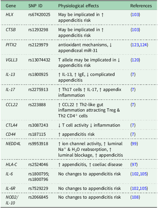
Several recent GWAS of acute appendicitis have identified additional genes, many of which are involved in immune and inflammatory pathways, including HLX, CTSB, NKX2-3, VGLL3, LTBR and DLEU1, IL-17, and CD44 (Table 3)(Reference Salminen, Paajanen and Rautio7,Reference Peeters, Martens and D’Onofrio89–Reference Kiss, Minderjahn and Reismann91,Reference King, Jeong and Underwood97,Reference Ricaño-Ponce, Peeters and Matzaraki103,Reference Zhou, Kanai and Wu105,Reference Ryoo, Hwang and Wright119,Reference Tartar, Akdeniz and Onalan120) . The IL-17 rs2275913 variant has been associated with an elevated risk of acute appendicitis and is thought to promote T helper 17 (Th17) cells to secrete the pro-inflammatory cytokine(Reference Salminen, Paajanen and Rautio7,Reference Appleby, Thorogood and McPherson35) . The CD44 variant rs187115 (A/G and G/G genotype) has been associated with a greater risk of perforated appendicitis(Reference Salminen, Paajanen and Rautio7). CD44 encodes an adhesion molecule expressed on various immune cell types that mediates interactions between Th1 and Th2 lymphocytes and the intestinal endothelium(Reference Goodison, Urquidi and Tarin121,Reference Bonder, Clark and Norman122) . These findings suggests that further exploration of regulatory mechanisms of these immune system-associated genes may be vital in understanding the development of appendicitis.
Single nucleotide polymorphisms (SNP) associated with acute appendicitis and related physiological effects
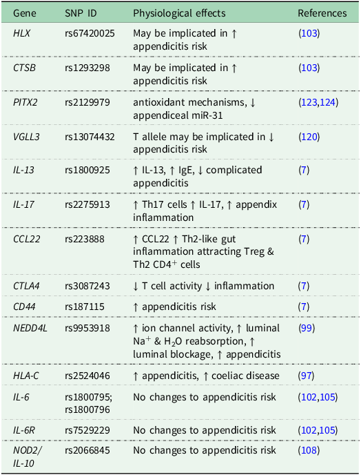
Table 3. Long description
A table listing single nucleotide polymorphisms (SNPs) associated with acute appendicitis and their physiological effects. The table has 15 rows and 4 columns. The columns are labeled Gene, SNP ID, Physiological effects, and References. Each row lists a specific gene, its corresponding SNP ID, the physiological effects associated with the SNP, and the references for the information. The table includes genes such as HLX, CTSB, PITX2, VGLL3, IL-13, IL-17, CCL22, CTLA4, CD44, NEDD4L, HLA-C, IL-6, IL-6R, and NOD2/IL-10, with their respective SNP IDs and physiological effects. The references column includes citations for the physiological effects listed.
Key for gene names: HLX (H2.0-like homeobox); CTSB (Cathepsin B); PITX2 (Pituitary Homeobox 2); VGLL3 (Vestigial like family member 3); IL-13 (Interleukin-13); IL-17 (Interleukin-17); CCL22 (C-C Motif Chemokine Ligand 22); CTLA4 (Cytotoxic T-Lymphocyte Associated Protein 4); CD44 (CD44 Molecule (Indian Blood Group)); NEDD4L (Neural Precursor Cell Expressed, Developmentally Down-Regulated 4-Like Ubiquitin-Protein Ligase); HLA-C (Major Histocompatibility Complex, Class I, C); IL-6 (Interleukin-6); IL-6R (Interleukin-6 Receptor); NOD2/Il-10 (Nucleotide-binding oligomerization domain-containing protein 2/Interleukin-10).
In addition, GWAS have identified several other genetic variants associated with acute appendicitis, including rs2129979 near PITX2 (Reference Kristjansson, Benonisdottir and Oddsson123,Reference Orlova, Yeh and Shi124) . Proposed roles of PITX2 include antioxidant and regenerative processes, potentially through the inhibition of appendiceal miR-31, which is already known to alleviate colonic inflammation(Reference Orlova, Yeh and Shi124,Reference Shi, Xie and Fu125) . Additional candidate genes implicated in appendicitis pathogenesis include RHOA (rs2247036), which is involved in maintaining intestinal epithelial cell integrity and regulating stem cell-mediated repair following injury, and MST1 (rs2247036), which has been linked to inflammatory bowel disease risk and the regulation of the innate immune response(Reference Orlova, Yeh and Shi124,Reference Chung, Kyoung and Rathor126–Reference Wu, Sun and Zuo129) . Further loci include variants in OSR-1, NCALD, and IRF8. OSR-1 and NCALD play roles in transmembrane ion transporter activity and regulation of G protein-coupled signal transduction, while IRF8 is linked to myeloid cell maturation. The implications of these three genes on the vermiform appendix inflammation are not yet understood(Reference King, Jeong and Underwood97).
Conclusion
There is a nutrition–microbiome–genetic axis in the pathogenesis of acute appendicitis driven by dietary habits associated to the western dietary pattern (WDP). These include mainly low consumption of fibre and water, and high levels of saturated fat, salt, processed meat and ultra-processed foods collectively, accounting for the main environmental risk of developing acute appendicitis. Specific dietary components including fats and redox-active metals were found as constituents of appendicoliths. The WDP may also explain, in part, the shifts in the microbiome observed in inflamed compared to non-inflamed appendices such as an increased abundance of Fusobacteria together with a lower level of Proteobacteria. Finally, there has been considerable progress over the past decade in identifying genetic risk factors implicated in appendicitis mainly pointing at allergic type of responses such as celiac disease and food allergy, most involving immune system driven inflammatory genes. Potential venues relating acute appendicitis to food allergic-like reactions warrant further investigation. The public health management of acute appendicitis risks requires a holistic multi-faceted approach that recognizes and addresses the complex interactions between nutrition and dietary patterns, appendiceal microbiome development, and genetic predisposition that equally play important roles in the development of the disease.
Acknowledgements
The authors would like to thank the Nutrition Society of Australia (NSA) for the invitation and opportunity to present this research at the Nutrition Society of Australia (NSA) Annual Scientific Meeting in December 2025.
Financial support
This research received no specific grant from any funding agency, commercial or not-for-profit sectors, but has been derived from the authors’ experience of researching the relationship between dietary intake and genetic risk factors with appendicitis over several years.
Author contributions
Original concept ER; writing original draft MR, ER; writing and editing ER, MR, DH; project management and supervision ER. All authors have read and agreed to the published version of the manuscript.
Competing interests
The authors declare no conflict of interest.


 Open access

