


Erdheim-Chester disease (ECD) is a rare non-Langerhans cell histiocytosis classified under the 2021 World Health Organization (WHO) classification of hematolymphoid tumors as a histiocytic neoplasm. The disease is characterized by infiltration of foamy histiocytes with associated fibrosis and predominantly harbors activating somatic mutations in the MAPK pathway, most notably the BRAF V600E mutation. Central nervous system (CNS) involvement occurs in almost half of patients and is associated with increased morbidity and poorer prognosis. Among CNS manifestations, extra-axial lesions account for approximately 30%–50% of cases and may radiographically mimic meningioma or subdural mass, thereby posing a significant diagnostic challenge. Reference Rai, Swartz and Soni1,Reference Goyal, Heaney and Collin2
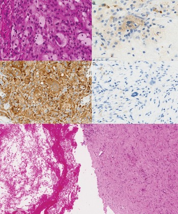
We report the case of a 76-year-old woman presenting with a two-month history of headache and left-sided weakness without antecedent head trauma. Neurological examination revealed mild left-sided motor weakness. Her past medical history was remarkable for rheumatoid arthritis, breast cancer and coronary artery disease, for which she was on dual antiplatelet therapy. Non-contrast CT of the brain demonstrated a large right fronto-parietal hypodense subdural collection with a hyperdense rim and significant mass effect (Figure 1(A)), leading to a presumptive diagnosis of chronic subdural hematoma (cSDH). The patient underwent burr-hole evacuation; intraoperatively, a thick, abnormal membrane was encountered beneath the dura and only a small quantity of “motor oil-like” fluid was evacuated under moderate pressure. Postoperative CT showed minimal evacuation and persistent mass effect (Figure 1(B)), prompting middle meningeal artery embolization. Nevertheless, the patient developed recurrent symptoms five months later, with interval enlargement of the collection on serial imaging (Figure 1(C)). Craniotomy was performed, during which the presumed hematoma was found to be a solid, fibrous mass requiring ultrasonic aspiration. Post-craniotomy CT showed removal of the lesion with relieved mass effect (Figure 1(D)). Histopathological analysis revealed dense histiocytic infiltration consisting of large aggregates of foamy histiocytes within a background of acute and chronic organizing hematoma. Immunohistochemistry (IHC) demonstrated CD68 positivity and CD1a negativity, with BRAF V600E positivity in the foamy histiocytes, consistent with a diagnosis of ECD (Figure 2). Next-generation sequencing confirmed the presence of the BRAF V600E mutation in the mass. Brain MRI (Figure 1(E, F)) was obtained only after the diagnosis of ECD, as the lesion had been presumed to represent a cSDH with organized hematoma. Subsequent systemic staging with FDG PET-CT and abdominal CT revealed bilateral symmetric metaphyseal and diaphyseal sclerosis around the knees and the characteristic “hairy kidney” pattern of perinephric infiltration, establishing multisystem disease. The patient achieved complete resolution of neurological symptoms and was referred for multidisciplinary review for consideration of targeted BRAF inhibitor therapy.
(A) The presenting CT brain showing the large right-sided subdural lesion causing mass effect and midline shift. (B) The CT scan post the burr-hole evacuation showing the significant residual lesion and persistent mass effect. (C) The 5-month follow-up CT scan demonstrating the residual subdural lesion. (D) Post-craniotomy CT showing the resection of the lesion and relieved mass effect over the adjacent brain. (E, F) axial and coronal MRI of the brain obtained after histological diagnosis demonstrating the residual pachymeningeal enhancement and mild mass effect.

Histopathological examination pictures 400x (A) Hematoxylin and Eosin (H&E) stain showing foamy histiocytes and Touton giant cells. (B) BRAF V600E immunohistochemistry (IHC) stain showing focal positivity. (C) IHC (CD68) stain positivity and (D) CD1a negativity. (E) 200x H&E stain demonstrating acute (left) and chronic (right) hemorrhage with granulation tissue within the resected lesion.

Dural-based ECD lesions associated with subdural hematoma represent an exceedingly rare presentation. To our knowledge, only two prior cases of confirmed ECD with subdural hematoma have been described in the literature. Noh et al. reported embolic infarction with subdural hemorrhage in a patient with ECD and noted that dural involvement may predispose patients to nontraumatic hemorrhage, without elaborating on the underlying mechanisms. Reference Noh and Kang3 Gupta et al. described a case of CNS-limited ECD presenting as a parafalcine dural mass associated with subdural hematoma and chronic superior sagittal sinus thrombosis. Reference Gupta, Haque, Reddy and Pérez4 Our case adds to this limited but important series by providing histopathological evidence of coexisting ECD infiltration and organizing hematoma.
The pathophysiological mechanism underlying SDH in ECD has not been established but is likely multifactorial. Histiocytic infiltration of the dura may disrupt the dural border cell layer. Furthermore, ECD lesions are characterized by a prominent inflammatory microenvironment with overexpression of cytokines, including tumor necrosis factor-α, interleukin-6 (IL-6) and interleukin-1 (IL-1). Reference Haroche, Arnaud and Cohen-Aubart5 These pro-inflammatory factors promote angiogenesis both directly and indirectly via upregulation of vascular endothelial growth factor (VEGF). Enhanced VEGF signaling may lead to the formation of fragile, immature neo-vessels within the infiltrated dura, predisposing to spontaneous hemorrhage. Moreover, perivascular and peri-adventitial infiltration – a hallmark of systemic ECD – may extend to intracranial vessels, producing local venous congestion and impaired outflow with secondary rupture of fragile vessels. The coexistence of venous sinus thrombosis in previously reported cases of dural ECD further supports a hemodynamic contribution. Reference Rai, Swartz and Soni1,Reference Gupta, Haque, Reddy and Pérez4 Of particular relevance, chronic inflammatory remodeling of the dura may create a pro-angiogenic and pro-fibrotic milieu analogous to the vascularized outer membrane seen in cSDH. Reference Arunachalam Sakthiyendran, Gonzalez-Salido, Perkins, Enriquez-Marulanda, Dasenbrock and Holsapple6 Within this inflammatory milieu, recurrent microhemorrhages from fragile capillaries could produce a mixed lesion containing both organizing hematoma and neoplastic histiocytic tissue, as observed in our case.
Our case underscores several clinical implications for the recognition and diagnosis of dural-based ECD. Alternative diagnoses should be considered in cases of atypical, recurrent or refractory subdural collections, particularly in the absence of trauma or when imaging reveals prominent or atypical membrane formation, nodularity or persistent mass effect after drainage. In retrospect, the thick membrane and scant “motor oil”-like fluid encountered at the initial burr-hole evacuation, together with minimal radiographic decompression, were early indicators of an underlying pathological process. In such cases, a mini-craniotomy may be preferable: the wider exposure ensures accurate placement of a subdural drain and permits sampling for histopathological analysis, potentially establishing the diagnosis at the index operation. Earlier contrast-enhanced MRI should likewise be considered, as it may reveal features suggestive of an infiltrative process rather than a simple hematoma.
Identification of the underlying molecular driver has direct therapeutic implications. Targeted therapy – including BRAF inhibitors for mutation-positive disease and MEK inhibitors for BRAF-negative or refractory cases – has transformed the prognosis of ECD. Reference Goyal, Heaney and Collin2,Reference Khoury, Solary and Abla7,Reference Go, Jacobsen and Baiocchi8 Surgical intervention remains essential for decompression and tissue diagnosis in cases presenting with significant mass effect; however, it is not curative. Optimal management therefore requires multidisciplinary collaboration among neurosurgery, neuropathology, hematology and neuro-oncology to guide both systemic therapy and long-term surveillance. Reference Go, Jacobsen and Baiocchi8
In conclusion, subdural hematoma may be a rare but clinically significant initial manifestation of dural-based ECD. Accumulating case reports suggest that histiocytic dural infiltration, inflammatory cytokine–mediated angiogenesis and vascular compromise collectively predispose to hemorrhage. Awareness of this association is critical to prevent delayed diagnosis and repeated ineffective interventions. When confronted with an atypical or recurrent SDH, particularly one with unusual intraoperative features, clinicians should consider an underlying neoplastic etiology, including ECD and pursue appropriate histopathological and molecular evaluation.
Author contributions
Conceptualization: WY, JM. Histopathological and molecular preparation and review: JH, YL, LA and CH. Manuscript writing and editing: WY, JM, LA and JH.
Funding statement
This study received no funding.
Competing interests
The authors declare no competing interest.


 Open access

