

1. Introduction
Crustaceans representing many of the extant groups of Malacostracans have been recorded from the Carboniferous rocks of Scotland since the mid-1800s and provide a small contribution to the overall palaeodiversity of that period. In the late 1970s, Euan Clarkson (University of Edinburgh) joined forces with Derek Briggs (then of Goldsmiths, University of London) to examine an unusual lagerstätte from the Firth of Forth at Muirhouse, near Edinburgh, Scotland (Briggs & Clarkson Reference Briggs, Clarkson, Briggs and Lane1983; Clarkson Reference Clarkson1985; Briggs et al. Reference Briggs, Clark and Clarkson1991). This lagerstätte contains many crustaceans, fish, soft-bodied animals, and the first examples of the famous conodont animal (Aldridge et al. Reference Aldridge, Briggs, Smith, Clarkson and Clark1993). Clarkson, with other researchers, continued the project throughout the 1980s, producing a comprehensive body of work on the palaeoenvironments, palaeoecology, taxonomy and taphonomy of these rarely preserved crustaceans that have served as a foundation to the continuing study of these fossils in the Carboniferous rocks of Scotland and elsewhere (Schram Reference Schram1979, Reference Schram1983; Briggs & Clarkson Reference Briggs, Clarkson, Briggs and Lane1983, Reference Briggs and Clarkson1985, Reference Briggs and Clarkson1989; Cater Reference Cater1987; Cater et al. Reference Cater, Briggs and Clarkson1989; Clark et al. Reference Clark, Miller and Ross2017). The higher taxonomy of these crustaceans has been widely debated, and some species have shifted between different groups depending on the interpretation of diagnostic structures. The taphonomy of the crustaceans and the process of moulting can influence the interpretation of certain structures (for example, whether the carapace is connected to all the thoracomeres could depend on whether the fossil represents a moult or a carcass (Clark Reference Clark2013).
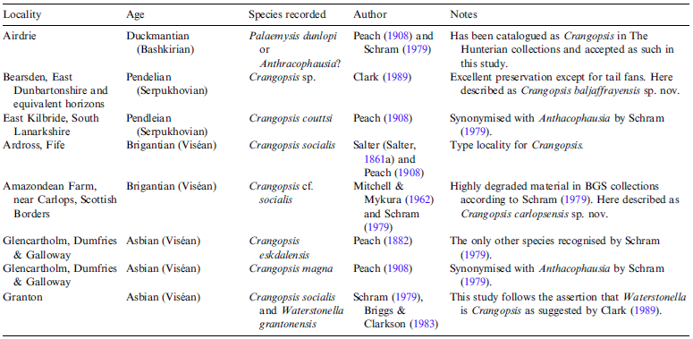
The earliest Carboniferous malacostracan crustaceans from Scotland are found in the Ballagan Formation of the Tournaisian in the Scottish Borders (Clark & Ross Reference Clark and Ross2024) and the latest are from the Duckmantian (Bashkirian) deposits of the Midland Valley of Scotland (Schram Reference Schram1979; Clark Reference Clark2024). Crangopsis has been recorded from rocks that span the range from the Asbian to the Duckmantian (Bashkirian) of the Airdrie Blackband Ironstone (Peach Reference Peach1908; Ross Reference Ross2010; McLean Reference McLean2018) (Fig. 1), although also from the earliest Carboniferous of Northumberland (Peach Reference Peach1908).
Map of Scotland showing the main localities and geological timescale showing the different occurrences of the genus Crangopsis in Scotland: 1 = Airdrie; 2 = Bearsden, East Kilbride and equivalent localities; 3 = Ardross and Carlops; 4 = Glencartholm and Granton. Ages of boundaries based on Cohen et al. (Reference Cohen, Finney, Gibbard and Fan2013).

The earliest description of Crangopsis was by Salter (Reference Salter1861a) where he described a small shrimp-like crustacean from the Carboniferous of Fife which he named Uronectes socialis. Later, he recognised that the name ‘Uronectes’ should not be applied to this crustacean as it was different in the number of exposed segments and suggested it represented a macrurous decapod giving it the name Palaeocrangon (Salter Reference Salter1861b). As this genus was previously occupied by an amphipod crustacean, Salter again renamed the crustacean as Crangopsis (Salter Reference Salter1863). Since then, ten species of Crangopsis have been recognised by Peach (Reference Peach1908) which were subsequently synonymised into two species by Schram (Reference Schram1979) – Crangopsis socialis (Salter) and Crangopsis eskdalensis (Peach). Peach had concluded that Pygocephalus huxleyi should not be placed within the genus Pygocephalus as originally described by Huxley (Reference Huxley1862) and Woodward (Reference Woodward1867) but should be included in the genus Crangopsis based on the woodcut images, and he named it Crangopsis huxleyi (Woodward). Although Huxley had the original specimen to study from the collections of the Reverend William Fraser of Paisley, only the woodcuts were available to Woodward, and Peach (Reference Peach1908) just mentioned this species and used the same illustration as Huxley. The specimen of Crangopsis huxleyi was still not available for Schram (Reference Schram1979) to study and is now considered to be lost. As the specimen of Crangopsis huxleyi comes from the Limestone Coal Formation (based on the description from Huxley Reference Huxley1862 and the stratigraphy documented in Hinxman et al. Reference Hinxman, Anderson and Carruthers1920 and Schram Reference Schram1979), it would appear likely that it came from an equivalent horizon to the strata at Bearsden recorded as ‘Manse Burn Formation’ by Clark (Reference Clark1989) and probably best renamed as the ‘Manse Burn Beds’ as part of the Limestone Coal Formation. This level is dated to within the lower Serpukhovian in the western Midland Valley of Scotland (Clark Reference Clark1989). Without the original specimen of C. huxleyi it is impossible to say if it is a species of Crangopsis or Palaemysis as the expanded second pleonal pleuron is a characteristic of both Crangopsis and Palaemysis and both these genera are often found together (Clark Reference Clark1989, Reference Clark1991). It is possible that C. huxleyi is synonymous with the new species C. baljaffrayensis sp. nov. described in this study, but without the original specimen it is impossible to be certain.
Peach (Reference Peach1908) split Crangopsis into two distinct groups. The first included species that had an enlarged pleuron on the second pleonal somite that overlapped the pleura of the first and third somites. This group included: C. socialis; C. eskdalensis; C. huxleyi; and C. elegans (Peach) (which were later grouped into two species by Schram (Reference Schram1979); C. socialis and C. eskdalensis). The second grouping was for species where the pleuron of the second pleonal somite is pointed and only slightly overlapped backwards on the succeeding pleuron. This second group included: C. couttsi Peach; C. rhodesi Peach; C. magna Peach; C. robusta Peach; C. minuta Peach; and C. hastata Peach. The second group was later synonymised with Anthracophausia by Schram (Reference Schram1979).
Brooks (Reference Brooks1962) suggested that Crangopsis and Anthracophausia could only be distinguished based on the length of the rostrum and that they should be classified together as ancestral euphausiacians. Schram (Reference Schram1979) went further and placed Crangopsis within the Hoplocarida and compared it with Aratidecthes on which he recorded the second maxilla as being stenopod (referring to it having the appearance of a walking limb). Crangopsis has more recently been assigned to the Kallidecthidae (another crustacean that has been assigned to the Hoplocarida (Stomatopoda)) based on the thoracopods shortening posteriorly (Hyzny et al. Reference Hyzny, Hoch, Schram and Rybar2014). This is not the case with well-preserved examples of Crangopsis from Granton where the limbs do not appear to reduce in length posteriorly. The number of paired thoracopods is ten, where the anterior two pairs appear slightly shorter than the posterior eight (and possibly modified as maxillipeds). This is more characteristic of the Mysidacea than the Stomatopoda although the pleuron of the second pleonal somite expands to partially cover those of the first and third somites. The expansion of the pleura seems to be a feature of some decapods rather than any other group, but this may represent a feature that evolved separately in the Decapoda but was lost in the extant Mysidacea. Schram & Koenemann (Reference Schram, Koenemann, Schram and Koenemann2021) placed Crangopsis in the Aeschronectida (an order of the Holpocarida); however, the diagnostic characters for this order include triflagellate antennules which none of the examples of Crangopsis studied here exhibit, having only two. The telson of Crangopsis is also bifid rather than sub-rectangular and flat, which is diagnostic of aschronectidans. Crangopsis is therefore here considered as a primitive mysidacean as previously suggested by Van Straelen (Reference Van Straelen1931) rather than a decapod as originally indicated by Salter (Reference Salter1861b); euphausiacean (Peach Reference Peach1908; Brooks Reference Brooks1962); or hoplocarid (Schram Reference Schram1979; Hyzny et al. Reference Hyzny, Hoch, Schram and Rybar2014; Schram & Koenemann Reference Schram, Koenemann, Schram and Koenemann2021) (Fig. 2).
Simplified relationships and taxonomic hierarchy of the living malacostracan crustaceans based on Richter and Scholtz (Reference Richter and Scholtz2001) with the groups in red that Crangopsis has previously been assigned to.

Waterstonella is a genus under consideration here as being a synonym of Crangopsis. It has been previously placed as the sole member of its own family (Waterstonellidae) by Schram (Reference Schram1979) and order Waterstonellidea (Schram Reference Schram1981). The type specimen of this genus is not well preserved but there are many hundreds of well-preserved examples assigned to this species that have been recovered from the same locality (Briggs & Clarkson Reference Briggs, Clarkson, Briggs and Lane1983). These are often closely associated with specimens identified as Crangopsis. The Waterstonellidea was considered by Schram (Reference Schram1981) to be a sister group to the Mysidacea within the Mysoida. The gross morphological similarity, as well as some diagnostic characters in common between these two genera, are here considered adequate to synonymise the two genera.
Only one other specimen of Waterstonella has been identified from another locality in Ireland (Orr & Briggs Reference Orr and Briggs1999). The illustration of the specimen also appears to be indistinguishable from specimens identified elsewhere as Crangopsis. This specimen will need to be re-examined in light of this study.
2. Material used in this study
The material used in this study are crustaceans in the collections of The Hunterian, University of Glasgow, that have been classified as Crangopsis and come from several localities around Scotland (Table 1). Some of these locations were examined by Euan Clarkson with Derek Briggs for their study on the Carboniferous crustaceans project in the 1980s. Other material used includes material mentioned previously by Schram (Reference Schram1979), Clark (Reference Clark1989) and Briggs et al. (Reference Briggs, Clark and Clarkson1991).
Scottish localities from which Crangopsis has been recorded that were used in this study.

3. Institutional abbreviations
GLAHM: The Hunterian, University of Glasgow, Glasgow, UK; NMS: National Museums of Scotland, Edinburgh, UK; BGS: British Geological Survey.
4. Taxonomy
Subclass EUMALACOSTRACA Grobben, Reference Grobben1892 sensu Martin & Davis, Reference Martin and Davis2001
Order MYSIDACEA Haworth, Reference Haworth1825
Genus Crangopsis Salter, Reference Salter1863
Diagnosis adapted from Schram Reference Schram1979 . Laterally compressed crustacean with a subtrapezoidal carapace with very short rostrum and a vertical cervical groove; antennal peduncles moderately large and long; antennules with paired flagella; pleon 2.5 times the length of the cephalothorax; pleonal pleura well developed; sixth pleonal somite especially elongate; pleuron of second pleonal somite expands anteriorly as well as posteriorly. Bifid telson associated with elongated uropodal exopods and short uropodal endopods.
Type species. Uronectes socialis Salter, Reference Salter1861a. Ardross, Fife; Brigantian.
Holotype. BGS GSM 87432/GSE 14002 (Schram Reference Schram1979; fig. 10) (Fig. 3a).
(a) Sketch of the type specimen of Crangopsis socialis (BGS GSI 87432) from the Lower Ardross Limestone showing the major subdivisions of the crustacean body. (b, c, d) Specimen GLAHM 163471 from Ardross showing the enlarged second pleonal pleuron and expanded view (d) of the raised ridge on the third pleonal somite. 1–6 – pleonal somites; an – antennules; as – antennal scale; c – carapace; cg – cervical groove; pl2 – second pleonal pleuron; r – ridge on pleonal somite; ro – rostrum; tf – tail fan; th – thoracomeres. Scale bar (a–c) = 5 mm; (d) = 1 mm.

Crangopsis socialis (Salter Reference Salter1861a)
(a, b) Crangopsis socialis from Granton showing partial ‘ghosting’ of the cuticle especially thinned on parts of the carapace and pleura of the pleonal somites where the limbs are showing through (GLAHM 152337). The second pleonal pleuron expands anteriorly and posteriorly to overlap those of the first and third pleonal pleura. (c, d) Moult of Crangopsis socialis with thinner cuticle that would have previously been classified as Waterstonella grantonensis. The carapace has folded and is misshapen and the pleonal somites and pleura are ‘ghosting’ but still discernible, including the expanded pleuron of the second pleonal somite. The biramous thoracopods are well preserved. 1–6 – pleonal somites; an – antennules; as – antennal scale; c – carapace; pl2 – second pleonal pleuron. Scale bar = 5 mm.

(a, b) Tail fan of Crangopsis socialis from Granton showing the bifid nature of the telson with short uropodal endopods and elongate uropodal exopods (GLAHM 163467). (c) Sketch of type specimen of Waterstonella grantonensis designated by Schram (Reference Schram1979) (BGS GSI 13053). (d) Sketch of large specimen identified by Briggs et al. 1991 as Waterstonella grantonensis with the type specimen below at the same scale. 1–6 – pleonal somites; an – antennules; as – antennal scale; c – carapace; en – uropodal endopod; ex – uropodal exopod; p – pleopods; t – telson; tf – tail fan; thp – thoracopods. Scale bar (a–b) = 2 mm; (c) = 3 mm; (d) = 5 mm.

(a, b, c) Dorso-ventrally preserved specimen of Crangopsis socialis from Granton showing bifid telson, shorter uropodal endopods and elongated blade-like uropodal exopods (GLAHM 163466). (b, c) white box on (a) enlarged. en – uropodal endopod; ex – uropodal exopod; t – telson. Scale bar (a) = 5 mm; (b–c) = 2 mm.

In addition to the extensive synonomy list provided by Schram (Reference Schram1979):
1979 Waterstonella grantonensis Schram (pp. 73–75; figs 30, 31)
1983 Crangopsis sp. Briggs & Clarkson (p. 165; text-fig. 5f–h; pl. 22, figs 6–8)
1983 Crangopsis socialis (Salter). Briggs & Clarkson (p. 165; text-fig. 5i; pl. 22, fig. 9)
1983 Waterstonella grantonensis Schram. Briggs & Clarkson (pp. 170–175; text-fig. 4a–f; text-fig. 5a–e; text-fig. 6; pl. 20, fig. 1; pl. 21, figs 2–9; pl. 22, figs 1–5)
Material. Holotype BGS GSM 87432/GSE 14002 designated by Schram (Reference Schram1979; p. 41 (Fig. 3a); GLAHM 163471 from the Lower Ardross Limestone (Brigantian); GLAHM 152337 (Fig. 4a–b), GLAHM 163462 (Fig. 4c–d), GLAHM 163467 (Fig. 5a–b), BGS GSE 13053 (Fig. 5c–d), NMS GY1989.2.9 (Fig. 5d), GLAHM 163466 (Fig. 6) from the Granton Shrimp Bed (Asbian). A total of 14 specimens from Ardross were examined from the collections of the BGS, NMS and GLAHM. Extensive collections of material from Granton were examined from the collections of the NMS and GLAHM.
Emended diagnosis (after Reference Schram Schram 1979 ). Second pleonal pleuron broadly rounded. Posterior pleonal somite pleura tapered posteroventrally.
Description. Carapace has a faint vertical cervical groove (Fig. 3a) which is very difficult to discern due to the wrinkled nature of the cuticle. This extends to the pleon where a short ridge appears on the second, third and fourth pleonal somites (Fig. 3b–d).
Remarks. The reason for including Waterstonella grantonensis in the synonymy list is that it is impossible to distinguish between Crangopsis and Waterstonella in the rocks at Granton on morphological grounds (Figs 3–5). The type specimen of Waterstonella (Fig. 5c–d) has a poorly preserved cuticle, as have many of the specimens that have previously been identified as Waterstonella. The thinning of the cuticle and the vast number of fragmentary parts to the crustaceans suggests that many of the specimens preserved at Granton represent moults. Both those identified as Crangopsis and Waterstonella have the same number of thoracopods, identical pleopods, elongated sixth pleonal somite, a bifid telson with short uropodal endopods and blade-like uropodal exopods. The main character that has been used to differentiate the two forms appears to be the thickness of the cuticle. The thinning of the cuticle is likely to be the result of the resorption of the cuticle during moulting (Corteel & Nauwynck Reference Corteel, Nauwynck and Alday-Sanz2010). The wrinkled nature of the cuticle may represent the moulted carcass of crustacean prior to the new cuticle hardening (Drage & Daley Reference Drage and Daley2016 and Drage et al. Reference Drage, Vandenbrouckem, Van Roy and Daley2019 describe similar structures in trilobites which are perhaps not the best animal for comparison due to the robustness of the calcified cuticle post ecdysis) or the demineralised remains of the shed cuticle. The separation of the carapace from the pleon in the examples of specimens with thin cuticle would suggest that the thinner cuticle fossils represent moults and the thicker cuticle fossils represent carcasses. The preservation, or not, of the pleura, even on specimens confidently identified as Crangopsis, is variable and probably related to postmortem or post-moulting taphonomy. The shape of the carapace and the tail fan also varies greatly amongst the thinner cuticle specimens. Where the loosely attached cuticle (such as the carapace) is thinner, it is more likely to fold and change shape, or more easily become damaged especially in crustaceans where the cuticle on the living crustacean was already quite thin prior to resorption of minerals during ecdysis (Corteel & Nauwynck Reference Corteel, Nauwynck and Alday-Sanz2010). As the major body features mentioned previously are indistinguishable between those specimens identified as one species or the other, the two species Crangopsis socialis and Waterstonella grantonensis are here considered to be synonymous.
Crangopsis eskdalensis (Peach Reference Peach1882)
(Fig. 7a–c)
(a) Sketch of the lectotype specimen of Crangopsis eskdalensis from Glencartholm showing the shape of the second pleonal pleuron (BGS GSE 5007). (b, c) tail fan from the same locality showing the bifid telson and elongate blade-like uropodal exopods (GLAHM 101117). 1–6 – pleonal somites; an - antennules; as – antennal scale; c – carapace; cg – cervical groove; ex – uropodal exopod; pl2 – second pleonal pleuron; ro – rostrum; t – telson. Scale bars = 5 mm.

Full synonomy list provided by Schram (Reference Schram1979).
Material. Lectotype BGS GSE 5007 (Fig. 7a) designated by Schram (Reference Schram1979, p, 42); GLAHM 101117 (Fig. 7b–c) from the Glencartholm Volcanic Beds (Asbian). Over 20 specimens were examined from the collections of the BGS, NMS and GLAHM.
Emended diagnosis (after Reference Schram Schram 1979 ). Second pleonal pleuron broadly rounded. Posterior pleonal somite pleura rounded posteroventrally.
Description. Carapace has a faint vertical cervical groove which is very difficult to discern due to the wrinkled nature of the cuticle similar to C. socialis. This species tends to be preserved less wrinkled but is more friable perhaps as a result of different taphonomic pathways, life environments, or environments of deposition. There is also a short ridge on the second, third and fourth pleonal somites. The second pleonal pleuron appears to be more inflated than C. socialis, but this may also be an artefact of the preservation of these crustaceans.
Remarks. Schram (Reference Schram1979) indicated that the main differences between C. eskdalensis and C. socialis were that C. socialis was bigger than C. eskdalensis, that the carapace of C. socialis extends further back, the first pleonal somite is longer in C. socialis, the posterior pleura attenuate posteriorly in C. socialis, and the pleuron of the second pleonal somite is flatter ventrally in C. eskdalensis. While the pleuron of the second pleonal somite is less rounded than that of C. socialis, it was found that the lectotype still appears to have rounded pleura rather than rectangular. The main difference appears to be that the pleura of the posterior somites are drawn posteriorly rather than anteriorly as they seem to be in C. socialis.
Crangopsis carlopsensis sp. nov.
(a, b) Holotype specimen of Crangopsis carlopsensis sp. nov., from the Brigantian shales near Carlops, showing the expanded pleuron of the second pleonal somite and the thoracomeres. A ridge is seen on the second pleonal somite but is not easily discernible due to the wrinkled preservation of the cuticle (GLAHM 163469). (c, d) Shows the anterior portion of the crustacean with thoracomeres extending dorsally, a short rostrum, as well as the antennal scales and antennules (GLAHM 163468). 1–2 – pleonal somites; an – antennules; as – antennal scale; c – carapace; cg – cervical groove; pl – pleonal pleura; pl2 – second pleonal pleuron; r – ridge on pleonal somite; ro – rostrum; th – thoracomeres. Scale bar = 5 mm.

(a, b) Tail fan of Crangopsis carlopsensis in ventral aspect showing the elongate blade-like uropodal exopods, shorter uropodal endopods, and the bifid telson (GLAHM 163470). en – uropodal endopod ; ex – uropodal exopod; t – telson. Scale bar = 2 mm.

Etymology. Named after the village close to where the type material was found.
Material. Holotype GLAHM 163469 (Fig. 8a–b); Syntypes GLAHM 163468 (Fig. 8c–d); GLAHM 163470 (Fig. 9) from Carlops Bridge near Amazondean Farm on the south bank of the River North Esk (NT16785646) (Brigantian).A total of eight specimens were examined for this study in the collections of GLAHM.
Diagnosis. Second pleonal pleuron broadly rounded. Posterior pleonal somite pleura with flatter termination posteroventrally.
Description. Carapace has a faint vertical cervical groove which is very difficult to discern due to the wrinkled nature of the cuticle. The rostrum is very short if present. The thoracic somites (thoracomeres) are long in lateral aspect suggesting that several of the posterior somites may be free from the carapace (Fig. 8c, d). The wrinkled cuticle also extends to the pleon where a short ridge appears on the second pleonal somite (Fig. 8a, b). Preservation at this locality is quite poor and Schram (Reference Schram1979) mentioned that the crustaceans in the collections of the Geological Survey were ‘disintegrating badly with time’ due to the pyrite content of the shales. The expansion of the second pleonal pleuron is preserved well however, as are the terminations of the other pleonal pleura which form a straight line along the pleon. The tail fans are well preserved (Fig. 9) to exhibit a characteristic bifid telson like that seen in Palaemysis (Clark Reference Clark1991). The blade-like uropodal exopods are about a third longer than the telson and curve outwards posterolaterally and have a medial groove. The uropodal endopods are about the same length as the telson and are elongated oval shaped.
Crangopsis baljaffrayensis sp. nov.
(a, b) Holotype specimen of Crangopsis baljaffrayensis sp. nov., from the Pendleian (Serpukhovian) shales near Bearsden, East Dunbartonshire showing the characteristic squared pleuron of the second pleonal somite and associated short ridge (GLAHM A2409.1). (c, d) Another example with an enlargement of the ridge structure on the second pleonal somite (GLAHM A2748). 1–6 – pleonal somites; an – antennules; as – antennal scale; c – carapace; cg – cervical groove; pl2 – second pleonal pleuron; r – ridge on pleonal somite; ro – rostrum; th – thoracomeres. Scale bar (a, b, c) = 5 mm; (d) = 0.5 mm.

(a, b, c) Crangopsis baljaffrayensis from Bearsden (GLAHM A2409.6) showing ridge structure on the 4th pleonal somite, expanded pleuron of the second pleonal somite, and carapace missing to reveal the thoracomeres ((c) = enlargement of white box seen in (a)). 1–6 – pleonal somites; pl2 – second pleonal pleuron; r – ridge on pleonal somite; th – thoracomeres. Scale bar (a, b) = 5 mm; (c) = 1 mm.

Etymology. Named after the Baljaffray Estate, the area in East Dunbartonshire within which the famous Bearsden fauna was discovered in 1982.
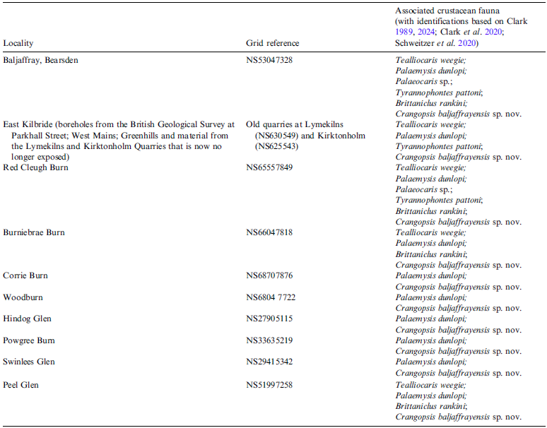
Material. Holotype GLAHM A2409.1 (Fig. 10a–b); Syntypes GLAHM A2409.6 (Fig. 11); GLAHM A2748 (Fig. 10b–c) from the type locality at Baljaffray, Bearsden, from an excavation at the Manse Burn conducted by Stan P. Wood under the auspices of the Hunterian Museum (Wood Reference Wood1982) in 1982–1983 (NS53047328). The equivalent horizon from shales above the Top Hosie Limestone (Pendleian, Serpukhovian) beyond the boundaries of Greater Glasgow (Table 2) also provided specimens of Crangopsis (Clark Reference Clark1989). Over 15 specimens of this species were studied from the collections of GLAHM.
List of localities (Carboniferous: Pendleian) from which Crangopsis baljaffrayensis sp. nov. has been found in the west of Scotland (Patton & Coutts Reference Patton and Coutts1885; Clark Reference Clark1989). The Peel Burn locality discovered by Patrick Gavin was mentioned in Clark (Reference Clark2013).

Diagnosis. Second pleonal pleuron laterally expanded with a ventrally flattened shape rather than rounded. Posterior pleonal somite pleura with tapered termination anteroventrally.
Description. The trapezoidal to triangular carapace has a faint vertical cervical groove and a very short rostrum. The antennules and the stretched oval-shaped antennal scales are occasionally preserved, as in the holotype (Fig. 10a–b). Thoracomeres can be seen where the carapace has been removed or angled away from the rest of the body as a result of ecdysis. The thoracomeres are long in lateral aspect suggesting that some (four or five) may be free from the carapace (Fig. 11a–c). The expansion of the second pleonal pleuron ends posteroventral in a flat edge. A short ridge appears on the second pleonal somite (Fig. 10a–d). The pleura of the other pleonal somites form a rounded but slightly tapered termination along the pleon. The tail fans are poorly preserved.
Crangopsis sp.
?1908 Palaemysis dunlopi Peach (pp. 66–67, pl. 8, figs 12-14)
(Fig. 12a, b)
(a, b) Tail fan of a specimen of Crangopsis sp. from the Duckmantian (Bashkirian) ironstone of Airdrie showing the characteristic Crangopsis bifid telson and elongated blade-like uropodal exopod (GLAHM 101102). (c, d) for comparison, a tail fan specimen previously identified as Crangopsis from East Kilbride with a triangular telson and shorter uropodal exopods characteristic of Anthracophausia. en – uropodal endopod; ex – uropodal exopod; t – telson. Scale bar = 5 mm.

Material. Tail fans from Airdrie, Lanarkshire from just above the Queenslie Marine Band (Duckmantian, Coal Measures Formation, Bashkirian). GLAHM 101102 contains multiple crustacean fragments and tail fans (Fig. 12a, b).
Description. The material has been catalogued within the collections of The Hunterian as representing the remains of Crangopsis. The bifid telson and long blade-like uropodal exopods are both characteristic of this genus. As the specimens lack the diagnostic pleonal and carapace structures, it is not possible to assign a species to these specimens at this stage. It is likely that these represent another new species due to the age difference (∼15 million years between the base of the Pendleian (Serpukhovian) and the Duckmantian (Bashkirian), Fig. 1) with the next youngest species Crangopsis baljaffrayensis.
Remarks. Schram (Reference Schram1979) recorded Pygocephalus dubius and ?Anthracophausia dunsiana from the Airdrie locality, but did not recognise Crangopsis. The specimen illustrated here does not belong to either of these genera due to the bifid telson (triangular in Anthracophausia, more square in Pygocephalus) and elongate uropodal exopods which are characteristics more aligned with Crangopsis. Until better specimens come to light, it is considered these specimens (GLAHM 101102) should be identified as Crangopsis sp. as there are no diagnostic characters that would distinguish them from any other known species of Crangopsis.
The type specimen of Palaemysis dunlopi first described by Peach (Reference Peach1908) came from this locality and his illustrations are identical in size and form to the specimen illustrated here (Fig. 12a, b). The uropodal exopods are about twice the length of the bifid telson which is similar to Crangopsis, whereas the uropodal exopods in Palaemysis are about 1.5 times the length of the telson and have a small terminal diaeresis (Clark Reference Clark1991). Further examination of other material from Airdrie may confirm this material as being Crangopsis which will necessitate a reassessment of Palaemysis dunlopi as described by Clark (Reference Clark1991). It is likely that Palaemysis from the Bearsden horizon would be best compared to Palaemysis couttsi Peach (Reference Peach1908), which was synonymised with Anthracophausia dunsiana by Schram (Reference Schram1979) and Palaemysis dunlopi by Clark (Reference Clark1991). Palaemysis couttsi was described from East Kilbride which is considered to be an equivalent horizon to the shales at Bearsden. Similar material of Palaemysis is also known from Granton (Briggs et al. Reference Briggs, Clark and Clarkson1991).
General remarks. The tail fans of the different species of Crangopsis all have a bifid structure to the posterior edge of the telson. This is in contrast to the triangular telson as seen in Anthracophausia from Glencartholm, East Kilbride (Fig. 12c, d) and elsewhere (Schram Reference Schram1979). The length of the uropodal exopods is much shorter relative to the telson in Anthracophausia although they are blade-like (see Fig. 12). The uropodal endopods are about the same length as the telson in Anthracophausia but are slightly shorter in Crangopsis. It should therefore be easy to distinguish these two genera from the tail fans alone if they are well enough preserved. Palaemysis (sensu Clark Reference Clark1991) has a very similar bifid telson to Crangopsis, but the uropodal endopods are the same length or slightly longer than the telson, and the uropodal exopods are more triangular than blade-like (Clark Reference Clark1989, Reference Clark1991). The second pleonal pleuron expanding to partially cover those of the first and third is also seen in Palaemysis but is absent in Anthracophausia.
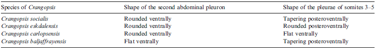
Of the eight thoracopods seen in Palaemysis the posterior seven appear to shorten posteriorly and the most anterior appear to be shorter than the others. This contrasts with the limbs of Crangopsis, which are best preserved in specimens from the Granton Shrimp Bed, where ten thoracopods can be counted. The posterior eight thoracopods are biramous and are of about the same length (see Fig. 3c–d) although the anterior two are shorter and appear uniramous (Fig. 5d). All the species of Crangopsis are very similar on basic body plan. The only consistent differences between them are the shape of the pleura (rounded anteriorly, rounded posteriorly or flat ventrally (Table 3). The main similarities that differentiate Crangopsis from other Carboniferous crustaceans include the expanded pleuron on the second pleonal somite and the elongated sixth pleonal somite.
Characteristics that differentiate the species of Crangopsis.

The length of the rostrum is the other main characteristic that differentiates these three genera. In Anthracophausia the rostrum is substantial and deep (Schram Reference Schram1979), whereas in both Crangopsis and Palaemysis the rostrum is very short relative to the size of the carapace (approximately one-thirtieth of the length of the carapace).
Crangopsis seems to be euryhaline as it occurs in the more marine sediments of Carlops and Granton, potentially less saline lagoonal facies of Ardross, Glencartholm and Bearsden and potentially freshwater environments represented by the deposits at Airdrie. It is a gregarious animal, often being found in large numbers, or at least relatively common where found, and may moult in numbers too (as seen at the Granton locality).
Although Crangopsis appears to be restricted to the United Kingdom, there is a record of a large Crangopsis from Czechia of about 30 mm in length (Hyzny et al. Reference Hyzny, Hoch, Schram and Rybar2014). The largest from Scotland is about 25 mm in length from Glencartholm, and the majority are about 15 mm. The thoracopods of the Czechia specimens appear to shorten posteriorly in a similar manner to Palaemysis and Kallidechthes (Schram Reference Schram1979; Clark Reference Clark1991). The antennae of Crangopsis are short and rarely observed, whereas the antennules are much more substantial with relatively long peduncles. The opposite is true of the specimens from Czechia (Hyzny et al. Reference Hyzny, Hoch, Schram and Rybar2014) and they are unlikely to be Crangopsis. This material should be re-examined in the light of these differences.
Crangopsis has also been recorded from northern England (Peach Reference Peach1908; Schram Reference Schram1979). Crangopsis huxleyi was synonymised with Crangopsis socialis by Schram (Reference Schram1979). Crangopsis rhodesi, Crangopsis magna, Crangopsis couttsi, Crangopsis hastata, as well as all species of Palaemysis, were synonymised with Anthracophausia dunsiana by Schram (Reference Schram1979). More recently, Palaemysis has been removed from the above synonymy (Clark Reference Clark1991). However, the type specimen from Airdrie that was used by Clark (Reference Clark1991) based on the original description by Peach (Reference Peach1908) may be a species of Crangopsis, as shown by the tail fan in Fig. 12 identified here as Crangopsis sp. from the same locality. Sorting the taxonomy of Palaemysis is beyond the scope of this current study.
Acknowledgements
Thanks go to the late Professor Euan N. K. Clarkson, who has contributed significantly to our understanding of Carboniferous crustaceans in Scotland, frequently visited Carlops after which Crangopsis carlopsensis is named, and started me off in my crustacean research. Many thanks to Drs Carrie Schweitzer and Pierre Guerriau who offered helpful reviews of this manuscript. Thanks also to NatureScot who provided the necessary permissions to access the Firth of Forth Special Protection Area and Geological Conservation Review Site at Ardross. Dr Yves Candela is thanked for the work involved in editing this manuscript for publication.


 Open access