


Thirty-seven-year-old John V.G.A. Durnin in 1960 published his first study in a series that fifteen years later was to culminate in the most cited British Journal of Nutrition (BJN) paper in its 75-year history (Fig. 1)(Reference Durnin and Taylor1,Reference Durnin and Womersley2) . Along with A. Taylor, Durnin critically examined the replicability of the underwater weighing body composition method in a meticulous study of ten men(Reference Durnin and Taylor1). The authors found that the error of a single measurement in 90 % of participants was about 0·004 density units, a level they judged to ‘be of a satisfactory degree of accuracy’ when estimating body fat.
Abstract of the classic 1974 Durnin–Womersley BNJ paper(Reference Durnin and Taylor1).

Interest in the underwater weighing method for estimating body fat was increasing in the 1950s, prior to Durnin and Taylor’s publication, following the technique’s introduction by Albert R. Behnke in 1942(Reference Behnke3). Behnke recognised that a person’s body density, measured by the underwater weighing method, reflects their level of adiposity. The underwater weighing method was placed on a solid footing in 1953 by Ancel Keys and Josef Brozek who proposed a series of initial body density equations that solved for two components of body mass: fat mass and fat-free mass (FFM)(Reference Keys and Brozek4). Just a few years later, in 1956, William E. Siri published an improved and simplified set of equations for deriving fat mass and FFM from measured body density that are still used today, almost seven decades later(Reference Siri, Lawrence and Tobias5). Durnin’s evaluation of the underwater weighing method was motivated by these early studies that provided a means by which estimates of energy expenditure could be referenced to metabolically active components such as FFM rather than to body weight(Reference Durnin6).
Durnin and his colleagues recognised, however, that weighing people under water was impractical in clinical and field settings. As an alternative, they turned to another, more feasible approach, skinfold anthropometry. First introduced in 1921 by the Czech anthropologist Jindrich Matiegka(Reference Matiegka7), publications related to skinfold anthropometry and body composition rapidly expanded in the 1950s. In an early study typical of this era, D.A.W. Edwards in 1950 reported a close quadratic relationship between skinfolds measured at fifty-three sites in young women and body weight(Reference Edwards8). One year later, in 1951, Brozek and Keys were the first to report in BJN a curvilinear relationship between skinfold thickness and body density, a proxy at the time for body fat content(Reference Brozek and Keys9). Studies published over the next decade established optimum skinfold measurement sites for estimating body density, the contributions of skin to the measured skinfolds, how skinfolds relate to subcutaneous and ‘internal’ body fat, and the associations between age and skinfold compressibility(Reference Keys and Brozek4,Reference Allen, Peng and Chen10–Reference Skerlj, Brozek and Hunt12) .
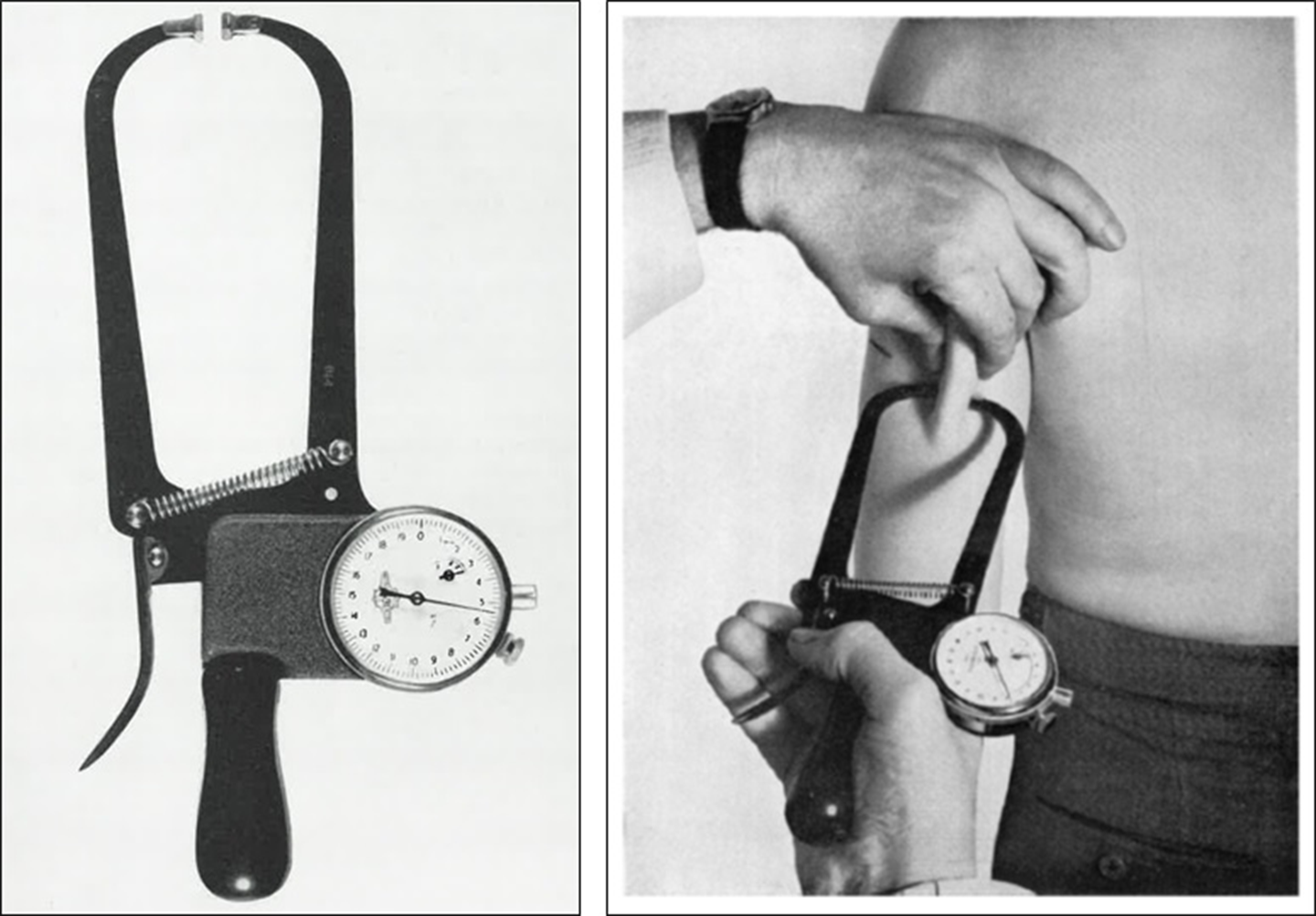
While publications on skinfolds proliferated during this period, there were two bottlenecks that limited further development of the skinfold body composition approach. First, until the late 1950s the field lacked an accessible reference method for quantifying body composition until underwater weighing and associated body composition equations were fully in place and accepted. Methods for estimating lean and fat body compartments such as whole-body counting for 40K were limited to only a few highly specialised centres(Reference Forbes, Gallup and Hursh13). Second, and importantly, skinfold calipers at the time were such rudimentary devices that Josef Brozek quipped that he and his colleagues had started a caliper ‘mausoleum’ to display the growing number of useless instruments they had evaluated(Reference Brozek, Brozek and Henschel14). Advances in technology and increasing interest among anthropologists and physiologists led to the introduction of the Lange caliper in the USA (Fig. 2) and the Harpenden caliper in Europe (Fig. 3) in the late 1950s(Reference Brozek, Brozek and Henschel14–Reference Tanner and Whitehouse16), two standardised and comparable devices still in widespread use today.
A portion of the 1961 Lange caliper patent (left) and an early prototype of the instrument (right; from 14, with permission).

Harpenden skinfold caliper illustration in the Brief Communication by Tanner and Whitehouse (left)(Reference Edwards, Hammond and Healy15) and in the BJN paper by Edwards etal. (Reference Tanner and Whitehouse16), both published in 1955.
Durnin and his colleagues were fully armed with these measurement tools by the mid-1960s. In a forerunner of what was to come, Durnin stated in his classic book with Reginald Passmore, Energy, Work, and Leisure (Reference Durnin and Passmore17), ‘Skinfold thicknesses have been shown to correlate reasonably well with body density, but as yet there is no established method for converting them into percentages of body fat. This will probably come’. That year, in 1967, Durnin and his associate Mohammed M. Rahaman published a paper in BJN(Reference Durnin and Rahaman18) that described skinfold–body composition relations in a sample of 191 young British adults and adolescents. Building on earlier studies(Reference Keys and Brozek4,Reference Allen, Peng and Chen10–Reference Skerlj, Brozek and Hunt12) , Durnin and Rahaman measured skinfold thicknesses at four sites (triceps, biceps, subscapular and suprailiac) with the Harpenden caliper and body density with underwater weighing; body fat was calculated using Siri’s equations(Reference Siri, Lawrence and Tobias5). Regression equations were then derived for estimating body density from a single skinfold or from a combination of skinfolds. Rather than using absolute values, the combined sum of skinfolds measured at the four sites was log transformed because of the curvilinear relationship between skinfolds and body density and the skew in skinfold distributions. A critical juncture at this point is that the author’s recognised sex and age differences in how skinfolds relate to body density and thus equations were derived separately for males and females in six age groups. Siri’s equations could be used by practitioners to convert body density to fat and FFM. To simplify, the authors also provided a table that gave the conversion of 4-skinfold sum to percentage fat. This BJN paper has been cited more than 1500 times since its publication in 1967 (Fig. 4), one of the journal’s top five. A follow-up abstract reported by Durnin and Womersley in 1969 expanded these analyses to a sample of ninety-six middle-aged British adults(Reference Durnin and Womersley19) and another abstract in 1973 reported an increase in the sample size to 571 people between the ages of 12 and 72 years(Reference Durnin and Womersley20).
Citations per year (upper panel) and cumulative citations (lower panel) since publication of the 1967 Durnin and Rahaman (D&R)(Reference Durnin and Passmore17) and 1974 Durnin and Womersley papers (D&W)(Reference Durnin and Taylor1). ![]() , D&R;
, D&R; ![]() , D&W.
, D&W.

In January 1974, John Womersley submitted his doctoral thesis at the Institute for Physiology at the University of Glasgow(Reference Womersley21). Womersley credited the conception of and experimental design of their recently ‘submitted’ (May, 1973) now-classic 1974 BJN paper(Reference Durnin and Womersley2) to ‘Dr. Durnin’, although he had done ‘virtually all of the experimental work and statistical analyses’. The Durnin–Womersley study, cited almost 5000 times since 1974 (Fig. 4), distilled data from the earlier reported samples(Reference Durnin and Rahaman18–Reference Durnin and Womersley20) and added new participants yielding 481 adolescents and adults between the ages of 16 and 72 years. The approach was the same as their earlier, more limited studies; body density prediction equations were developed from the log of individual skinfolds and various combinations of the four skinfolds across males and females distributed in five age groups. Both the Lange and Harpenden calipers were used in this study to measure skinfold thicknesses. Again, the authors provided a convenient table that gave %fat estimates directly from the sum of four skinfolds. Here, we see the coming together in a single study of a large sex, age and weight-diverse sample; use of validated measuring instruments and models; and an analysis approach making the findings easily usable by other investigators. Moreover, the findings of the study would soon be used in the growing fitness industry by practitioners evaluating the ever-increasing number of patients with obesity and in remote field settings that lacked the power and other resources needed for evaluating nutritional status.
Although in retrospect this is a remarkable study and publication, the authors at the time were appropriately self-critical, and they probed deeply into questions raised by their findings. Notably, as a centrepiece of the study Durnin and Womersley reported twelve sex- and age-specific body density prediction equations. In their discussion section, the authors questioned why these prediction models had differing slopes and intercepts, thus necessitating their separate development. Their main hypothesis was that the distribution of adipose tissue into subcutaneous and internal components varied across sex and age groups. Skinfolds at the time were seen as measures of subcutaneous adipose tissue and, by contrast, body density was viewed as a means of quantifying total body adipose tissue or fat mass(Reference Brozek and Keys9,Reference Allen, Peng and Chen10,Reference Skerlj, Brozek and Hunt12,Reference Brozek, Brozek and Henschel14) . While Durnin and Womersley thought that differing subcutaneous and internal adipose tissue relations across sex and age groups provided a good explanation for why they needed multiple body density prediction equations, at the time they had no way to directly measure adipose tissue distribution. In a temporal coincidence, Godfrey Hounsfield working at Atkinson Morley Hospital near Wimbledon, South-West of London, had reported his first computed tomography measurements in 1971 and published his findings in 1973(Reference Hounsfield22), the same year as Durnin and Womersley submitted their manuscript to BJN; by the end of the decade subcutaneous and visceral adipose tissue measurements by computed tomography were being reported(Reference Heymsfield and Noel23). Multiple studies since then have laid out the differing sex and age adipose tissue distribution patterns as described by computed tomography and in the 1980s by MRI(Reference Hayes, Sowood and Belyavin24).
A second hypothesis advanced by the authors to explain their sex- and age-specific body density models is that the density of FFM varies with sex and age. Accurate estimates of body fat by underwater weighing rely on the assumption that fat and FFM have respective densities of 0·9007 and 1·100 kg/l at body temperature(Reference Keys and Brozek4,Reference Siri, Lawrence and Tobias5) . However, the density of FFM is not a physical constant but can vary according to the compartment’s proportions of bone and other minerals, water, protein and other minor molecular components(Reference Keys and Brozek4,Reference Siri, Lawrence and Tobias5) . Durnin and Womersley hypothesised that the proportion of FFM as bone mineral varies across males and females and with age. Limited data were available at the time to test their hypothesis and the concept of dual-photon absorptiometry for bone mineral measurement had just been introduced a few years earlier by Richard Mazess and his group in 1970(Reference Mazess, Ort and Judy25), a method advanced for body composition assessment in the early 1980s(Reference Mazess, Peppler and Gibbons26); what we recognise today as whole-body dual-energy X-ray absorptiometry became available to clinical investigators in the late 1980s. Durnin and Womersley correctly surmised, based on contemporary studies, that variation in the proportion of FFM as bone across males and females and with age has only a small influence on the validity of Siri’s two-component body density model(Reference Siri, Lawrence and Tobias5).
The measurement of skinfolds for quantifying adiposity is still in widespread use today in settings that lack more advanced body composition methods such as computed tomography, MRI and dual-energy X-ray absorptiometry(Reference Kasper, Langan-Evans and Hudson27). Underwater weighing is less in use today, giving way to newer methods of evaluating body density such as air-displacement plethysmography(Reference Fields, Goran and McCrory28), but skinfold methods also have contemporary competition in non-research settings from techniques such as bioimpedance analysis, ultrasound, photogrammetry and three-dimensional optical imaging(Reference Kasper, Langan-Evans and Hudson27).
Has the skinfold caliper approach thus reached its nadir in development? In fact, new possibilities abound with the introduction of modern digital and Internet technology. Envision clinical investigators all over the globe, trained in anthropometry, uploading skinfolds, circumferences and lengths along with reference body composition measurements taken on their unique populations to a specially designed cloud storage site. Such a site could screen and house thousands of carefully acquired measurements. Body composition prediction models could then be developed at investigator request using artificial intelligence and machine learning software housed within the cloud site. These prediction models could be updated at frequent intervals as more data become available. Early versions of these kinds of opportunities are already active(Reference Sobhiyeh, Kennedy and Dunkel29), signalling a promising future for the kinds of measurement methods stimulated by the success of Durnin and Womersley’s classic 1974 BJN paper(Reference Durnin and Womersley2).
Acknowledgements
This work was partially supported by National Institutes of Health NORC Center Grants P30DK072476, Pennington/Louisiana and P30DK040561, Harvard University.
Concept and design: S. B. H., B. J. G. S., acquisition, analysis or interpretation of data: S. B. H., B. J. G. S., drafting of the manuscript: S. B. H., B. J. G. S., critical revision of the manuscript for important intellectual content: S. B. H., B. J. G. S., administrative, technical or material support: S. B. H., B. J. G. S.
S. B. H. is on the Tanita Medical Advisory Board.
Dr. Heymsfield is a member of the Medical Advisory Board of Tanita Corporation.


