


Solute Carrier Family 6 Member 3 (SLC6A3) gene encodes the sodium-dependent dopamine transporter, terminating dopamine action by high-affinity reuptake into presynaptic terminals. Pathogenic SLC6A3 variants cause dopamine transporter deficiency syndrome (DTDS), an autosomal recessive complex movement disorder (MD). Classic early-onset and atypical later-onset forms are known. Reference Ng, Zhen, Meyer, Erreger, Li and Kakar1,Reference Kurian2 In classic DTDS, infants manifest non-specific findings (irritability, feeding difficulties, axial hypotonia, and delayed motor development), followed by hyperkinetic MD (chorea, dystonia, ballismus, and orolingual dyskinesia). Over time, parkinsonism-dystonia develops. Secondary orthopedic, gastrointestinal, and respiratory complications are common.
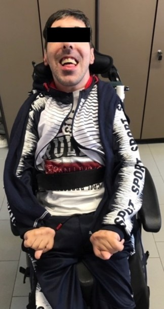
We present a male patient, now aged 27 years (Figure 1), born at term after uneventful pregnancy, to healthy first cousins. Abnormal movements were noticed at birth: rigidity and hyperkinetic MD (combining chorea and dystonia). At 6 months, spontaneous motility was severely reduced and episodes of opisthotonic posturing occurred. Aged 2 years he presented global developmental delay, absent speech, generalized dystonia, hand postural tremor, and orolingual and facial dyskinesia. He subsequently developed hypomimia and progressive feeding difficulties. At follow-up, neurological signs displayed a stable course, but secondary orthopedic complications occurred (kyphosis and feet deformity). Raised sweating was present. Despite absent expressive language, he developed non-verbal communication strategies.
A picture of our patient, now aged 27 years, showing generalized dystonia in his upper and lower limbs, trunk and face (with hypomymia).
Brain magnetic resonance imaging and electroneurography (ENG) were normal. Serial electroencephalograms showed poor background activity, without epileptic discharges. Cerebral tomoscintigraphy (13 years) demonstrated non-specific low perfusion in the left temporoparietal and right temporal areas.
Muscle biopsy documented mild increase in endomysial connective tissue.
Cerebrospinal fluid (CSF) neurotransmitters profile (16 years) showed raised homovanillic to 5-hydroxyindolacetic acid ratio (8.3 nmol/l, normal range: 1.5–3.5), raised 3,4-Dihydroxyphenylacetic acid (DOPAC) and levo-dopa.
Array comparative genomic hybridization (Array-CGH, Human Genome CGH Microarray Kit 8x60K –Agilent- 100–150 kb resolution), dystonia protein (DYT)-torsin family 1 member A (TOR1A), THAP domain-containing protein 1 (DYT-THAP1), GTP cyclohydrolase 1 (DYT-GCH1), tyrosine hydroxylase (DYT-TH), epsilon (ε)-sarcoglycan (DYT-SGCE), parkin RBR E3 ubiquitin protein ligase (PRKN/PARK2), leucine rich repeat kinase 2 (LRRK2/PARK8) direct sequencing and targeted next-generation sequencing panel for 37 mitochondrial DNA genes were negative.
Whole exome sequencing targeting all protein coding exons, exon–intron boundaries (±20 bps) and selected non-coding deep intronic variants coupled with whole exome deletion/duplication (copy number variation, CNV) analysis was performed. Given the absence of familial cases of neurological disorder and the presence of parental consanguinity, exome data were analyzed for heterozygous (potentially de novo) variants and variants following a recessive mode of inheritance. We identified the homozygous missense SLC6A3 gene variant c.655C > A, p.(Arg219Ser), classified as of unknown significance. Parents were heterozygous carriers.
Muscle relaxants, dopaminergic, and gabaergic agents were either ineffective or gave temporary clinical response. Dopaminergic precursors and agonists exacerbated dystonic crises.
The c.655C > A p.(Arg219Ser) variant has not been reported in the literature or disease-related variation databases (ClinVar, The Human Gene Mutation Database, HGMD). However, several elements support pathogenicity: absence from large reference population cohorts (gnomAD), involvement of a highly conserved amino acid, moderate physicochemical difference between arginine and serine (Grantham score: 110), in silico prediction (damaging by the Sorting Intolerant from Tolerant, SIFT and MutationTaster, probably damaging by Polyphen), strong association between phenotype and SLC6A3-related disorder, Reference Kurian, Li, Zhen, Meyer, Hai and Christen3 and segregation analysis. CSF elevation in homovanillic acid to 5-hydroxyindoleacetic acid (HVA:5HIAA) ratio is a supportive laboratory finding for typical DTDS. Reference Ng, Zhen, Meyer, Erreger, Li and Kakar1,Reference Kurian, Li, Zhen, Meyer, Hai and Christen3,Reference Rodan, Gibson and Pearl4
The majority of patients were reported on in infancy or childhood (mean: 6.7 years, range: 8 months-35 years). A number of patients with classic DTDS die in late childhood/early adolescence. Reference Kurian2 Five patients (5/19, 26.3%) were reported to have died (mean age: 12.9 years, range: 8.9–16.2). Reference Ng, Zhen, Meyer, Erreger, Li and Kakar1,Reference Kurian, Li, Zhen, Meyer, Hai and Christen3,Reference Yildiz, Pektas and Tokatli5,Reference Heidari, Razmara, Hosseinpour, Tavasoli and Garshasbi6 Currently, a 35- year-old woman Reference Ng, Zhen, Meyer, Erreger, Li and Kakar1 is the oldest patient in the medical literature.
We describe a second patient, alive in his third decade, in order to raise awareness on this condition, which might mimic other neurological infantile-onset syndromes (cerebral palsy, neurometabolic disorders, and other dystonic syndromes). Reference Kurian, Li, Zhen, Meyer, Hai and Christen3
In conclusion, this is one of the few reports describing classic DTDS long-term evolution until adulthood.
Acknowledgements
The authors would like to thank the patient and his family.
Conflict of Interest
The authors have no conflicts of interest to disclose.
Statement of Authorship
MB and LS wrote the first manuscript draft, collected the clinical data, and approved the manuscript in its final form. CS contributed to manuscript writing, critically reviewed the manuscript, and approved it in its final form. SR, GGS, DF, and FP critically reviewed the manuscript and approved it in its final form. CF conceived the study design, critically reviewed the draft for intellectual content, and approved the manuscript in its final form.

